ISSN 1671-3710
CN 11-4766/R
主办:中国科学院心理研究所
出版:科学出版社
CN 11-4766/R
主办:中国科学院心理研究所
出版:科学出版社
心理科学进展 ›› 2021, Vol. 29 ›› Issue (10): 1783-1795.doi: 10.3724/SP.J.1042.2021.01783 cstr: 32111.14.2021.01783
那宇亭, 赵宇雯, 关丽丽( )
)
收稿日期:2020-11-14
出版日期:2021-10-15
发布日期:2021-08-23
基金资助:
NA Yuting, ZHAO Yuwen, GUAN Lili()
Received:2020-11-14
Online:2021-10-15
Published:2021-08-23
摘要:
自我面孔识别反映了个体通过自我与他人的区分识别出自我面孔的过程。本文采用ALE元分析的方法, 对自我面孔识别的fMRI研究进行系统的定量分析, 探究自我面孔识别的神经基础。结果显示, 自我面孔识别的关键脑区包括顶上小叶、额中回、额下回、脑岛、梭状回、楔前叶和枕叶皮层。另外, 自我面孔识别可能包括两个层面的加工过程:知觉层面的加工整合过程以及由知觉引发的评价和情绪反应过程。知觉加工整合涵盖了自我面孔识别的各个加工阶段, 主要涉及枕叶、梭状回和楔前叶的功能; 而评价加工及情绪反应过程则发生在自我面孔识别的中晚期, 主要涉及顶上小叶、额中回、额下回及脑岛的功能。未来研究可结合时间和空间数据并关注脑区间的协同功能, 考察与内感受的神经关联, 开展临床研究并探索威胁信息的影响机制。
中图分类号:
那宇亭, 赵宇雯, 关丽丽. (2021). 自我面孔识别的神经机制:基于fMRI研究的ALE元分析. 心理科学进展 , 29(10), 1783-1795.
NA Yuting, ZHAO Yuwen, GUAN Lili. (2021). The neural mechanism of self-face recognition: An ALE meta-analysis of fMRI studies. Advances in Psychological Science, 29(10), 1783-1795.

图1 元分析文献筛选流程图
| 研究 | 被试人数 | 平均年龄 | 提取坐标数 | 坐标空间 | 对比条件 |
|---|---|---|---|---|---|
| Apps et al., | 16 | 24.31 | 19 | MNI | Self > Familiar |
| Arzy et al., | 12 | 33.70 | 15 | MNI | Self > Familiar |
| Devue et al., | 20 | 22.10 | 2 | MNI | Self > Colleague |
| 5 | Colleague > Self | ||||
| Guan et al., | 69 | 20.75 | 7 | MNI | Self > Friend |
| Kircher et al., | 6 | —— | 17 | Talairach | Self > Unknown |
| Kircher et al., | 20 | 31.00 | 8 | Talairach | Self > Partner |
| Ma & Han, | 20 | 22.40 | 3 | MNI | Self > Friend |
| Morita et al., | 19 | 26.00 | 8 | MNI | Self > Unfamiliar |
| 4 | Unfamiliar > Self | ||||
| Morita et al., | 32 | 21.30 | 17 | MNI | Self > Unfamiliar |
| Morita et al., | 22 | 18~47 | 22 | MIN | Self > Unfamiliar |
| Morita et al., | 20 | 20.90 | 24 | MNI | Self > Unfamiliar |
| Morita et al., | 50 | 21.60 | 17 | MNI | Self > Unfamiliar |
| Oikawa et al., | 28 | 20.80 | 6 | MNI | Self > Friend |
| Platek et al., | 5 | —— | 1 | Talairach | Self > Famous |
| Platek et al., | 14 | 24.79 | 16 | Talairach | Self > Unfamiliar |
| Platek et al., | 12 | 19.36 | 7 | MNI | Self > Familiar |
| 1 | Familiar >Self | ||||
| Platek & Kemp, | 12 | 27.58 | 3 | MNI | Self > Friend |
| 1 | Friend > Self | ||||
| Scheepers et al., | 41 | 21.00 | 11 | MNI | Self > Familiar |
| 4 | Familiar > Self | ||||
| Sugiura et al., | 34 | 18~26 | 3 | Talairach | Self > Unfamiliar |
| Sugiura et al., | 42 | 18~24 | 6 | Talairach | Self > Other |
| Sugiura et al., | 29 | 18~24 | 3 | MNI | Self > Friend |
| 3 | Friend > Self | ||||
| Sugiura et al., | 23 | 18~24 | 6 | MNI | Self > Friend |
| 2 | Friend > Self | ||||
| Sugiura et al., | 27 | 19~25 | 15 | MNI | Self > Unfamiliar |
| Sui & Han, | 12 | 24.30 | 1 | Talairach | Self > Familiar |
| Taylor et al., | 10 | 35.40 | 7 | MNI | Self > Unfamiliar |
| Uddin et al., | 10 | 26.90 | 4 | MNI | Self > Familiar |
| 8 | Familiar >Self | ||||
| Verosky & Todorov, | 30 | 22.00 | 5 | Talairach | Self > Unfamiliar |
表1 纳入研究的详细信息
| 研究 | 被试人数 | 平均年龄 | 提取坐标数 | 坐标空间 | 对比条件 |
|---|---|---|---|---|---|
| Apps et al., | 16 | 24.31 | 19 | MNI | Self > Familiar |
| Arzy et al., | 12 | 33.70 | 15 | MNI | Self > Familiar |
| Devue et al., | 20 | 22.10 | 2 | MNI | Self > Colleague |
| 5 | Colleague > Self | ||||
| Guan et al., | 69 | 20.75 | 7 | MNI | Self > Friend |
| Kircher et al., | 6 | —— | 17 | Talairach | Self > Unknown |
| Kircher et al., | 20 | 31.00 | 8 | Talairach | Self > Partner |
| Ma & Han, | 20 | 22.40 | 3 | MNI | Self > Friend |
| Morita et al., | 19 | 26.00 | 8 | MNI | Self > Unfamiliar |
| 4 | Unfamiliar > Self | ||||
| Morita et al., | 32 | 21.30 | 17 | MNI | Self > Unfamiliar |
| Morita et al., | 22 | 18~47 | 22 | MIN | Self > Unfamiliar |
| Morita et al., | 20 | 20.90 | 24 | MNI | Self > Unfamiliar |
| Morita et al., | 50 | 21.60 | 17 | MNI | Self > Unfamiliar |
| Oikawa et al., | 28 | 20.80 | 6 | MNI | Self > Friend |
| Platek et al., | 5 | —— | 1 | Talairach | Self > Famous |
| Platek et al., | 14 | 24.79 | 16 | Talairach | Self > Unfamiliar |
| Platek et al., | 12 | 19.36 | 7 | MNI | Self > Familiar |
| 1 | Familiar >Self | ||||
| Platek & Kemp, | 12 | 27.58 | 3 | MNI | Self > Friend |
| 1 | Friend > Self | ||||
| Scheepers et al., | 41 | 21.00 | 11 | MNI | Self > Familiar |
| 4 | Familiar > Self | ||||
| Sugiura et al., | 34 | 18~26 | 3 | Talairach | Self > Unfamiliar |
| Sugiura et al., | 42 | 18~24 | 6 | Talairach | Self > Other |
| Sugiura et al., | 29 | 18~24 | 3 | MNI | Self > Friend |
| 3 | Friend > Self | ||||
| Sugiura et al., | 23 | 18~24 | 6 | MNI | Self > Friend |
| 2 | Friend > Self | ||||
| Sugiura et al., | 27 | 19~25 | 15 | MNI | Self > Unfamiliar |
| Sui & Han, | 12 | 24.30 | 1 | Talairach | Self > Familiar |
| Taylor et al., | 10 | 35.40 | 7 | MNI | Self > Unfamiliar |
| Uddin et al., | 10 | 26.90 | 4 | MNI | Self > Familiar |
| 8 | Familiar >Self | ||||
| Verosky & Todorov, | 30 | 22.00 | 5 | Talairach | Self > Unfamiliar |
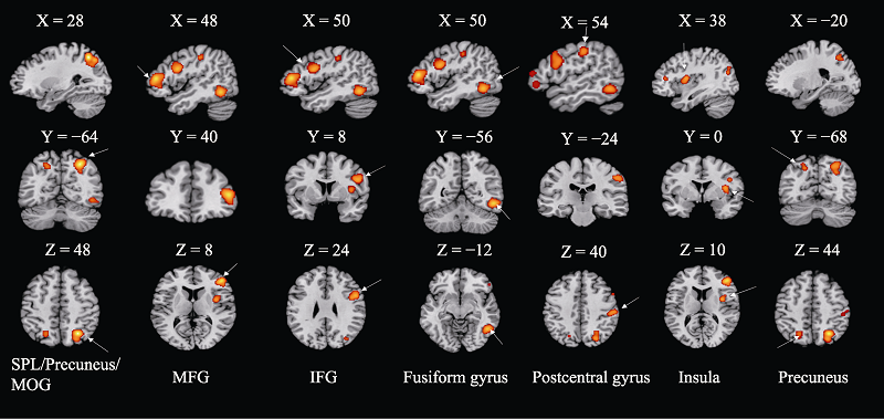
图2 元分析结果图 注:图为元分析结果, 簇群水平的FWE校正, p < 0.05, 图中所激活的脑区达到了显著激活水平。图中所激活的脑区从左到右分别为:右侧顶上小叶(SPL)/楔前叶(Precuneus)/枕中回(MOG)、额中回(MFG)、额下回(IFG)、梭状回(Fusiform Gyrus)、中央后回(Postcentral Gyrus)、脑岛(Insula)和左侧楔前叶。
| 脑区 | 半球 | BA区 | 中心坐标 | 体积 (mm3) | ALE值 (×10-2) | ||
|---|---|---|---|---|---|---|---|
| x | y | z | |||||
| 顶上小叶/ | 右 | 7 | 28 | -64 | 48 | 5208 | 4.33 |
| 楔前叶/ | 7 | 26 | -72 | 44 | 3.51 | ||
| 枕中回 | 19 | 38 | -76 | 22 | 1.77 | ||
| 额中回 | 右 | 46 | 48 | 40 | 8 | 4256 | 3.87 |
| 额下回 | 右 | 9 | 50 | 8 | 24 | 3376 | 3.53 |
| 梭状回 | 右 | 37 | 50 | -56 | -12 | 2880 | 3.58 |
| 中央后回 | 右 | 2 | 54 | -24 | 40 | 1488 | 2.39 |
| 脑岛 | 右 | 13 | 38 | 0 | 10 | 1320 | 2.74 |
| 楔前叶 | 左 | 7 | -20 | -68 | 44 | 856 | 1.84 |
表2 ALE元分析激活簇结果
| 脑区 | 半球 | BA区 | 中心坐标 | 体积 (mm3) | ALE值 (×10-2) | ||
|---|---|---|---|---|---|---|---|
| x | y | z | |||||
| 顶上小叶/ | 右 | 7 | 28 | -64 | 48 | 5208 | 4.33 |
| 楔前叶/ | 7 | 26 | -72 | 44 | 3.51 | ||
| 枕中回 | 19 | 38 | -76 | 22 | 1.77 | ||
| 额中回 | 右 | 46 | 48 | 40 | 8 | 4256 | 3.87 |
| 额下回 | 右 | 9 | 50 | 8 | 24 | 3376 | 3.53 |
| 梭状回 | 右 | 37 | 50 | -56 | -12 | 2880 | 3.58 |
| 中央后回 | 右 | 2 | 54 | -24 | 40 | 1488 | 2.39 |
| 脑岛 | 右 | 13 | 38 | 0 | 10 | 1320 | 2.74 |
| 楔前叶 | 左 | 7 | -20 | -68 | 44 | 856 | 1.84 |
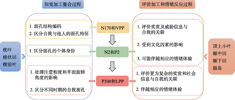
图3 自我面孔识别的知觉加工整合过程及评价加工和情绪反应过程
| *表示元分析用到的文献。 | |
| [1] | 常荷, 董毅, 汪凯, 张凤凤, 李慧, 赵子丛, … 陈学权. (2012). 精神分裂症患者自我面孔识别与自我监控能力的相关性研究. 中国健康心理学杂志, 20(9), 1281-1283. |
| [2] | 高强. (2012). 自闭症儿童自我面孔识别与加工研究 (硕士学位论文). 漳州师范学院. |
| [3] | 关丽丽. (2013). 威胁性信息影响自我面孔优势效应的认知神经机制探究 (硕士学位论文). 西南大学, 重庆. |
| [4] | 关丽丽, 齐铭铭, 侯艳, 杨娟. (2011). 自我面孔识别的脑机制. 心理科学进展, 19(9), 1313-1318. |
| [5] | 关丽丽, 张庆林, 齐铭铭, 侯燕, 杨娟. (2012). 自我概念威胁以及与重要他人的比较共同削弱自我面孔优势效应. 心理学报, 44(6), 789-796. |
| [6] | 贾竑晓, 尹梦雅, 朱虹. (2013). 躁狂发作患者的自我面孔识别速度研究. 首都医科大学学报, 34(2), 204-206. |
| [7] | 雷旭, 尧德中. (2014). 同步脑电-功能磁共振(EEG-fMRI)原理与技术. 科学出版社. |
| [8] | 李稳. (2009). 探讨区分自我-他人面孔的神经基础 (硕士学位论文). 首都师范大学, 北京. |
| [9] | 刘珊珊, 马辛, 尹梦雅, 朱虹, 赵永厚, Nelson, B., … 贾竑晓. (2014). 神经心理学损害: 重性抑郁障碍患者自我加工效应的异常. 科学通报, 59(13), 1223-1229. |
| [10] | 谭群, 尹月阳, 刘燊, 韩尚锋, 徐强, 张林. (2018). 自我积极表情加工优势效应:来自ERPs的证据. 心理学报, 50(10), 1120-1130. |
| [11] | 徐蕊, 杨敬铭, 朱虹, 刘珊珊, 贾竑晓. (2017). 人格解体障碍病人的自我面孔识别速度的研究. 首都医科大学学报, 38(2), 192-196. |
| [12] | 徐圆圆, 周菘, 朱虹, 耿海燕, 贾竑晓. (2018). 精神分裂症自知力与无意识自我面孔识别的关系. 首都医科大学学报, 39(2), 209-212. |
| [13] | 杨敬铭, 刘珊珊, 贾竑晓, 朱虹, 尹梦雅, 刘嘉. (2014). 强迫性障碍患者对自我和名人面孔识别速度异常的实验研究. 中国心理卫生杂志, 28(5), 339-343. |
| [14] | 尹梦雅, 贾竑晓, 朱虹, 刘嘉. (2013). 抑郁症患者的自我面孔识别研究. 首都医科大学学报, 34(2), 207-210. |
| [15] | 张兆青. (2014). 恋人概念威胁对面孔优势效应的影响 (硕士学位论文). 天津师范大学. |
| [16] | 郑莹灿. (2015). 重要他人概念威胁影响自我面孔识别优势 (硕士学位论文). 西南大学, 重庆. |
| [17] | 钟毅平, 李琎, 占友龙, 范伟, 杨子鹿. (2016). 自我旋转面孔识别的ERPs研究. 心理学报, 48(11), 1379-1389. |
| [18] | 周爱保, 谢佩, 潘超超, 田喆, 谢君伟, 刘炯. (2020). 寻找丢失的自我——精神分裂症患者的自我面孔识别. 心理学报, 52(2), 184-196. |
| [19] | 朱虹, 贾竑晓. (2014). 以阴性与阳性症状为主的精神分裂症患者的自我面孔识别速度研究. 首都医科大学学报, 35(2), 200-204. |
| [20] | Alarcon, G., Sauder, M., Teoh, J. Y., Forbes, E. E., & Quevedo, K. (2019). Amygdala functional connectivity during self-face processing in depressed adolescents with recent suicide attempt. Journal of the American Academy of Child and Adolescent Psychiatry, 58(2), 221-231. |
| [21] | Alzueta, E., Melcon, M., Jensen, O., & Capilla, A. (2020). The 'Narcissus Effect': Top-down alpha-beta band modulation of face-related brain areas during self-face processing. Neuroimage, 213, Article 116754. |
| [22] |
Alzueta, E., Melcon, M., Poch, C., & Capilla, A. (2019). Is your own face more than a highly familiar face? Biological Psychology, 142, 100-107.
doi: 10.1016/j.biopsycho.2019.01.018 URL |
| [23] |
* Apps, M. A. J., Tajadura-Jimenez, A., Turley, G., & Tsakiris, M. (2012). The different faces of one's self: An fMRI study into the recognition of current and past self-facial appearances. Neuroimage, 63(3), 1720-1729.
doi: 10.1016/j.neuroimage.2012.08.053 URL |
| [24] |
* Arzy, S., Collette, S., Ionta, S., Fornari, E., & Blanke, O. (2009). Subjective mental time: The functional architecture of projecting the self to past and future. European Journal of Neuroscience, 30(10), 2009-2017.
doi: 10.1111/ejn.2009.30.issue-10 URL |
| [25] |
Bortolon, C., Capdevielle, D., Altman, R., Macgregor, A., Attal, J., & Raffard, S. (2017). Mirror self-face perception in individuals with schizophrenia: Feelings of strangeness associated with one's own image. Psychiatry Research, 253, 205-210.
doi: 10.1016/j.psychres.2017.03.055 URL |
| [26] |
Bortolon, C., Capdevielle, D., Salesse, R. N., & Raffard, S. (2016a). Further insight into self-face recognition in schizophrenia patients: Why ambiguity matters. Journal of Behavior Therapy and Experimental Psychiatry, 50, 215-222.
doi: 10.1016/j.jbtep.2015.09.006 URL |
| [27] | Bortolon, C., Capdevielle, D., Salesse, R. N., & Raffard, S. (2016b). Self-face recognition in schizophrenia: Preliminary eye-tracking study. Frontiers in Human Neuroscience, 10, Article 3. |
| [28] |
Cassia, V. M., Turati, C., & Simion, F. (2004). Can a nonspecific bias toward top-heavy patterns explain newborns' face preference? Psychological Science, 15, 379-383.
doi: 10.1111/j.0956-7976.2004.00688.x URL |
| [29] | Chakraborty, A., & Chakrabarti, B. (2018). Looking at my own face: Visual processing strategies in self-other face recognition. Frontiers in Psychology, 9, Article 121. |
| [30] |
Chein, J. M., Fissell, K., Jacobs, S., & Fiez, J. A. (2002). Functional heterogeneity within Broca's area during verbal working memory. Physiology & Behavior, 77, 635-639.
doi: 10.1016/S0031-9384(02)00899-5 URL |
| [31] |
* Devue, C., Collette, F., Balteau, E., Dequeldre, C., Luxen, A., Maquet, P., & Bredart, S. (2007). Here I am: The cortical correlates of visual self-recognition. Brain Research, 1143, 169-182.
doi: 10.1016/j.brainres.2007.01.055 URL |
| [32] | Dor-Ziderman, Y., Lutz, A., & Goldstein, A. (2019). Prediction-based neural mechanisms for shielding the self from existential threat. Neuroimage, 202, Article 116080. |
| [33] |
Eickhoff, S. B., Laird, A. R., Grefkes, C., Wang, L. E., Zilles, K., & Fox, P. T. (2009). Coordinate-based activation likelihood estimation meta-analysis of neuroimaging data: A random-effects approach based on empirical estimates of spatial uncertainty. Human Brain Mapping, 30(9), 2907-2926.
doi: 10.1002/hbm.20718 pmid: 19172646 |
| [34] |
Geng, H., Zhang, S., Li, Q., Tao, R., & Xu, S. (2012). Dissociations of subliminal and supraliminal self-face from other-face processing: Behavioral and ERP evidence. Neuropsychologia, 50(12), 2933-2942.
doi: 10.1016/j.neuropsychologia.2012.07.040 URL |
| [35] |
Gobbini, M. I., & Haxby, J. V. (2007). Neural systems for recognition of familiar faces. Neuropsychologia, 45(1), 32-41.
pmid: 16797608 |
| [36] | Gonzalez-Franco, M., Bellido, A. I., Blom, K. J., Slater, M., & Rodriguez-Fornells, A. (2016). The neurological traces of look-alike avatars. Frontiers in Human Neuroscience, 10, Article 392. |
| [37] |
Guan, L. L., Chen, Y., Xu, X. F., Qiao, L., Wei, J., Han, S. H., … Liu, Y. J. (2015). Self-esteem buffers the mortality salience effect on the implicit self-face processing. Personality and Individual Differences, 85, 77-85.
doi: 10.1016/j.paid.2015.04.032 URL |
| [38] |
Guan, L. L., Qi, M., Li, H., Hitchman, G., Yang, J., & Liu, Y. (2015). Priming with threatening faces modulates the self-face advantage by enhancing the other-face processing rather than suppressing the self-face processing. Brain Research, 1608, 97-107.
doi: 10.1016/j.brainres.2015.03.002 URL |
| [39] |
Guan, L. L., Qi, M., Zhang, Q., & Yang, J. (2014). The neural basis of self-face recognition after self-concept threat and comparison with important others. Social Neuroscience, 9(4), 424-435.
doi: 10.1080/17470919.2014.920417 URL |
| [40] | * Guan, L. L., Wu, T., Yang, J., Xie, X., Han, S., & Zhao, Y. (2020). Self-esteem and cultural worldview buffer mortality salience effects on responses to self-face: Distinct neural mediators. Biological Psychology, 155, Article 107944. |
| [41] | Guan, L. L., Zhao, Y., Wang, Y., Chen, Y., & Yang, J. (2017). Self-esteem modulates the P3 component in response to the self-face processing after priming with emotional faces. Frontiers in Psychology, 8, Article 1399. |
| [42] |
Hirot, F., Lesage, M., Pedron, L., Meyer, I., Thomas, P., Cottencin, O., & Guardia, D. (2016). Impaired processing of self-face recognition in anorexia nervosa. Eating and Weight Disorders-Studies on Anorexia Bulimia and Obesity, 21(1), 31-40.
doi: 10.1007/s40519-015-0223-y URL |
| [43] |
Joyce, C., & Rossion, B. (2005). The face-sensitive N170 and VPP components manifest the same brain processes: The effect of reference electrode site. Clinical Neurophysiology, 116(11), 2613-2631.
doi: 10.1016/j.clinph.2005.07.005 URL |
| [44] |
Kaplan, J. T., Aziz-Zadeh, L., Uddin, L. Q., & Iacoboni, M. (2008). The self across the senses: An fMRI study of self-face and self-voice recognition. Social Cognitive and Affective Neuroscience, 3(3), 218-223.
doi: 10.1093/scan/nsn014 pmid: 19015113 |
| [45] |
Keyes, H., Brady, N., Reilly, R. B., & Foxe, J. J. (2010). My face or yours? Event-related potential correlates of self-face processing. Brain and Cognition, 72(2), 244-254.
doi: 10.1016/j.bandc.2009.09.006 URL |
| [46] |
Kim, M.-K., Yoon, H.-J., Shin, Y.-B., Lee, S.-K., & Kim, J.-J. (2016). Neural basis of distorted self-face recognition in social anxiety disorder. Neuroimage-Clinical, 12, 956-964.
doi: 10.1016/j.nicl.2016.04.010 URL |
| [47] |
* Kircher, T. T., Senior, C., Phillips, M. L., Benson, P. J., Bullmore, E. T., Brammer, M., … David, A. S. (2000). Towards a functional neuroanatomy of self processing: Effects of faces and words. Cognitive Brain Research, 10(1), 133-144.
doi: 10.1016/S0926-6410(00)00036-7 URL |
| [48] |
* Kircher, T. T., Senior, C., Phillips, M. L., Rabe-Hesketh, S., Benson, P. J., Bullmore, E. T., … David, A. S. (2001). Recognizing one's own face. Cognition, 78(1), 1-15.
pmid: 11062324 |
| [49] |
Ma, Y., & Han, S. (2009). Self-face advantage is modulated by social threat - Boss effect on self-face recognition. Journal of Experimental Social Psychology, 45(4), 1048-1051.
doi: 10.1016/j.jesp.2009.05.008 URL |
| [50] |
Ma, Y., & Han, S. (2010). Why we respond faster to the self than to others? An implicit positive association theory of self-advantage during implicit face recognition. Journal of Experimental Psychology: Human Perception and Performance, 36(3), 619-633.
doi: 10.1037/a0015797 URL |
| [51] |
* Ma, Y., & Han, S. (2012). Functional dissociation of the left and right fusiform gyrus in self-face recognition. Human Brain Mapping, 33(10), 2255-2267.
doi: 10.1002/hbm.v33.10 URL |
| [52] |
Mele, G., Cavaliere, C., Alfano, V., Orsini, M., Salvatore, M., & Aiello, M. (2019). Simultaneous EEG-fMRI for functional neurological assessment. Frontiers in Neurology, 10, 848-859.
doi: 10.3389/fneur.2019.00848 URL |
| [53] |
Menon, V., & Uddin, L. Q. (2010). Saliency, switching, attention and control: A network model of insula function. Brain Structure and Function, 214(5), 655-667.
doi: 10.1007/s00429-010-0262-0 URL |
| [54] |
* Morita, T., Asada, M., & Naito, E. (2020). Right-hemispheric dominance in self-body recognition is altered in left-handed individuals. Neuroscience, 425, 68-89.
doi: 10.1016/j.neuroscience.2019.10.056 URL |
| [55] |
* Morita, T., Itakura, S., Saito, D. N., Nakashita, S., Harada, T., Kochiyama, T., & Sadato, N. (2008). The role of the right prefrontal cortex in self-evaluation of the face: A functional magnetic resonance imaging study. Journal of Cognitive Neuroscience, 20(2), 342-355.
doi: 10.1162/jocn.2008.20024 URL |
| [56] |
Morita, T., Kosaka, H., Saito, D. N., Fujii, T., Ishitobi, M., Munesue, T., … Sadato, N. (2016). Neural correlates of emotion processing during observed self-face recognition in individuals with autism spectrum disorders. Research in Autism Spectrum Disorders, 26, 16-32.
doi: 10.1016/j.rasd.2016.02.011 URL |
| [57] |
Morita, T., Kosaka, H., Saito, D. N., Ishitobi, M., Munesue, T., Itakura, S., … Sadato, N. (2012). Emotional responses associated with self-face processing in individuals with autism spectrum disorders: An fMRI study. Social Neuroscience, 7(3), 223-239.
doi: 10.1080/17470919.2011.598945 URL |
| [58] |
* Morita, T., Saito, D. N., Ban, M., Shimada, K., Okamoto, Y., Kosaka, H., … Naito, E. (2017). Self-face recognition shares brain regions active during proprioceptive illusion in the right inferior fronto-parietal superior longitudinal fasciculus III network. Neuroscience, 348, 288-301.
doi: 10.1016/j.neuroscience.2017.02.031 URL |
| [59] |
* Morita, T., Saito, D. N., Ban, M., Shimada, K., Okamoto, Y., Kosaka, H., … Naito, E. (2018). Self-face recognition begins to share active region in right inferior parietal lobule with proprioceptive illusion during adolescence. Cerebral Cortex, 28(4), 1532-1548.
doi: 10.1093/cercor/bhy027 URL |
| [60] |
* Morita, T., Tanabe, H. C., Sasaki, A. T., Shimada, K., Kakigi, R., & Sadato, N. (2014). The anterior insular and anterior cingulate cortices in emotional processing for self-face recognition. Social Cognitive and Affective Neuroscience, 9(5), 570-579.
doi: 10.1093/scan/nst011 URL |
| [61] |
Northoff, G., Heinzel, A., de Greck, M., Bermpohl, F., Dobrowolny, H., & Panksepp, J. (2006). Self-referential processing in our brain--a meta-analysis of imaging studies on the self. Neuroimage, 31(1), 440-457.
pmid: 16466680 |
| [62] |
* Oikawa, H., Sugiura, M., Sekiguchi, A., Tsukiura, T., Miyauchi, C. M., Hashimoto, T., … Kawashima, R. (2012). Self-face evaluation and self-esteem in young females: An fMRI study using contrast effect. Neuroimage, 59(4), 3668-3676.
doi: 10.1016/j.neuroimage.2011.10.098 URL |
| [63] | Panagiotopoulou, E., Filippetti, M. L., Tsakiris, M., & Fotopoulou, A. (2017). Affective touch enhances self-face recognition during multisensory integration. Scientific Reports, 7, Article 12883. |
| [64] |
Pitcher, D., Walsh, V., & Duchaine, B. (2011). The role of the occipital face area in the cortical face perception network. Experiment Brain Research, 209(4), 481-493.
doi: 10.1007/s00221-011-2579-1 URL |
| [65] |
* Platek, S. M., Keenan, J. P., Gallup, G. G., & Mohamed, F. B. (2004). Where am I? The neurological correlates of self and other. Cognitive Brain Research, 19(2), 114-122.
doi: 10.1016/j.cogbrainres.2003.11.014 URL |
| [66] |
* Platek, S. M., Keenan, J. P., & Mohamed, F. B. (2005). Sex differences in the neural correlates of child facial resemblance: An event-related fMRI study. Neuroimage, 25(4), 1336-1344.
doi: 10.1016/j.neuroimage.2004.12.037 URL |
| [67] |
* Platek, S. M., & Kemp, S. M. (2009). Is family special to the brain? An event-related fMRI study of familiar, familial, and self-face recognition. Neuropsychologia, 47(3), 849-858.
doi: 10.1016/j.neuropsychologia.2008.12.027 URL |
| [68] |
Platek, S. M., Krill, A. L., & Kemp, S. M. (2008). The neural basis of facial resemblance. Neuroscience Letters, 437(2), 76-81.
doi: 10.1016/j.neulet.2008.03.040 URL |
| [69] |
Platek, S. M., Krill, A. L., & Wilson, B. (2009). Implicit trustworthiness ratings of self-resembling faces activate brain centers involved in reward. Neuropsychologia, 47(1), 289-293.
doi: 10.1016/j.neuropsychologia.2008.07.018 URL |
| [70] |
* Platek, S. M., Loughead, J. W., Gur, R. C., Busch, S., Ruparel, K., Phend, N., … Langleben, D. D. (2006). Neural substrates for functionally discriminating self-face from personally familiar faces. Human Brain Mapping, 27(2), 91-98.
doi: 10.1002/(ISSN)1097-0193 URL |
| [71] |
Quevedo, K., Harms, M., Sauder, M., Scott, H., Mohamed, S., Thomas, K. M., … Smyda, G. (2018). The neurobiology of self face recognition among depressed adolescents. Journal of Affective Disorders, 229, 22-31.
doi: S0165-0327(17)31787-1 pmid: 29304386 |
| [72] | Quevedo, K., Liu, G., Teoh, J. Y., Ghosh, S., Zeffiro, T., Ahrweiler, N., … Paret, C. (2019). Neurofeedback and neuroplasticity of visual self-processing in depressed and healthy adolescents: A preliminary study. Developmental Cognitive Neuroscience, 40, Article 100707. |
| [73] |
Quevedo, K., Ng, R., Scott, H., Martin, J., Smyda, G., Keener, M., & Oppenheimer, C. W. (2016). The neurobiology of self-face recognition in depressed adolescents with low or high suicidality. Journal of Abnormal Psychology, 125(8), 1185-1200.
pmid: 27618278 |
| [74] |
Rossion, B., Campanella, S., Gomez, C., Delinte, A., Debatisse, D., Liard, L., … Guerit, J.-M. (1999). Task modulation of brain activity related to familiar and unfamiliar face processing: An ERP study. Clinical Neurophysiology, 110(3), 449-462.
pmid: 10363769 |
| [75] |
Rousselet, G. A., Macé, M. J.-M., & Fabre-Thorpe, M. (2003). Is it an animal? Is it a human face? Fast processing in upright and inverted natural scenes. Journal of Vision, 3(6), 440-455.
doi: 10.1167/3.9.440 URL |
| [76] | Rubianes, M., Munoz, F., Casado, P., Hernandez-Gutierrez, D., Jimenez-Ortega, L., Fondevila, S., … Martin-Loeches, M. (2021). Am I the same person across my life span? An event-related brain potentials study of the temporal perspective in self-identity. Psychophysiology, 58(1), Article e13692. |
| [77] | * Scheepers, D., Derks, B., Nieuwenhuis, S., Lelieveld, G.-J., van Nunspeet, F., Rombouts, S. A. R. B., & de Rover, M. (2013). The neural correlates of in-group and self-face perception: Is there overlap for high identifiers? Frontiers in Human Neuroscience, 7, Article 528. |
| [78] |
Seeley, W. W., Menon, V., Schatzberg, A. F., Keller, J., Glover, G. H., Kenna, H., … Greicius, M. D. (2007). Dissociable intrinsic connectivity networks for salience processing and executive control. Journal of Neuroscience, 27(9), 2349-2356.
doi: 10.1523/JNEUROSCI.5587-06.2007 URL |
| [79] | Sel, A., Azevedo, R. T., & Tsakiris, M. (2017). Heartfelt self: Cardio-visual integration affects self-face recognition and interoceptive cortical processing. Cerebral Cortex, 27(11), 5144-5155. |
| [80] |
Sereno, M. I., & Huang, R.-S. (2006). A human parietal face area contains aligned head-centered visual and tactile maps. Nature Neuroscience, 9(10), 1337-1343.
doi: 10.1038/nn1777 URL |
| [81] |
* Sugiura, M., Miyauchi, C. M., Kotozaki, Y., Akimoto, Y., Nozawa, T., Yomogida, Y., … Kawashima, R. (2014). Neural mechanism for mirrored self-face recognition. Cerebral Cortex, 25(9), 2806-2814.
doi: 10.1093/cercor/bhu077 URL |
| [82] |
* Sugiura, M., Sassa, Y., Jeong, H., Horie, K., Sato, S., & Kawashima, R. (2008). Face-specific and domain-general characteristics of cortical responses during self-recognition. Neuroimage, 42(1), 414-422.
doi: 10.1016/j.neuroimage.2008.03.054 URL |
| [83] |
* Sugiura, M., Sassa, Y., Jeong, H., Wakusawa, K., Horie, K., Sato, S., & Kawashima, R. (2012). Self-face recognition in social context. Human Brain Mapping, 33(6), 1364-1374.
doi: 10.1002/hbm.v33.6 URL |
| [84] |
* Sugiura, M., Sassa, Y., jeong, H. J., Miura, N., Akitsuki, Y., Horie, K., … Kawashima, R. (2006). Multiple brain networks for visual self-recognition with different sensitivity for motion and body part. Neuroimage, 32(4), 1905-1917.
doi: 10.1016/j.neuroimage.2006.05.026 URL |
| [85] |
* Sugiura, M., Watanabe, J., Maeda, Y., Matsue, Y., Fukuda, H., & Kawashima, R. (2005). Cortical mechanisms of visual self-recognition. Neuroimage, 24(1), 143-149.
doi: 10.1016/j.neuroimage.2004.07.063 URL |
| [86] |
* Sui, J., & Han, S. (2007). Self-construal priming modulates neural substrates of self-awareness. Psychological Science, 18, 861-866.
doi: 10.1111/j.1467-9280.2007.01992.x URL |
| [87] |
Sui, J., Hong, Y.-Y., Liu, C. H., Humphreys, G. W., & Han, S. (2013). Dynamic cultural modulation of neural responses to one's own and friend's faces. Social Cognitive and Affective Neuroscience, 8(3), 326-332.
doi: 10.1093/scan/nss001 URL |
| [88] |
Sui, J., Liu, C. H., & Han, S. (2009). Cultural difference in neural mechanisms of self-recognition. Social Neuroscience, 4(5), 402-411.
doi: 10.1080/17470910802674825 URL |
| [89] |
Tacikowski, P., & Nowicka, A. (2010). Allocation of attention to self-name and self-face: An ERP study. Biological Psychology, 84(2), 318-324.
doi: 10.1016/j.biopsycho.2010.03.009 pmid: 20298741 |
| [90] |
Tanaka, J. W., Curran, T., Porterfield, A. L., & Collins, D. (2006). Activation of preexisting and acquired face representations: The N250 event-related potential as an index of face familiarity. Journal of Cognitive Neuroscience, 18, 1488-1497.
pmid: 16989550 |
| [91] |
* Taylor, M. J., Arsalidou, M., Bayless, S. J., Morris, D., Evans, J. W., & Barbeau, E. J. (2009). Neural correlates of personally familiar faces: Parents, partner and own faces. Human Brain Mapping, 30(7), 2008-2020.
doi: 10.1002/hbm.v30:7 URL |
| [92] |
Turkeltaub, P. E., Eickhoff, S. B., Laird, A. R., Fox, M., Wiener, M., & Fox, P. (2012). Minimizing within-experiment and within-group effects in Activation Likelihood Estimation meta-analyses. Human Brain Mapping, 33(1), 1-13.
doi: 10.1002/hbm.21186 pmid: 21305667 |
| [93] |
* Uddin, L. Q., Kaplan, J. T., Molnar-Szakacs, I., Zaidel, E., & Iacoboni, M. (2005). Self-face recognition activates a frontoparietal "mirror" network in the right hemisphere: An event-related fMRI study. Neuroimage, 25(3), 926-935.
doi: 10.1016/j.neuroimage.2004.12.018 URL |
| [94] |
* Verosky, S. C., & Todorov, A. (2010). Differential neural responses to faces physically similar to the self as a function of their valence. Neuroimage, 49(2), 1690-1698.
doi: 10.1016/j.neuroimage.2009.10.017 pmid: 19837179 |
| [95] |
Yamawaki, R., Nakamura, K., Aso, T., Shigemune, Y., Fukuyama, H., & Tsukiura, T. (2017). Remembering my friends: Medial prefrontal and hippocampal contributions to the self-reference effect on face memories in a social context. Human Brain Mapping, 38(8), 4256-4269.
doi: 10.1002/hbm.23662 pmid: 28548263 |
| [96] | Zhan, Y. L., Chen, J., Xiao, X., Li, J., Yang, Z. L., Fan, W., & Zhong, Y. P. (2016). Reward promotes self-face processing: An event-related potential study. Frontiers in Psychology, 7, Article 735. |
| [97] | Zhan, Y. L., Xiao, X., Chen, J., Li, J., Fan, W., & Zhong, Y. P. (2017). Consciously over unconsciously perceived rewards facilitate self-face processing: An ERP study. Scientific Reports, 7, Article 7836. |
| [98] | Zhou, S., Xu, Y. Y., Wang, N. B., Zhang, S., Geng, H. Y., & Jia, H. X. (2020). Deficits of subliminal self-face processing in schizophrenia. Consciousness and Cognition, 79, Article 102896. |
| [1] | 程晓荣, 仇式明, 定险峰, 范炤. 动作如何影响元认知?——基于认知模型和神经机制的探讨[J]. 心理科学进展, 2025, 33(3): 425-438. |
| [2] | 巩芳颍, 孙逸梵, 贺琴, 石可, 刘伟, 陈宁. 教学互动中师生脑间同步性及其调节因素[J]. 心理科学进展, 2025, 33(3): 452-464. |
| [3] | 夏熠, 张婕, 张火垠, 雷怡, 窦皓然. 焦虑个体趋避冲突失调的认知神经机制[J]. 心理科学进展, 2025, 33(3): 477-493. |
| [4] | 刘月月, 何文广. 书写认知老化发生机制及神经机理[J]. 心理科学进展, 2024, 32(9): 1502-1513. |
| [5] | 雷怡, 梅颖, 王金霞, 袁子昕. 焦虑青少年无意识恐惧的神经机制及干预[J]. 心理科学进展, 2024, 32(8): 1221-1232. |
| [6] | 丁颖, 汪紫滢, 李卫东. 抑郁症疼痛加工的行为特点及神经机制[J]. 心理科学进展, 2024, 32(8): 1315-1327. |
| [7] | 曾庆贺, 崔晓宇, 唐为, 李娟. 记忆辨别力受老化影响的认知神经机制及其应用[J]. 心理科学进展, 2024, 32(7): 1138-1151. |
| [8] | 刘海宁, 董现玲, 刘海虹, 刘艳丽, 李现文. 老年遗忘型轻度认知障碍执行功能的神经机制及数字干预[J]. 心理科学进展, 2024, 32(6): 873-885. |
| [9] | 冯攀, 赵恒越, 姜雨矇, 张悦彤, 冯廷勇. 催产素影响条件化恐惧情绪加工的认知机制及神经基础[J]. 心理科学进展, 2024, 32(4): 557-567. |
| [10] | 郑好, 陈荣荣, 买晓琴. 第三方惩罚行为的认知神经机制[J]. 心理科学进展, 2024, 32(2): 398-412. |
| [11] | 孙丽君, 杨玉芳. 预期视角下音乐节拍结构的认知与神经机制[J]. 心理科学进展, 2024, 32(10): 1567-1577. |
| [12] | 潘晗希, 陈泽锋, 许楠, 高在峰. 社会工作记忆的脑机制:来自fMRI的证据[J]. 心理科学进展, 2023, 31(suppl.): 107-107. |
| [13] | 龚政鑫, 周明, 戴宇萱, 文雨珊, 甄宗雷. 真实场景视觉加工的功能核磁共振大型公开数据集[J]. 心理科学进展, 2023, 31(suppl.): 156-156. |
| [14] | 周明, 龚政鑫, 戴宇萱, 文雨珊, 刘友谊, 甄宗雷. 自然场景下人类动作识别的大规模功能核磁共振数据集[J]. 心理科学进展, 2023, 31(suppl.): 165-165. |
| [15] | 杨以诺, 刘星宇, 甄宗雷. 基于自然视频刺激的多维度动作表征空间研究[J]. 心理科学进展, 2023, 31(suppl.): 166-166. |
| 阅读次数 | ||||||
|
全文 |
|
|||||
|
摘要 |
|
|||||