ISSN 0439-755X
CN 11-1911/B
CN 11-1911/B
Acta Psychologica Sinica ›› 2021, Vol. 53 ›› Issue (1): 38-54.doi: 10.3724/SP.J.1041.2021.00038
• Reports of Empirical Studies • Previous Articles Next Articles
JIN Hua1,2,3( ), LIANG Ziping1,2,3, ZHU Ziliang1,2,3, YAN Shizhen1,2,3, LIN Lin1,2,3, AISIKAER Aikedan4, YIN Jianzhong4, JIANG Yunpeng1,2,3, TIAN Xin5
Received:2020-06-05
Published:2021-01-25
Online:2020-11-24
Contact:
JIN Hua
E-mail:jinhua@tjnu.edu.cn
Supported by:JIN Hua, LIANG Ziping, ZHU Ziliang, YAN Shizhen, LIN Lin, AISIKAER Aikedan, YIN Jianzhong, JIANG Yunpeng, TIAN Xin. (2021). Aging of global motion perception is accompanied by the changes of resting-state functional activity in the middle temporal gyrus. Acta Psychologica Sinica, 53(1), 38-54.
Add to citation manager EndNote|Ris|BibTeX
URL: https://journal.psych.ac.cn/acps/EN/10.3724/SP.J.1041.2021.00038
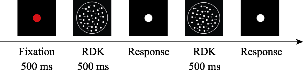
Figure 1. Experimental procedure for determining the global motion direction.
| AAL | BA | Cluster size | Peak t value | MNI coordination (x, y, z) | |||
|---|---|---|---|---|---|---|---|
| ReHo | Old > young | Cerebelum_6_L | 19 | visual associated cortex | 44 | 5.82 | -18, -60, -24 |
| Cerebelum_6_R | 18 | secondary visual cortex | 32 | 4.30 | 10, -62, -22 | ||
| Lingual_R | 19 | visual associated cortex | 38 | 4.37 | 22, -60, -4 | ||
| Old < young | Occipital_Inf_R | 19 | visual associated cortex | 783 | -10.34 | 36, -86, -2 | |
| Lingual_R | 19 | visual associated cortex | 712 | ||||
| Lingual_L | 19 | visual associated cortex | 670 | ||||
| Occipital_Inf_L | 19 | visual associated cortex | 709 | ||||
| Temporal_Mid_R | 21 | middle temporal gyrus | 916 | -6.40 | 50, -56, 20 | ||
| Temporal_Mid_L | 21 | middle temporal gyrus | 126 | -5.71 | -66, -28, -6 | ||
| ALFF | Old < young | Temporal_Sup_R | 21 | middle temporal gyrus | 208 | -8.74 | 50, -56, 22 |
| Temporal_Mid_L | 21 | middle temporal gyrus | 1371 | -9.67 | -58, -54, 8 | ||
Table 1 The brain areas that showed significant group differences in ReHo and ALFF
| AAL | BA | Cluster size | Peak t value | MNI coordination (x, y, z) | |||
|---|---|---|---|---|---|---|---|
| ReHo | Old > young | Cerebelum_6_L | 19 | visual associated cortex | 44 | 5.82 | -18, -60, -24 |
| Cerebelum_6_R | 18 | secondary visual cortex | 32 | 4.30 | 10, -62, -22 | ||
| Lingual_R | 19 | visual associated cortex | 38 | 4.37 | 22, -60, -4 | ||
| Old < young | Occipital_Inf_R | 19 | visual associated cortex | 783 | -10.34 | 36, -86, -2 | |
| Lingual_R | 19 | visual associated cortex | 712 | ||||
| Lingual_L | 19 | visual associated cortex | 670 | ||||
| Occipital_Inf_L | 19 | visual associated cortex | 709 | ||||
| Temporal_Mid_R | 21 | middle temporal gyrus | 916 | -6.40 | 50, -56, 20 | ||
| Temporal_Mid_L | 21 | middle temporal gyrus | 126 | -5.71 | -66, -28, -6 | ||
| ALFF | Old < young | Temporal_Sup_R | 21 | middle temporal gyrus | 208 | -8.74 | 50, -56, 22 |
| Temporal_Mid_L | 21 | middle temporal gyrus | 1371 | -9.67 | -58, -54, 8 | ||

Figure 2. The brain areas that showed significant group differences in regional functional activity: ReHo (left) and ALFF (right).
| Seed ROI | AAL | BA | Cluster size | Peak t value | MNI coordinate (x, y, z) | ||
|---|---|---|---|---|---|---|---|
| V1 | Old > young | Paracentral_Lobule_R | 4 | primary motor cortex | 721 | 6.95 | 4, -28, 66 |
| V2 | Old > young | Paracentral_Lobule_L | 4 | primary motor cortex | 1174 | 6.51 | -8, -22, 60 |
| V3 | Old > young | Calcarine_L | 18 | secondary visual cortex | 315 | 6.43 | -26, -64, 12 |
| Old < young | Occipital_Sup_R | 19 | visual associated cortex | 129 | -6.91 | 28, -76, 20 | |
| MT/V5 | Old > young | Supp_Motor_Area_L | 6 | pre-motor and supple-mentary motor cortex | 1347 | 7.13 | -8, -12, 56 |
| Old < young | Angular_R | 39 | angular gyrus | 72 | -6.32 | 42, -52, 24 | |
Table 2 The brain areas that showed significant group differences in voxel-wise FC
| Seed ROI | AAL | BA | Cluster size | Peak t value | MNI coordinate (x, y, z) | ||
|---|---|---|---|---|---|---|---|
| V1 | Old > young | Paracentral_Lobule_R | 4 | primary motor cortex | 721 | 6.95 | 4, -28, 66 |
| V2 | Old > young | Paracentral_Lobule_L | 4 | primary motor cortex | 1174 | 6.51 | -8, -22, 60 |
| V3 | Old > young | Calcarine_L | 18 | secondary visual cortex | 315 | 6.43 | -26, -64, 12 |
| Old < young | Occipital_Sup_R | 19 | visual associated cortex | 129 | -6.91 | 28, -76, 20 | |
| MT/V5 | Old > young | Supp_Motor_Area_L | 6 | pre-motor and supple-mentary motor cortex | 1347 | 7.13 | -8, -12, 56 |
| Old < young | Angular_R | 39 | angular gyrus | 72 | -6.32 | 42, -52, 24 | |
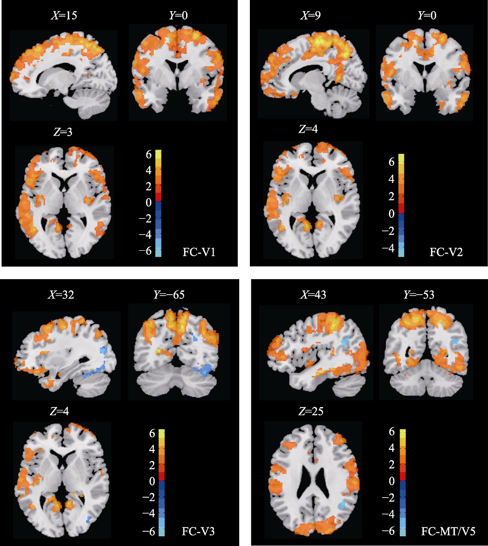
Figure 3. The brain areas that showed significant group differences in voxel-wise FC between the two groups.

Figure 4. The brain areas that showed significant group differences in ROI-wise FC between the two groups.
| AAL | BA | t value | |||
|---|---|---|---|---|---|
| K | Enodal | b | |||
| Old > young | |||||
| Calcarine_L | 17/18 | primary/secondary visual cortex | 3.12** | 1.89 | 1.04 |
| Calcarine_R | 17/18 | primary/secondary visual cortex | 4.34*** | 2.91** | 2.59* |
| Cuneus_L | 18/19 | secondary/associated visual cortex | 3.09** | 1.69 | 1.13 |
| Lingual_L | 18/19 | secondary/associated visual cortex | 2.27* | 0.67 | 0.14 |
| Lingual_R | 18/19 | secondary/association visual cortex | 3.47*** | 1.58 | 1.33 |
| Occipital_Mid_L | 19/39 | visual associate cortex/angular gyrus | 2.25* | -0.54 | -2.95** |
| Occipital_Mid_R | 19/39 | visual associate cortex/angular gyrus | 2.39* | 0.37 | -0.42 |
| Occipital_Inf_R | 19 | visual associate cortex | 3.02** | 0.85 | 2.42* |
| Old < young | |||||
| Heschl_R | 48 | retrosubicular area | -2.27* | -3.73*** | -1.26 |
| Temporal_Sup_R | 22 | superior temporal gyrus | -0.50 | -2.01* | -0.33 |
| Temporal_Pole_Sup_L | 38/21 | temporopolar area/middle temporal gyrus | -2.97** | -4.06*** | -3.32** |
| Temporal_Pole_Sup_R | 38/21 | temporopolar area/middle temporal gyrus | -1.79 | -2.92** | -1.97 |
| Temporal_Mid_R | 21/20 | middle/inferior temporal gyrus | 0.18 | -1.50 | -2.28* |
| Temporal_Pole_Mid_L | 38/20/21 | temporopolar area/inferior/middle temporal gyrus | -2.33* | -3.24* | -1.52 |
| Temporal_Pole_Mid_R | 38/20/21 | temporopolar area/inferior/middle temporal gyrus | -2.08* | -2.81** | 0.05 |
| Temporal_Inf_R | 20 | inferior temporal gyrus | 0.27 | -1.35 | -2.07* |
Table 3 Inter-group differences in nodal centralities in ROI regions
| AAL | BA | t value | |||
|---|---|---|---|---|---|
| K | Enodal | b | |||
| Old > young | |||||
| Calcarine_L | 17/18 | primary/secondary visual cortex | 3.12** | 1.89 | 1.04 |
| Calcarine_R | 17/18 | primary/secondary visual cortex | 4.34*** | 2.91** | 2.59* |
| Cuneus_L | 18/19 | secondary/associated visual cortex | 3.09** | 1.69 | 1.13 |
| Lingual_L | 18/19 | secondary/associated visual cortex | 2.27* | 0.67 | 0.14 |
| Lingual_R | 18/19 | secondary/association visual cortex | 3.47*** | 1.58 | 1.33 |
| Occipital_Mid_L | 19/39 | visual associate cortex/angular gyrus | 2.25* | -0.54 | -2.95** |
| Occipital_Mid_R | 19/39 | visual associate cortex/angular gyrus | 2.39* | 0.37 | -0.42 |
| Occipital_Inf_R | 19 | visual associate cortex | 3.02** | 0.85 | 2.42* |
| Old < young | |||||
| Heschl_R | 48 | retrosubicular area | -2.27* | -3.73*** | -1.26 |
| Temporal_Sup_R | 22 | superior temporal gyrus | -0.50 | -2.01* | -0.33 |
| Temporal_Pole_Sup_L | 38/21 | temporopolar area/middle temporal gyrus | -2.97** | -4.06*** | -3.32** |
| Temporal_Pole_Sup_R | 38/21 | temporopolar area/middle temporal gyrus | -1.79 | -2.92** | -1.97 |
| Temporal_Mid_R | 21/20 | middle/inferior temporal gyrus | 0.18 | -1.50 | -2.28* |
| Temporal_Pole_Mid_L | 38/20/21 | temporopolar area/inferior/middle temporal gyrus | -2.33* | -3.24* | -1.52 |
| Temporal_Pole_Mid_R | 38/20/21 | temporopolar area/inferior/middle temporal gyrus | -2.08* | -2.81** | 0.05 |
| Temporal_Inf_R | 20 | inferior temporal gyrus | 0.27 | -1.35 | -2.07* |

Figure 5. The relationships between ReHo and individuals’ MCT.
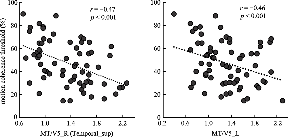
Figure 6. The relationships between ALFF and individuals’ MCT.
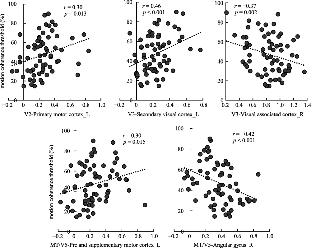
Figure 7. The relationships between voxel-wise FC and individuals’ MCT.
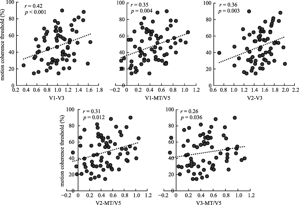
Figure 8. The relationships between ROI-wise FC and individuals’ MCT.
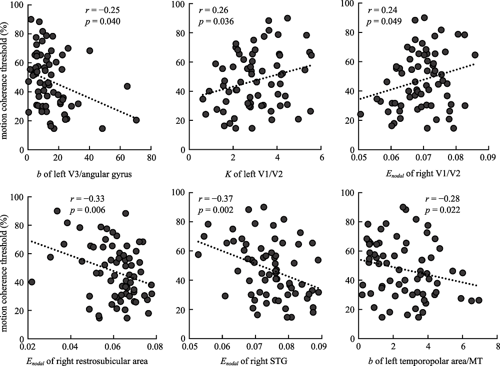
Figure 9. The relationships between nodal centralities in ROI regions and individuals’ MCT.
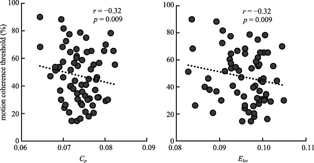
Figure 10. The relationships between global network metrics and individuals’ MCT.
| [1] |
Achard, S., & Bullmore, E. (2007). Efficiency and cost of economical brain functional networks. PLoS Computational Biology, 3(2), e17.
URL pmid: 17274684 |
| [2] |
Ajilore, O., Lamar, M., & Kumar, A. (2014). Association of brain network efficiency with aging, depression, and cognition. The American Journal of Geriatric Psychiatry, 22(2), 102-110.
URL pmid: 24200596 |
| [3] |
Antonenko, D., Meinzer, M., Lindenberg, R., Witte, A. V., & Flöel, A. (2012). Grammar learning in older adults is linked to white matter microstructure and functional connectivity. NeuroImage, 62(3), 1667-1674.
doi: 10.1016/j.neuroimage.2012.05.074 URL |
| [4] |
Barthelemy, M. (2010). Spatial networks. Physics Reports, 499(1-3), 1-101.
doi: 10.1016/j.physrep.2010.11.002 URL |
| [5] |
Bennett, P. J., Sekuler, R., & Sekuler, A. B. (2007). The effects of aging on motion detection and direction identification. Vision Research, 47(6), 799-809.
doi: 10.1016/j.visres.2007.01.001 URL |
| [6] |
Biehl, S. C., Andersen, M., Waiter, G. D., & Pilz, K. S. (2017). Neural changes related to motion processing in healthy aging. Neurobiology of Aging, 57, 162-169.
URL pmid: 28648917 |
| [7] |
Billino, J., & Pilz, K. S. (2019). Motion perception as a model for perceptual aging. Journal of Vision, 19(4), 1-28.
URL pmid: 30933237 |
| [8] | Braddick, O., & Qian, N. (2001). The organization of global motion and transparency. In: J. M. Zanker & J. Zeil (Eds.), Motion Vision (pp. 85-112). Springer Berlin Heidelberg. |
| [9] |
Cabeza, R. (2002). Hemispheric asymmetry reduction in older adults: the HAROLD model. Psychology and Aging, 17(1), 85-100.
doi: 10.1037/0882-7974.17.1.85 URL |
| [10] |
Cai, P., Chen, N., Zhou, T., Thompson, B., & Fang, F. (2014). Global versus local: double dissociation between mt+ and v3a in motion processing revealed using continuous theta burst transcranial magnetic stimulation. Experimental Brain Research, 232(12), 4035-4041.
doi: 10.1007/s00221-014-4084-9 URL |
| [11] |
Cao, M., Wang, J.-H., Dai, Z.-J., Cao, X.-Y., Jiang, L.-L., Fan, F.-M., ... He, Y. (2014). Topological organization of the human brain functional connectome across the lifespan. Developmental Cognitive Neuroscience, 7, 76-93.
doi: 10.1016/j.dcn.2013.11.004 URL |
| [12] |
Chen, N., Cai, P., Zhou, T., Thompson, B., & Fang, F. (2016). Perceptual learning modifies the functional specializations of visual cortical areas. Proceedings of the National Academy of Sciences of the United States of America, 113(20), 5724-5729.
URL pmid: 27051066 |
| [13] |
Damoiseaux, J. S. (2017). Effects of aging on functional and structural brain connectivity. NeuroImage, 160, 32-40.
URL pmid: 28159687 |
| [14] |
Damoiseaux, J. S., Beckmann, C. F., Arigita, E. J. S., Barkhof, F., Scheltens, P., Stam, C. J., ... Rombouts, S. A. R. B. (2008). Reduced resting-state brain activity in the "default network" in normal aging. Cerebral Cortex, 18(8), 1856-1864.
URL pmid: 18063564 |
| [15] |
Farràs-Permanyer, L., Mancho-Fora, N., Montalà-Flaquer, M., Bartrés- Faz, D., Vaqué-Alcázar, L., Peró-Cebollero, M., & Guàrdia-Olmos, J. (2019). Age-related changes in resting-state functional connectivity in older adults. Neural Regeneration Research, 14(9), 1544-1555.
URL pmid: 31089053 |
| [16] |
Ferreira, L. K., & Busatto, G. F. (2013). Resting-state functional connectivity in normal brain aging. Neuroscience & Biobehavioral Reviews, 37(3), 384-400.
URL pmid: 23333262 |
| [17] |
Fountain-Zaragoza, S., Samimy, S., Rosenberg, M. D., & Prakash, R. S. (2019). Connectome-based models predict attentional control in aging adults. Neuroimage, 186, 1-13.
URL pmid: 30394324 |
| [18] |
Friston. K., J. (1994). Functional and effective connectivity in neuroimaging: a synthesis. Human Brain Mapping, 2(1-2), 56-78.
doi: 10.1002/hbm.v2:1/2 URL |
| [19] |
Friston, K. J. (2011). Functional and effective connectivity: a review. Brain Connectivity, 1(1), 13-36.
doi: 10.1089/brain.2011.0008 URL |
| [20] |
Geerligs, L., Renken, R. J., Saliasi, E., Maurits, N. M., & Lorist, M. M. (2015). A brain-wide study of age-related changes in functional connectivity. Cerebral Cortex, 25(7), 1987-1999.
doi: 10.1093/cercor/bhu012 URL |
| [21] |
Grill-Spector, K., & Malach, R. (2004). The human visual cortex. Annual Review of Neuroscience, 27(1), 649-677.
doi: 10.1146/annurev.neuro.27.070203.144220 URL |
| [22] |
He, Y., D……agher, A., Chen, Z., Charil, A., Zijdenbos, A., Worsley, K., & Evans, A. (2009). Impaired small-world efficiency in structural cortical networks in multiple sclerosis associated with white matter lesion load. Brain, 132(12), 3366-3379.
doi: 10.1093/brain/awp089 URL |
| [23] |
Hutchinson, C. V., Arena, A., Allen, H. A., & Ledgeway, T. (2012). Psychophysical correlates of global motion processing in the aging visual system: a critical review. Neuroscience and biobehavioral reviews, 36(4), 1266-1272.
doi: 10.1016/j.neubiorev.2012.02.009 URL |
| [24] |
Itahashi, T., Yamada, T., Watanabe, H., Nakamura, M., Jimbo, D., Shioda, S., ... Hashimoto, R. (2014). Altered network topologies and hub organization in adults with autism: a resting-state fMRI study. PloS ONE, 9(4), e94115.
URL pmid: 24714805 |
| [25] |
Jacob, Y., Morris, L. S., Huang, K.-H., Schneider, M., Rutter, S., Verma, G., … Balchandani, P. (2020). Neural correlates of rumination in major depressive disorder: a brain network analysis. NeuroImage: Clinical, 25, 102142.
doi: 10.1016/j.nicl.2019.102142 URL |
| [26] | Jia, B., Liu, Z., Min, B., Wang, Z., Zhou, A., Li, Y., … Jia, J. (2015). The effects of acupuncture at real or sham acupoints on the intrinsic brain activity in mild cognitive impairment patients. Evidence- Based Complementary and Alternative Medicine, 2015, 1-9. |
| [27] |
Kavcic, V., Martin, T., & Zalar, B. (2013). Aging effects on visual evoked potentials (VEPs) for motion direction discrimination. International journal of psychophysiology, 89(1), 78-87.
doi: 10.1016/j.ijpsycho.2013.05.012 URL |
| [28] |
Kong, X.-M., Xu, S.-X., Sun, Y., Wang, K.-Y., Wang, C., Zhang, J., … Xie, X.-H. (2017). Electroconvulsive therapy changes the regional resting state function measured by regional homogeneity (ReHo) and amplitude of low frequency fluctuations (ALFF) in elderly major depressive disorder patients: An exploratory study. Psychiatry Research: Neuroimaging, 264, 13-21.
URL pmid: 28412557 |
| [29] |
Kravitz, D. J., Saleem, K. S., Baker, C. I., & Mishkin, M. (2011). A new neural framework for visuospatial processing. Nature Reviews Neuroscience, 12(4), 217-230.
URL pmid: 21415848 |
| [30] |
Kunchulia, M., Kotaria, N., Pilz, K., Kotorashvili, A., & Herzog, M. H. (2019). Associations between genetic variations and global motion perception. Experimental brain research, 237(10), 2729-2734.
URL pmid: 31432227 |
| [31] |
Lacherez, P., Turner, L., Lester, R., Burns, Z., & Wood, J. M. (2014). Age-related changes in perception of movement in driving scenes. Ophthalmic & physiological optics, 34(4), 445-451.
URL pmid: 24845410 |
| [32] |
Lee, H.-H., & Hsieh, S. (2017). Resting-state fMRI associated with stop- signal task performance in healthy middle-aged and elderly people. Frontiers in Psychology, 8, 766.
URL pmid: 28553253 |
| [33] |
Li, M., Chen, H., Wang, J., Liu, F., Long, Z., Wang, Y., ... Chen, H. (2014). Handedness-and hemisphere-related differences in small- world brain networks: a diffusion tensor imaging tractography study. Brain connectivity, 4(2), 145-156.
URL pmid: 24564422 |
| [34] |
Li, Y., Guo, S., Wang, Y., & Chen, H. (2017). Altered motion repulsion in alzheimer’s disease. Scientific Reports, 7(1), 1-13.
doi: 10.1038/s41598-016-0028-x URL |
| [35] |
Liao, W., Zhang, Z., Mantini, D., Xu, Q., Wang, Z., Chen, G., ... Lu, G. (2013). Relationship between large-scale functional and structural covariance networks in idiopathic generalized epilepsy. Brain connectivity, 3(3), 240-254.
doi: 10.1089/brain.2012.0132 URL |
| [36] |
Lindenberger, U., & Baltes, P. B. (1994). Sensory functioning and intelligence in old age: a strong connection. Psychology and aging, 9(3), 339-355.
doi: 10.1037/0882-7974.9.3.339 URL |
| [37] |
Liu, X., Si, S., Hu, B., Zhao, H., & Zhu, J. (2020). A generative network model of the human brain normal aging process. Symmetry, 12(1), 91.
doi: 10.3390/sym12010091 URL |
| [38] | Lu, Z. L., & Dosher, B. A. (1999). Characterizing human perceptual inefficiencies with equivalent internal noise. Journal of the Optical Society of America, 16(3), 764-778. |
| [39] |
Meier, T. B., Desphande, A. S., Vergun, S., Nair, V. A., Song, J., Biswal, B. B., … Prabhakaran, V. (2012). Support vector machine classification and characterization of age-related reorganization of functional brain networks. NeuroImage, 60(1), 601-613.
URL pmid: 22227886 |
| [40] | Narasimhan, S., & Giaschi, D. (2012). The effect of dot speed and density on the development of global motion perception. Vision Research, 62, 102-107. |
| [41] | Newsome, W. T., & Paré, E. B. (1988). A selective impairment of motion perception following lesions of the middle temporal visual area (mt). Journal of Neuroscience the Official Journal of the Society for Neuroscience, 8(6), 2201-2011. |
| [42] |
Ng, K. K., Lo, J. C., Lim, J. K. W., Chee, M. W. L., & Zhou, J. (2016). Reduced functional segregation between the default mode network and the executive control network in healthy older adults: A longitudinal study. NeuroImage, 133, 321-330.
URL pmid: 27001500 |
| [43] |
Owsley, C. (2011). Aging and vision. Vision Research, 51(13), 1610-1622.
URL pmid: 20974168 |
| [44] |
Peters, R., White, D. J., & Scholey, A. (2020). Resting state fmri reveals differential effects of glucose administration on central appetite signalling in young and old adults. Journal of Psychopharmacology, 34(3), 304-314.
URL pmid: 31909672 |
| [45] |
Pilz, K. S., Miller, L., & Agnew, H. C. (2017). Motion coherence and direction discrimination in healthy aging. Journal of Vision, 17(1), 31.
URL pmid: 28129415 |
| [46] |
Pitzalis, S., Fattori, P., & Galletti, C. (2013). The functional role of the medial motion area V6. Frontiers in behavioral neuroscience, 6, 91.
URL pmid: 23335889 |
| [47] |
Porter, G., Wattam-Bell, J., Bayer, A., Haworth, J., Braddick, O., Atkinson, J., & Tales, A. (2017). Different trajectories of decline for global form and global motion processing in aging, mild cognitive impairment and Alzheimer's disease. Neurobiology of aging, 56, 17-24.
doi: 10.1016/j.neurobiolaging.2017.03.004 URL |
| [48] |
Raichle, M. E., & Snyder, A. Z. (2007). A default mode of brain function: a brief history of an evolving idea. Neuroimage, 37(4), 1083-1090.
doi: 10.1016/j.neuroimage.2007.02.041 URL |
| [49] |
Sala-Llonch, R., Junqué, C., Arenaza-Urquijo, E. M., Vidal-Piñeiro, D., Valls-Pedret, C., Palacios, E. M., … Bartrés-Faz, D. (2014). Changes in whole-brain functional networks and memory performance in aging. Neurobiology of Aging, 35(10), 2193-2202.
doi: 10.1016/j.neurobiolaging.2014.04.007 URL |
| [50] | Seidler, R. D., Bernard, J. A., Burutolu, T. B., Fling, B. W., Gordon, M. T., Gwin, J. T., … Lipps, D. B. (2010). Motor control and aging: Links to age-related brain structural, functional, and biochemical effects. Neuroscience & Biobehavioral Reviews, 34(5), 721-733. |
| [51] |
Smith, C. D., Umberger, G. H., Manning, E. L., Slevin, J. T., Wekstein, D. R., Schmitt, F. A., … Gash, D. M. (1999). Critical decline in fine motor hand movements in human aging. Neurology, 53(7), 1458-1458.
URL pmid: 10534251 |
| [52] |
Sporns, O., Tononi, G., & Kötter, R. (2005). The human connectome: A structural description of the human brain. PLoS Computational Biology, 1(4), e42.
URL pmid: 16201007 |
| [53] |
Taylor, C. M., Olulade, O. A., Luetje, M. M., & Eden, G. F. (2018). An fmri study of coherent visual motion processing in children and adults. Neuroimage, 173, 223-239.
URL pmid: 29477442 |
| [54] |
Vertes P., E., Alexander-Bloch, A. F., Gogtay, N., Giedd, J. N., Rapoport, J. L., & Bullmore, E. T. (2012). Simple models of human brain functional networks. Proceedings of the National Academy of Sciences of the United States of America, 109(15), 5868-5873.
URL pmid: 22467830 |
| [55] |
Wang, J., Wang, X., Xia, M., Liao, X., Evans, A., & He, Y. (2015). GRETNA: a graph theoretical network analysis toolbox for imaging connectomics. Frontiers in human neuroscience, 9, 386.
URL pmid: 26175682 |
| [56] |
Wang, Q., Wei, H., Liu, D., Han, B., Jiang, Q., Niu, J., & Ding, Y. (2020). Functional connectivity in parkinson's disease patients with mild cognitive impairment. Research Square. Under review.
URL pmid: 32995765 |
| [57] |
Ward, L. M., Morison, G., Simmers, A. J., & Shahani, U. (2018). Age- related changes in global motion coherence: conflicting haemodynamic and perceptual responses. Scientific Reports, 8(1), 10013.
URL pmid: 29968729 |
| [58] | Wen, W., Zhu, W., He, Y., Kochan, N. A., Reppermund, S., Slavin, M. J., … Sachdev, P. (2011). Discrete neuroanatomical networks are associated with specific cognitive abilities in old age. Journal of Neuroscience, 31(4), 1204-1212. |
| [59] | Willis, A., & Anderson, S. J. (2000). Effects of glaucoma and aging on photopic and scotopic motion perception. Investigative Ophthalmology & Visual Science, 41(1), 325-335. |
| [60] | Woodard, J. L., Seidenberg, M., Nielson, K. A., Smith, J. C., Antuono, P., Durgerian, S., … Rao, S. M. (2010). Prediction of cognitive decline in healthy older adults using fMRI. Journal of Alzheimer’s Disease, 21(3), 871-885. |
| [61] | Yan, C.-G., Wang, X.-D., Zuo, X.-N., Zang, Y.-F. (2016). DPABI: Data processing & analysis for (resting-state) brain imaging. Neuroinformatics 14, 339-351. |
| [62] | Yan, L., Zhuo, Y., Wang, B., & Wang, D. J. (2011). Loss of coherence of low frequency fluctuations of bold fmri in visual cortex of healthy aged subjects. The Open Neuroimaging Journal, 5(Suppl 1), 105-111. |
| [63] | Yap, P.-T., Wu, G., & Shen, D. (2010). Human brain connectomics: networks, techniques, and applications [life sciences]. IEEE Signal Processing Magazine, 27(4), 131-134. |
| [64] |
Zang, Y., Jiang, T., Lu, Y., He, Y., & Tian, L. (2004). Regional homogeneity approach to fMRI data analysis. Neuroimage, 22(1), 394-400.
URL pmid: 15110032 |
| [65] |
Zang, Y.-F., He, Y., Zhu, C.-Z., Cao, Q.-J., Sui, M.-Q., Liang, M., … Wang, Y. (2007). Altered baseline brain activity in children with ADHD revealed by resting-state functional MRI. Brain and Development, 29(2), 83-91.
URL pmid: 16919409 |
| [66] | Zhang, J., Wang, J., Wu, Q., Kuang, W., Huang, X., He, Y., & Gong, Q. (2011). Disrupted brain connectivity networks in drug-naive, first- episode major depressive disorder. Biological Psychiatry, 70(4), 334-342. |
| [1] | QI Yapeng, WANG Yixuan, ZHU Hua, ZHOU Chenglin, WANG Yingying. Effects associated with long-term training in sports requiring high levels of strategy on brain white matter structure in expert athletes: A DTI study [J]. Acta Psychologica Sinica, 2021, 53(7): 798-806. |
| [2] | XU Ran, ZHANG Baoshan, LIN Yao. Effects of family affective involvement on aging self-stereotypes: An analysis based on latent growth model [J]. Acta Psychologica Sinica, 2021, 53(11): 1215-1227. |
| [3] | CUI Fang, YANG Jiamiao, GU Ruolei, LIU Jie. Functional connectivities of the right temporoparietal junction and moral network predict social framing effect: Evidence from resting-state fMRI [J]. Acta Psychologica Sinica, 2021, 53(1): 55-66. |
| [4] | ZHOU Heng, HE Hua, YU Wei, WANG Aijun, ZHANG Ming. Sound-induced flash illusion in older adults: Evidence from low-frequency fluctuation amplitudes in resting-state fMRI [J]. Acta Psychologica Sinica, 2020, 52(7): 823-834. |
| [5] | WU Hanlin, YU Zhou, WANG Xuejiao, ZHANG Qingfang. Language processing in normal aging: Contributions of information-universal and information-specific factors [J]. Acta Psychologica Sinica, 2020, 52(5): 541-561. |
| [6] | XIAO Rong,LIANG Dandan,LI Shanpeng. Effects of aging on the Mandarin lexical tone perception: Evidence from ERPs [J]. Acta Psychologica Sinica, 2020, 52(1): 1-11. |
| [7] | ZHAO Ruiying,LOU Hao,OUYANG Mingkun,ZHANG Qingfang. Aging of the tip of the tongue in daily life: A diary study [J]. Acta Psychologica Sinica, 2019, 51(5): 598-611. |
| [8] | YANG Qun, ZHANG Qingfang. Aging effect of picture naming in Chinese: The influence of the non-selective inhibition ability [J]. Acta Psychologica Sinica, 2019, 51(10): 1079-1090. |
| [9] | Qi JIANG, Lulu HOU, Jiang QIU, Changran LI, Huanzhen WANG. The relationship between the caudate nucleus-orbitomedial prefrontal cortex connectivity and reactive aggression: A resting-state fMRI study [J]. Acta Psychologica Sinica, 2018, 50(6): 655-666. |
| [10] | ZHOU Chu, SU Man, ZHOU Chong, YANG Yan, XI Yaqi, DONG Qun. Imagination inflation effect in older adults [J]. Acta Psychologica Sinica, 2018, 50(12): 1369-1380. |
| [11] | ZHANG Lanlan, SHEN Cheng, ZHU Hua, LI Xuepei, DAI Wen, WU Yin, ZHANG Jian. The effects of motor skill level and somatosensory input on motor imagery: An fMRI study on basketball free shot [J]. Acta Psychologica Sinica, 2017, 49(3): 307-316. |
| [12] | REN Zhihong, RUAN Yijun, ZHAO Qingbai, ZHANG Wei, LAI Lizu, JIANG Guangrong. The neuropsychological mechanism of therapy in depression and anxiety disorder: A meta-analysis of functional neuroimaging studies [J]. Acta Psychologica Sinica, 2017, 49(10): 1302-1321. |
| [13] | HUANG Tingting, LIU Liqian, WANG Dahua, ZHANG Wenhai. Socioeconomic status and sociometric status: Age differences on the effects of social comparison on subjective well-Being [J]. Acta Psychologica Sinica, 2016, 48(9): 1163-1174. |
| [14] | LI WenFu, TONG DanDan, QIU Jiang, ZHANG QingLin. The neural basis of scientific innovation problems solving [J]. Acta Psychologica Sinica, 2016, 48(4): 331-342. |
| [15] | LUO Li; HUANG Min-Er. The age differences of the mediation effect of emotion regulation between traits and emotion [J]. Acta Psychologica Sinica, 2016, 48(11): 1455-1466. |
| Viewed | ||||||
|
Full text |
|
|||||
|
Abstract |
|
|||||