ISSN 0439-755X
CN 11-1911/B
主办:中国心理学会
中国科学院心理研究所
出版:科学出版社
CN 11-1911/B
主办:中国心理学会
中国科学院心理研究所
出版:科学出版社
心理学报 ›› 2021, Vol. 53 ›› Issue (1): 15-25.doi: 10.3724/SP.J.1041.2021.00015 cstr: 32110.14.2021.00015
雷震1, 毕蓉2, 莫李澄2, 于文汶2, 张丹丹1,2( )
)
收稿日期:2020-05-03
发布日期:2020-11-24
出版日期:2021-01-25
基金资助:
LEI Zhen1, BI Rong2, MO Licheng2, YU Wenwen2, ZHANG Dandan1,2()
Received:2020-05-03
Online:2020-11-24
Published:2021-01-25
摘要:
准确识别言语中的情绪韵律信息对社会交往非常重要。本研究采用功能近红外成像技术, 探索外显和内隐情绪加工条件下愤怒、恐惧、快乐三种情绪韵律加工过程中的大脑皮层神经活动。结果表明, 对愤怒、恐惧、快乐韵律进行特异性加工的脑区分别为左侧额极/眶额叶、右侧缘上回、左侧额下回, 其中右侧缘上回脑区同时受到情绪和任务的调控。此外, 右侧颞中回、颞下回和颞极在情绪外显任务中的激活明显强于内隐任务。本研究的结果部分支持了情绪韵律的层次模型, 也对该模型的第三层次, 即“额区对语音情绪信息的精细加工需要外显性情绪加工任务参与”提出了质疑。
中图分类号:
雷震, 毕蓉, 莫李澄, 于文汶, 张丹丹. (2021). 外显和内隐情绪韵律加工的脑机制:近红外成像研究. 心理学报, 53(1), 15-25.
LEI Zhen, BI Rong, MO Licheng, YU Wenwen, ZHANG Dandan. (2021). The brain mechanism of explicit and implicit processing of emotional prosodies: An fNIRS study. Acta Psychologica Sinica, 53(1), 15-25.
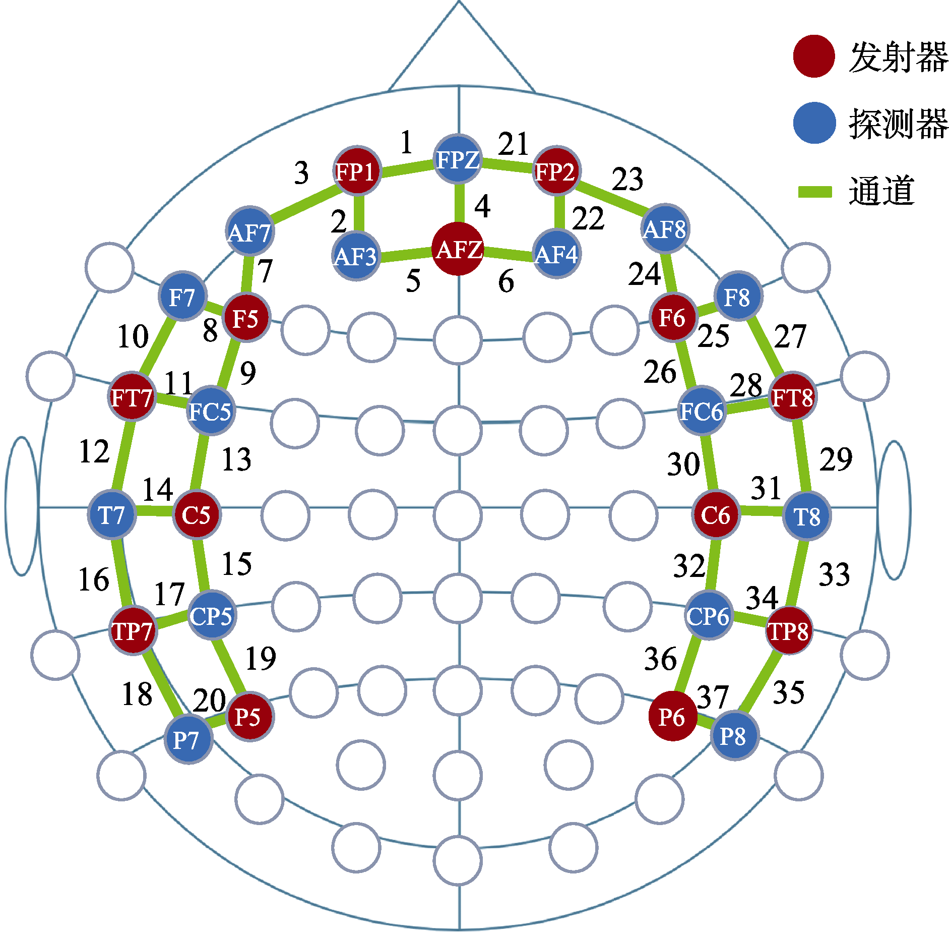
图1 NIRS通道排布图
| 通道 | 发射器-探测器 | MNI坐标 | Brodmann分区及脑区重合度* | ||
|---|---|---|---|---|---|
| x | y | z | |||
| 1 | Fp1-Fpz | -10 | 68 | -5 | 10 - Frontopolar area (0.62) |
| 2 | Fp1-AF3 | -25 | 66 | 4 | 10 - Frontopolar area (1.00) |
| 3 | Fp1-AF7 | -32 | 62 | -8 | 10 - Frontopolar area (0.58) 11 - Orbitofrontal area (0.42) |
| 4 | AFz-Fpz | 3 | 66 | 11 | 10 - Frontopolar area (1.00) |
| 5 | AFz-AF3 | -12 | 65 | 20 | 10 - Frontopolar area (1.00) |
| 6 | AFz-AF4 | 16 | 65 | 20 | 10 - Frontopolar area (1.00) |
| 7 | F5-AF7 | -46 | 48 | 0 | 10 - Frontopolar area (0.46) 47 - Inferior prefrontal gyrus (0.34) |
| 8 | F5-F7 | -52 | 39 | 0 | 47 - Inferior prefrontal gyrus (0.62) |
| 9 | F5-FC5 | -56 | 27 | 16 | 45 - pars triangularis, part of Broca’s area (0.64) |
| 10 | FT7-F7 | -57 | 21 | -13 | 38 - Temporopolar area (0.68) |
| 11 | FT7-FC5 | -61 | 8 | 2 | 22 - Superior temporal gyrus (0.61) |
| 12 | FT7-T7 | -66 | -7 | -14 | 21 - Middle temporal gyrus (1.00) |
| 13 | C5-FC5 | -64 | -2 | 24 | 6 - Pre-motor and supplementary motor cortex (0.67) |
| 14 | C5-T7 | -68 | -17 | 8 | 42 - Primary and auditory association cortex (0.51) |
| 15 | C5-CP5 | -66 | -30 | 28 | 40 - Supramarginal gyrus, part of Wernicke’s area (0.73) |
| 16 | TP7-T7 | -69 | -31 | -9 | 21 - Middle temporal gyrus (1.00) |
| 17 | TP7-CP5 | -67 | -44 | 11 | 22 - Superior temporal gyrus (0.92) |
| 18 | TP7-P7 | -64 | -55 | -4 | 21 - Middle temporal gyrus (0.58) 37 - Fusiform gyrus (0.42) |
| 19 | P5-CP5 | -60 | -56 | 28 | 40 - Supramarginal gyrus, part of Wernicke’s area (0.58) |
| 20 | P5-P7 | -58 | -68 | 13 | 39 - Angular gyrus, part of Wernicke’s area (0.42) |
| 21 | Fp2-Fpz | 14 | 68 | -5 | 10 - Frontopolar area (0.66) |
| 22 | Fp2-AF4 | 28 | 66 | 4 | 10 - Frontopolar area (1.00) |
| 23 | Fp2-AF8 | 35 | 63 | -8 | 10 - Frontopolar area (0.63) |
| 24 | F6-AF8 | 49 | 48 | 1 | 10 - Frontopolar area (0.45) |
| 25 | F6-F8 | 54 | 39 | 1 | 47 - Inferior prefrontal gyrus (0.56) |
| 26 | F6-FC6 | 58 | 25 | 16 | 45 - pars triangularis, part of Broca’s area (0.69) |
| 27 | FT8-F8 | 59 | 21 | -12 | 38 - Temporopolar area (0.62) |
| 28 | FT8-FC6 | 63 | 7 | 3 | 22 - Superior temporal gyrus (0.63) |
| 29 | FT8-T8 | 67 | -7 | -12 | 21 - Middle temporal gyrus (1.00) |
| 30 | C6-FC6 | 66 | -3 | 24 | 6 - Pre-motor and supplementary motor cortex (0.66) |
| 31 | C6-T8 | 70 | -17 | 8 | 42 - Primary and auditory association cortex (0.50) |
| 32 | C6-CP6 | 67 | -30 | 28 | 40 - Supramarginal gyrus, part of Wernicke’s area (0.78) |
| 33 | TP8-T8 | 70 | -30 | -9 | 21 - Middle temporal gyrus (0.98) |
| 34 | TP8-CP6 | 68 | -43 | 11 | 22 - Superior temporal gyrus (0.92) |
| 35 | TP8-P8 | 64 | -54 | -4 | 37 - Fusiform gyrus (0.54) 21 - Middle temporal gyrus (0.46) |
| 36 | P6-CP6 | 61 | -56 | 28 | 40 - Supramarginal gyrus, part of Wernicke’s area (0.61) |
| 37 | P6-P8 | 57 | -67 | 13 | 39 - Angular gyrus, part of Wernicke’s area (0.54) |
表1 实验中37个NIRS通道的空间配准信息
| 通道 | 发射器-探测器 | MNI坐标 | Brodmann分区及脑区重合度* | ||
|---|---|---|---|---|---|
| x | y | z | |||
| 1 | Fp1-Fpz | -10 | 68 | -5 | 10 - Frontopolar area (0.62) |
| 2 | Fp1-AF3 | -25 | 66 | 4 | 10 - Frontopolar area (1.00) |
| 3 | Fp1-AF7 | -32 | 62 | -8 | 10 - Frontopolar area (0.58) 11 - Orbitofrontal area (0.42) |
| 4 | AFz-Fpz | 3 | 66 | 11 | 10 - Frontopolar area (1.00) |
| 5 | AFz-AF3 | -12 | 65 | 20 | 10 - Frontopolar area (1.00) |
| 6 | AFz-AF4 | 16 | 65 | 20 | 10 - Frontopolar area (1.00) |
| 7 | F5-AF7 | -46 | 48 | 0 | 10 - Frontopolar area (0.46) 47 - Inferior prefrontal gyrus (0.34) |
| 8 | F5-F7 | -52 | 39 | 0 | 47 - Inferior prefrontal gyrus (0.62) |
| 9 | F5-FC5 | -56 | 27 | 16 | 45 - pars triangularis, part of Broca’s area (0.64) |
| 10 | FT7-F7 | -57 | 21 | -13 | 38 - Temporopolar area (0.68) |
| 11 | FT7-FC5 | -61 | 8 | 2 | 22 - Superior temporal gyrus (0.61) |
| 12 | FT7-T7 | -66 | -7 | -14 | 21 - Middle temporal gyrus (1.00) |
| 13 | C5-FC5 | -64 | -2 | 24 | 6 - Pre-motor and supplementary motor cortex (0.67) |
| 14 | C5-T7 | -68 | -17 | 8 | 42 - Primary and auditory association cortex (0.51) |
| 15 | C5-CP5 | -66 | -30 | 28 | 40 - Supramarginal gyrus, part of Wernicke’s area (0.73) |
| 16 | TP7-T7 | -69 | -31 | -9 | 21 - Middle temporal gyrus (1.00) |
| 17 | TP7-CP5 | -67 | -44 | 11 | 22 - Superior temporal gyrus (0.92) |
| 18 | TP7-P7 | -64 | -55 | -4 | 21 - Middle temporal gyrus (0.58) 37 - Fusiform gyrus (0.42) |
| 19 | P5-CP5 | -60 | -56 | 28 | 40 - Supramarginal gyrus, part of Wernicke’s area (0.58) |
| 20 | P5-P7 | -58 | -68 | 13 | 39 - Angular gyrus, part of Wernicke’s area (0.42) |
| 21 | Fp2-Fpz | 14 | 68 | -5 | 10 - Frontopolar area (0.66) |
| 22 | Fp2-AF4 | 28 | 66 | 4 | 10 - Frontopolar area (1.00) |
| 23 | Fp2-AF8 | 35 | 63 | -8 | 10 - Frontopolar area (0.63) |
| 24 | F6-AF8 | 49 | 48 | 1 | 10 - Frontopolar area (0.45) |
| 25 | F6-F8 | 54 | 39 | 1 | 47 - Inferior prefrontal gyrus (0.56) |
| 26 | F6-FC6 | 58 | 25 | 16 | 45 - pars triangularis, part of Broca’s area (0.69) |
| 27 | FT8-F8 | 59 | 21 | -12 | 38 - Temporopolar area (0.62) |
| 28 | FT8-FC6 | 63 | 7 | 3 | 22 - Superior temporal gyrus (0.63) |
| 29 | FT8-T8 | 67 | -7 | -12 | 21 - Middle temporal gyrus (1.00) |
| 30 | C6-FC6 | 66 | -3 | 24 | 6 - Pre-motor and supplementary motor cortex (0.66) |
| 31 | C6-T8 | 70 | -17 | 8 | 42 - Primary and auditory association cortex (0.50) |
| 32 | C6-CP6 | 67 | -30 | 28 | 40 - Supramarginal gyrus, part of Wernicke’s area (0.78) |
| 33 | TP8-T8 | 70 | -30 | -9 | 21 - Middle temporal gyrus (0.98) |
| 34 | TP8-CP6 | 68 | -43 | 11 | 22 - Superior temporal gyrus (0.92) |
| 35 | TP8-P8 | 64 | -54 | -4 | 37 - Fusiform gyrus (0.54) 21 - Middle temporal gyrus (0.46) |
| 36 | P6-CP6 | 61 | -56 | 28 | 40 - Supramarginal gyrus, part of Wernicke’s area (0.61) |
| 37 | P6-P8 | 57 | -67 | 13 | 39 - Angular gyrus, part of Wernicke’s area (0.54) |
| 通道 | 脑区 | F | p* | 愤怒β值 | 恐惧β值 | 快乐β值 |
|---|---|---|---|---|---|---|
| 3 | L Frontopolar/orbitofrontal area | 12.51 | 0.001 | 0.21 ± 0.20 | 0.12 ± 0.23 | 0.06 ± 0.20 |
| 9 | L pars triangularis/Broca’s area | 24.24 | < 0.001 | 0.10 ± 0.16 | 0.10 ± 0.15 | 0.21 ± 0.15 |
| 32 | R Supramarginal gyrus | 12.48 | 0.001 | 0.10 ± 0.56 | 0.36 ± 0.51 | 0.11 ± 0.45 |
表2 情绪主效应结果
| 通道 | 脑区 | F | p* | 愤怒β值 | 恐惧β值 | 快乐β值 |
|---|---|---|---|---|---|---|
| 3 | L Frontopolar/orbitofrontal area | 12.51 | 0.001 | 0.21 ± 0.20 | 0.12 ± 0.23 | 0.06 ± 0.20 |
| 9 | L pars triangularis/Broca’s area | 24.24 | < 0.001 | 0.10 ± 0.16 | 0.10 ± 0.15 | 0.21 ± 0.15 |
| 32 | R Supramarginal gyrus | 12.48 | 0.001 | 0.10 ± 0.56 | 0.36 ± 0.51 | 0.11 ± 0.45 |
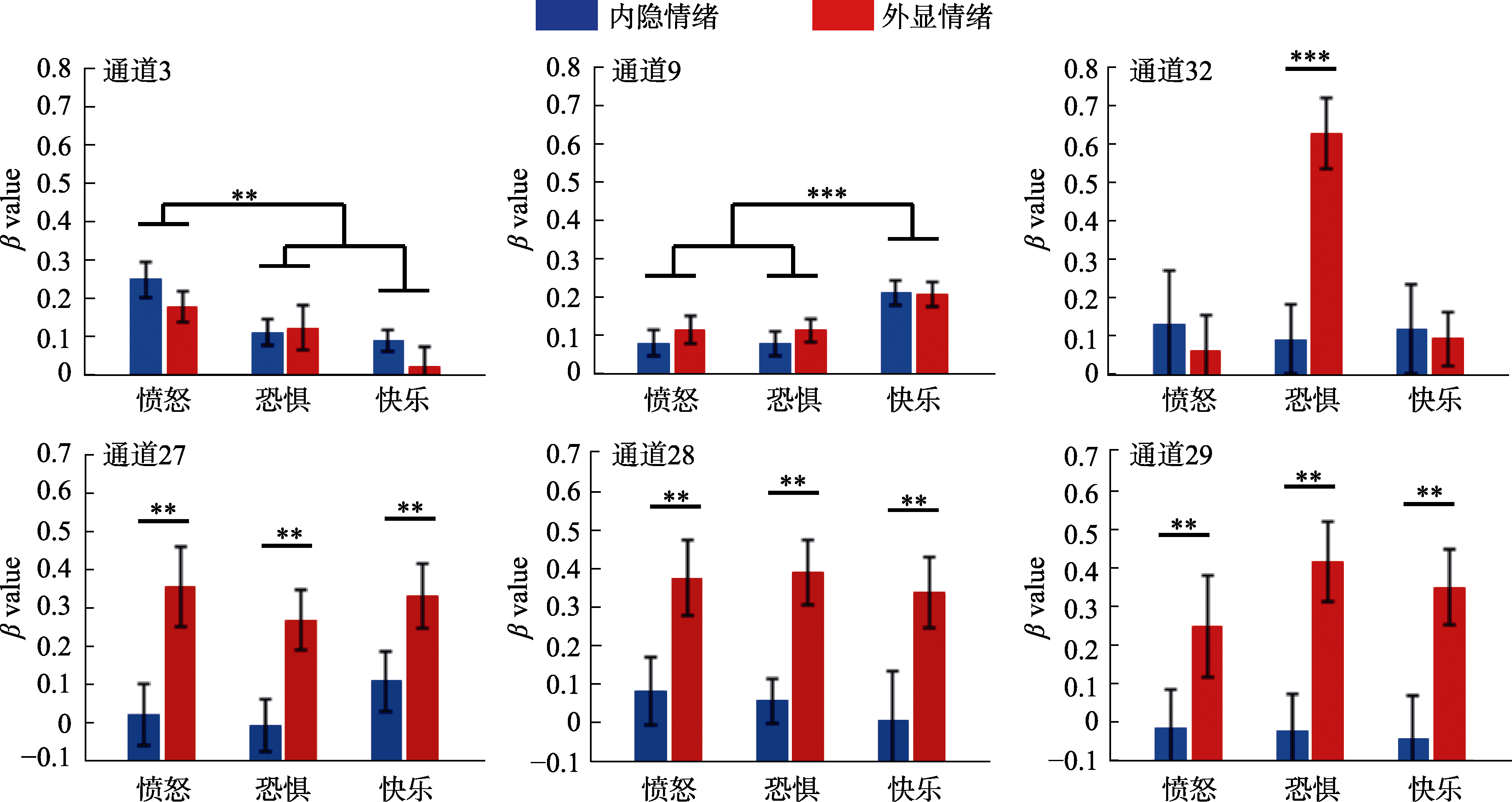
图2 不同脑区在情绪和任务条件中的激活(仅显示出现显著效应的通道)。图中的errorbar表示均值的标准误。

图3 脑区激活的成像图。
| 通道 | 脑区 | F | p* | 内隐β值 | 外显β值 |
|---|---|---|---|---|---|
| 27 | R Temporopolar area | 11.62 | 0.004 | 0.04 ± 0.36 | 0.32 ± 0.42 |
| 28 | R Superior temporal gyrus | 26.17 | < 0.001 | 0.05 ± 0.45 | 0.37 ± 0.43 |
| 29 | R Middle temporal gyrus | 15.84 | 0.003 | -0.03 ± 0.49 | 0.34 ± 0.53 |
表3 任务主效应结果
| 通道 | 脑区 | F | p* | 内隐β值 | 外显β值 |
|---|---|---|---|---|---|
| 27 | R Temporopolar area | 11.62 | 0.004 | 0.04 ± 0.36 | 0.32 ± 0.42 |
| 28 | R Superior temporal gyrus | 26.17 | < 0.001 | 0.05 ± 0.45 | 0.37 ± 0.43 |
| 29 | R Middle temporal gyrus | 15.84 | 0.003 | -0.03 ± 0.49 | 0.34 ± 0.53 |
| [1] |
Adolphs, R., Damasio, H., Tranel, D., Cooper, G., & Damasio, A. R. (2000). A role for somatosensory cortices in the visual recognition of emotion as revealed by three-dimensional lesion mapping. The Journal of Neuroscience, 20(7), 2683-2690.
URL pmid: 10729349 |
| [2] |
Alba-Ferrara, L., Kochen, S., & Hausmann, M. (2018). Emotional prosody processing in epilepsy: Some insights on brain reorganization. Frontiers in Human Neuroscience, 12, 92.
URL pmid: 29593517 |
| [3] |
Aryani, A., Hsu, C.-T., & Jacobs, A. M. (2018). The sound of words evokes affective brain responses. Brain Sciences, 8(6), 94.
doi: 10.3390/brainsci8060094 URL |
| [4] |
Bach, D. R., Grandjean, D., Sander, D., Herdener, M., Strik, W. K., & Seifritz, E. (2008). The effect of appraisal level on processing of emotional prosody in meaningless speech. Neuroimage, 42(2), 919-927.
doi: 10.1016/j.neuroimage.2008.05.034 URL pmid: 18586524 |
| [5] |
Beaucousin, V., Zago, L., Herve, P.-Y., Strelnikov, K., Crivello, F., Mazoyer, B., & Tzourio-Mazoyer, N. (2011). Sex-dependent modulation of activity in the neural networks engaged during emotional speech comprehension. Brain Research, 1390, 108-117.
URL pmid: 21439268 |
| [6] |
Belyk, M., & Brown, S. (2014). Perception of affective and linguistic prosody: An ALE meta-analysis of neuroimaging studies. Social Cognitive and Affective Neuroscience, 9(9), 1395-1403.
doi: 10.1093/scan/nst124 URL pmid: 23934416 |
| [7] |
Ben-David, B. M., Multani, N., Shakuf, V., Rudzicz, F., & van Lieshout, P. H. H. M. (2016). Prosody and semantics are separate but not separable channels in the perception of emotional speech: test for rating of emotions in speech. Journal of Speech Language and Hearing Research, 59(1), 72-89.
doi: 10.1044/2015_JSLHR-H-14-0323 URL |
| [8] |
Beyer, F., Munte, T. F., Gottlich, M., & Kramer, U. M. (2014). Orbitofrontal cortex reactivity to angry facial expression in a social interaction correlates with aggressive behavior. Cerebral Cortex, 25(9), 3057-3063.
URL pmid: 24842782 |
| [9] |
Brück, C., Kreifelts, B., & Wildgruber, D. (2011). Emotional voices in context: A neurobiological model of multimodal affective information processing. Physics of Life Reviews, 8(4), 383-403.
doi: 10.1016/j.plrev.2011.10.002 URL pmid: 22035772 |
| [10] |
Calvo, M. G., & Nummenmaa, L. (2016). Perceptual and affective mechanisms in facial expression recognition: An integrative review. Cognition and Emotion, 30(6), 1081-1106.
URL pmid: 26212348 |
| [11] |
Dieler, A. C., Tupak, S. V., & Fallgatter, A. J. (2012). Functional near-infrared spectroscopy for the assessment of speech related tasks. Brain and Language, 121(2), 90-109.
URL pmid: 21507475 |
| [12] |
Enea, V., & Iancu, S. (2016). Processing emotional body expressions: state-of-the-art. Social Neuroscience, 11(5), 495-506.
URL pmid: 26513592 |
| [13] |
Ethofer, T., Anders, S., Erb, M., Herbert, C., Wiethoff, S., Kissler, J., … Wildgruber, D. (2006). Cerebral pathways in processing of affective prosody: a dynamic causal modeling study. Neuroimage, 30(2), 580-587.
URL pmid: 16275138 |
| [14] |
Ethofer, T., Bretscher, J., Gschwind, M., Kreifelts, B., Wildgruber, D., & Vuilleumier, P. (2012). Emotional voice areas: Anatomic location, functional properties, and structural connections revealed by combined fMRI/DTI. Cerebral Cortex, 22(1), 191-200.
URL pmid: 21625012 |
| [15] |
Ethofer, T., Kreifelts, B., Wiethoff, S., Wolf, J., Grodd, W., Vuilleumier, P., & Wildgruber, D. (2009b). Differential influences of emotion, task, and novelty on brain regions underlying the processing of speech melody. Journal of Cognitive Neuroscience, 21(7), 1255-1268.
URL pmid: 18752404 |
| [16] |
Ethofer, T., van de Ville, D., Scherer, K., & Vuilleumier, P. (2009a). Decoding of emotional information in voice- sensitive cortices. Current Biology, 19(12), 1028-1033.
URL pmid: 19446457 |
| [17] |
Fox, K. C. R., Yih, J., Raccah, O., Pendekanti, S. L., Limbach, L. E., Maydan, D. D., & Parvizi, J. (2018). Changes in subjective experience elicited by direct stimulation of the human orbitofrontal cortex. Neurology, 91(16), e1519-e1527.
URL pmid: 30232252 |
| [18] |
Frühholz, S., Ceravolo, L., & Grandjean, D. (2012). Specific brain networks during explicit and implicit decoding of emotional prosody. Cerebral Cortex, 22(5), 1107-1117.
URL pmid: 21750247 |
| [19] |
Frühholz, S., & Grandjean, D. (2013a). Multiple subregions in superior temporal cortex are differentially sensitive to vocal expressions: a quantitative meta-analysis. Neuroscience and Biobehavioral Reviews, 37(1), 24-35.
URL pmid: 23153796 |
| [20] |
Frühholz, S., & Grandjean, D. (2013b). Processing of emotional vocalizations in bilateral inferior frontal cortex. Neuroscience and Biobehavioral Reviews, 37(10), 2847-2855.
doi: 10.1016/j.neubiorev.2013.10.007 URL |
| [21] |
Frühholz, S., Hofstetter, C., Cristinzio, C., Saj, A., Seeck, M., & Vuilleumier, P., & Grandjean, D.(2015). Asymmetrical effects of unilateral right or left amygdala damage on auditory cortical processing of vocal emotions. Proceedings of the National Academy of Sciences of the United States of America, 112(5), 1583-1588.
URL pmid: 25605886 |
| [22] |
Frühholz, S., Trost, W., & Kotz, S. A. (2016). The sound of emotions-Towards a unifying neural network perspective of affective sound processing. Neuroscience and Biobehavioral Reviews, 68, 96-110.
URL pmid: 27189782 |
| [23] |
Goucha, T., & Friederici, A. D. (2015). The language skeleton after dissecting meaning: A functional segregation within Broca’s Area. Neuroimage, 114, 294-302.
URL pmid: 25871627 |
| [24] |
Hartwigsen, G., Baumgaertner, A., Price, C. J., Koehnke, M., Ulmer, S., & Siebner, H. R. (2010). Phonological decisions require both the left and right supramarginal gyri. Proceedings of the National Academy of Sciences of the United States of America, 107(38), 16494-16499.
URL pmid: 20807747 |
| [25] |
Hensel, L., Bzdok, D., Müller, V. I., Zilles, K., & Eickhoff, S. B. (2015). Neural correlates of explicit social judgments on vocal stimuli. Cerebral Cortex, 25(5), 1152-1162.
URL pmid: 24243619 |
| [26] |
Herpertz, S. C., Nagy, K., Ueltzhöffer, K., Schmitt, R., Mancke, F., Schmahl, C., & Bertsch, K. (2017). Brain mechanisms underlying reactive aggression in borderline personality disorder-sex matters. Biological Psychiatry, 82(4), 257-266.
URL pmid: 28388995 |
| [27] |
Hinojosa, J. A., Mercado, F., & Carretié, L. (2015). N170 sensitivity to facial expression: A meta-analysis. Neuroscience and Biobehavioral Reviews, 55, 498-509.
URL pmid: 26067902 |
| [28] |
Johnstone, T., van Reekum, C. M., Oakes, T. R., & Davidson, R. J. (2006). The voice of emotion: an FMRI study of neural responses to angry and happy vocal expressions. Social Cognitive and Affective Neuroscience, 1(3), 242-249.
URL pmid: 17607327 |
| [29] |
Kirby, L. A. J., & Robinson, J. L. (2017). Affective mapping: An activation likelihood estimation (ALE) meta-analysis. Brain and Cognition, 118, 137-148.
doi: 10.1016/j.bandc.2015.04.006 URL pmid: 26074298 |
| [30] |
Knight, M. J., & Baune, B. T. (2019). Social cognitive abilities predict psychosocial dysfunction in major depressive disorder. Depression and Anxiety, 36(1), 54-62.
URL pmid: 30211966 |
| [31] |
Kotz, S. A., Kalberlah, C., Bahlmann, J., Friederici, A. D., & Haynes, J.-D. (2013). Predicting vocal emotion expressions from the human brain. Human Brain Mapping, 34(8), 1971-1981.
URL pmid: 22371367 |
| [32] |
Kotz, S. A., Meyer, M., Alter, K., Besson, M., von Cramon, D. Y., & Friederici, A. D. (2003). On the lateralization of emotional prosody: an event-related functional MR investigation. Brain and Language, 86(3), 366-376.
URL pmid: 12972367 |
| [33] | Köchel, A., Schöngassner, F., & Schienle, A. (2013). Cortical activation during auditory elicitation of fear and disgust: a near-infrared spectroscopy (NIRS) study. Neuroscience Letters, 9(549), 197-200. |
| [34] |
Lancaster, J. L., Woldorff, M. G., Parsons, L. M., Liotti, M., Freitas, C. S., Rainey, L.,… Fox, P. (2000). Automated Talairach atlas labels for functional brain mapping. Human Brain Mapping, 10(3), 120-131.
doi: 10.1002/1097-0193(200007)10:3<120::aid-hbm30>3.0.co;2-8 URL pmid: 10912591 |
| [35] | Liebenthal, E., Silbersweig, D. A., & Stern, E. (2016). The Language, Tone and prosody of emotions: neural substrates and dynamics of spoken-word emotion perception. Frontiers in Aging Neuroscience, 10, 506. |
| [36] | Lin, Y., Ding, H., & Zhang, Y. (2018). Emotional prosody processing in schizophrenic patients: A selective review and meta-analysis. Journal of Clinical Medicine, 7(10), 363. |
| [37] | Lindquist, K. A., Wager, T. D., Kober, H., Bliss-Moreau, E., & Barrett, L. F. (2012). The brain basis of emotion: a meta- analytic review. Behavioral and Brain Sciences, 35(3), 121-143. |
| [38] |
Liu, P., & Pell, M. D. (2012). Recognizing vocal emotions in Mandarin Chinese: a validated database of Chinese vocal emotional stimuli. Behavior Research Methods, 44, 1042-1051.
URL pmid: 22539230 |
| [39] |
Matsui, T., Nakamura, T., Utsumi, A., Sasaki, A. T., Koike, T., Yoshida, Y., … Sadato, N. (2016). The role of prosody and context in sarcasm comprehension: Behavioral and fMRI evidence. Neuropsychologia, 87, 74-84.
URL pmid: 27157883 |
| [40] |
Mitchell, R. L. C. (2007). fMRI delineation of working memory for emotional prosody in the brain: commonalities with the lexico-semantic emotion network. Neuroimage, 36(3), 1015-1025.
URL pmid: 17481919 |
| [41] |
Mitchell, R. L. C., & Xu, Y. (2015). What is the value of embedding artificial emotional prosody in human-computer interactions? Implications for theory and design in psychological science. Frontiers in Psychology, 6, 1750.
URL pmid: 26617563 |
| [42] |
Molavi, B., & Dumont, G. A. (2012). Wavelet-based motion artifact removal for functional near-infrared spectroscopy. Physiological Measurement, 33(2), 259-270.
URL pmid: 22273765 |
| [43] |
Mothes-Lasch, M., Mentzel, H.-J., Miltner, W. H. R., & Straube, T. (2011). Visual attention modulates brain activation to angry voices. Journal of Neuroscience, 31(26), 9594-9598.
URL pmid: 21715624 |
| [44] |
Patel, S., Oishi, K., Wright, A., Sutherland-Foggio, H., Saxena, S., Sheppard, S. M., & Hillis, A. E. (2018). Right hemisphere regions critical for expression of emotion through prosody. Frontiers in Neurology, 9, 224.
URL pmid: 29681885 |
| [45] |
Patterson, R. D., Uppenkamp, S., Johnsrude, I. S., & Griffiths, T. D. (2002). The processing of temporal pitch and melody information in auditory cortex. Neuron, 36(4), 767-776.
URL pmid: 12441063 |
| [46] |
Paulmann, S., Seifert, S., & Kotz, S. A. (2010). Orbito-frontal lesions cause impairment during late but not early emotional prosodic processing. Social Neuroscience, 5(1), 59-75.
URL pmid: 19658025 |
| [47] |
Quadflieg, S., Mohr, A., Mentzel, H.-J., Miltner, W. H. R., & Straube, T. (2008). Modulation of the neural network involved in the processing of anger prosody: the role of task-relevance and social phobia. Biological Psychology, 78(2), 129-137.
URL pmid: 18353521 |
| [48] |
Ross, E. D. (1981). The aprosodias. Functional-anatomic organization of the affective components of language in the right hemisphere. Archives of Neurology, 38(9), 561-569.
URL pmid: 7271534 |
| [49] |
Schirmer, A., & Kotz, S. A. (2006). Beyond the right hemisphere: brain mechanisms mediating vocal emotional processing. Trends in Cognitive Sciences, 10(1), 24-30.
URL pmid: 16321562 |
| [50] |
Steber, S., König, N., Stephan, F., & Rossi, S. (2020). Uncovering electrophysiological and vascular signatures of implicit emotional prosody. Scientific Reports, 10(1), 5807.
URL pmid: 32242032 |
| [51] |
Tong, Y., Hocke, L. M., & Frederick, B. B., (2011). Isolating the sources of widespread physiological fluctuations in functional near-infrared spectroscopy signals. Journal of Biomedical Optics, 16(10), 106005.
URL pmid: 22029352 |
| [52] |
Witteman, J., van Heuven, V. J., & Schiller, N. O. (2012). Hearing feelings: a quantitative meta-analysis on the neuroimaging literature of emotional prosody perception. Neuropsychologia, 50(12), 2752-2763.
URL pmid: 22841991 |
| [53] |
Zhang, D., Chen, Y., Hou, X., & Wu, Y. J. (2019). Near-infrared spectroscopy reveals neural perception of vocal emotions in human neonates. Human Brain Mapping, 40(8), 2434-2448.
URL pmid: 30697881 |
| [54] |
Zhang, D., Zhou, Y., Hou, X., Cui, Y., & Zhou, C. (2017). Discrimination of emotional prosodies in human neonates: A pilot fNIRS study. Neuroscience Letters, 658, 62-66.
URL pmid: 28842278 |
| [55] |
Zhang, D., Zhou, Y., & Yuan, J. (2018). Speech prosodies of different emotional categories activate different brain regions in adult cortex: an fNIRS study. Scientific Reports, 8(1), 218.
URL pmid: 29317758 |
| [1] | 张环, 秦锡权, 刘雨, 林琳, 吴捷. 不同强度趋近动机积极情绪对基于语义关联性错误记忆的影响及其神经机制[J]. 心理学报, 2025, 57(3): 349-362. |
| [2] | 夏莲香, 刘凯歌, 李新宇, 叶群. 编码方式与叙事连贯性调节情绪对时间顺序记忆的影响[J]. 心理学报, 2025, 57(1): 1-17. |
| [3] | 姚雨佳, 颜之悦, 林慧慧, 陈静全, 宣雨阳. 人际距离和策略创造性对人际情绪调节的影响[J]. 心理学报, 2025, 57(1): 125-134. |
| [4] | 张环, 王晨, 李俊霞, 林琳, 吴捷. 情绪效价和动机强度对社会分享型提取诱发遗忘的影响[J]. 心理学报, 2024, 56(8): 999-1014. |
| [5] | 王丹, 付雨佳, 陈文锋. 社会情境对情绪感染的影响:一项基于EMG的超扫描研究[J]. 心理学报, 2024, 56(8): 1047-1060. |
| [6] | 张明杨, 杨盈, 包寒吴霜, 蔡华俭. 中国人的积极理想情绪:近几十年来的变迁[J]. 心理学报, 2024, 56(7): 847-858. |
| [7] | 黄顺森, 来枭雄, 张彩, 赵心媚, 代欣然, 祁梦迪, 王欢蕾, 王文荣, 王耘. 青少年手机压力与心理健康的关系:基于多元宇宙样分析和密集追踪方法[J]. 心理学报, 2024, 56(6): 745-758. |
| [8] | 孟现鑫, 罗怡, 韩晨媛, 吴国伟, 常姣, 袁加锦, 千坤, 傅小兰. 社会接纳调节社会排斥对情绪冲突适应的影响[J]. 心理学报, 2024, 56(5): 577-593. |
| [9] | 童廷婷, 白幼玲, 冯廷勇. 情绪调节改善拖延行为的认知机制:任务厌恶中介作用[J]. 心理学报, 2024, 56(4): 458-468. |
| [10] | 王伟晗, 曹斐臻, 余林伟, 曾珂, 杨鑫超, 徐强. 群体信息对面部表情识别的影响[J]. 心理学报, 2024, 56(3): 268-280. |
| [11] | 尹华站, 肖春花, 夏安妮, 袁中静, 崔晓冰, 李丹. 基本情绪对时距知觉的影响: 来自三水平元分析和网络元分析的证据[J]. 心理学报, 2024, 56(12): 1676-1690. |
| [12] | 张瑾, 许销冰, 庄晓涵. 产品包装的医药符号线索对产品偏好的影响: 感知有效性和规避情绪的中介作用[J]. 心理学报, 2024, 56(12): 1836-1850. |
| [13] | 肖程元, 赵世瑞, 袁加锦. 积极认知重评对负性信息传播的调控及多维证据[J]. 心理学报, 2024, 56(11): 1471-1487. |
| [14] | 李其容, 王春淼, 孙明慧. 创业激情的“错位”对创业努力和创业成瘾的影响机制[J]. 心理学报, 2024, 56(11): 1568-1584. |
| [15] | 田杨阳, 李东, 闫向博, 李曌, 崔倩, 蒋重清. 动态高兴表情评价中的表征动量效应和参照依赖效应[J]. 心理学报, 2024, 56(1): 29-43. |
| 阅读次数 | ||||||
|
全文 |
|
|||||
|
摘要 |
|
|||||