ISSN 1671-3710
CN 11-4766/R
主办:中国科学院心理研究所
出版:科学出版社
CN 11-4766/R
主办:中国科学院心理研究所
出版:科学出版社
心理科学进展 ›› 2022, Vol. 30 ›› Issue (2): 255-274.doi: 10.3724/SP.J.1042.2022.00255 cstr: 32111.14.2022.00255
陈幸明1, 付彤1, 刘昌3, 张宾1, 伏云发4, 李恩泽5, ZHANG Jian6, 陈盛强2, 党彩萍1,2( )
)
收稿日期:2021-02-17
出版日期:2022-02-15
发布日期:2021-12-24
基金资助:
CHEN Xingming1, FU Tong1, LIU Chang3, ZHANG Bin1, FU Yunfa4, LI Enze5, ZHANG Jian6, CHEN Shengqiang2, DANG Caiping1,2()
Received:2021-02-17
Online:2022-02-15
Published:2021-12-24
摘要:
工作记忆训练(Working Memory Training, WMT)诱发神经可塑性, 但其具体机制尚不明晰。为探索WMT改变正常人群大脑功能的时空特性, 以“扩展的智力顶额整合理论”和“神经效率假说”为依据, 采用逐层递进的5种方法, 分6个步骤来查究近20年来正常人群WMT的37篇fMRI文献。第一步, 用叙述性综述、频数分析和卡方检验法比较脑区激活模式和脑网络功能连接在WMT前后发生的改变, 发现WMT改变了大脑的5个联合区、7个宏观区和3个子区。其中, 额上回、顶下小叶和扣带回这3个子区各自激活减弱的报道文献数量多于其激活增强的, 且这种差异分别具有统计学意义。第二步, 采用激活似然估计法对其中26篇开展元分析, 发现大脑的3个子区激活减弱水平在WMT前后的差异具有统计学意义, 即额中回(BA6和8)、额上回(BA6)和前扣带回(BA24和32)。第三步, 综合定性和定量分析结果, 提出WMT脑区分布递减时空模型, 产生5个结果和讨论。第四步, 采用非参数检验进一步追踪WMT效应的调节因素, 发现训练的任务类型和时间分别对脑区激活的影响具有统计学意义。第五步, 针对正常人群WMT诱发神经可塑性的时空特性, 得出3个结论:第一, WMT改变了正常人群相应脑区的神经活动, 表现为减弱或增强, 但减弱更加突出, 且更新和较短时间的WMT倾向于诱发较多减弱; 第二, 这些神经活动变化主要发生在额顶叶联合区, 但也包括分别以颞叶、枕叶、扣带回及纹状体为主的联合区, 在一定范围内体现了整脑功能联合。这体现了WMT诱发神经可塑性的空间特性, 且符合“扩展的智力顶额整合理论”; 第三, 额中回、额上回、顶下小叶和扣带回(尤其前扣带回)这4个子区在激活减弱水平上重点展示了WMT神经可塑性的时间特性, 且符合“神经效率假说”, 恰好体现出“聪明的大脑更懒惰”。第六步, 指出WMT诱发神经可塑性的未来研究可能关注脑可塑性中的低活跃性、辨析额中回、额上回、顶下小叶和扣带回(尤其前扣带回)这4个子区在激活减弱水平上体现的时间特性、找寻训练减弱或增强大脑活动的综合性影响因素。
中图分类号:
陈幸明, 付彤, 刘昌, 张宾, 伏云发, 李恩泽, ZHANG Jian, 陈盛强, 党彩萍. (2022). 工作记忆训练诱发的神经可塑性——基于系列fMRI实验的脑区分布递减时空模型. 心理科学进展 , 30(2), 255-274.
CHEN Xingming, FU Tong, LIU Chang, ZHANG Bin, FU Yunfa, LI Enze, ZHANG Jian, CHEN Shengqiang, DANG Caiping. (2022). Neuroplasticity induced by working memory training: A spatio-temporal model of decreased distribution in brain regions based on fMRI experiments. Advances in Psychological Science, 30(2), 255-274.
 |
表1 WMT改变脑区激活模式汇总(32篇文献,36个实验)
|
 |
表2 WMT 改变脑网络FC汇总(6篇文献, 7个实验)
|
 |
表3 WMT改变频数较多的8个大脑子区分布(28篇文献,32个实验)(全部脑区的频数分布见网络版附表1)
|
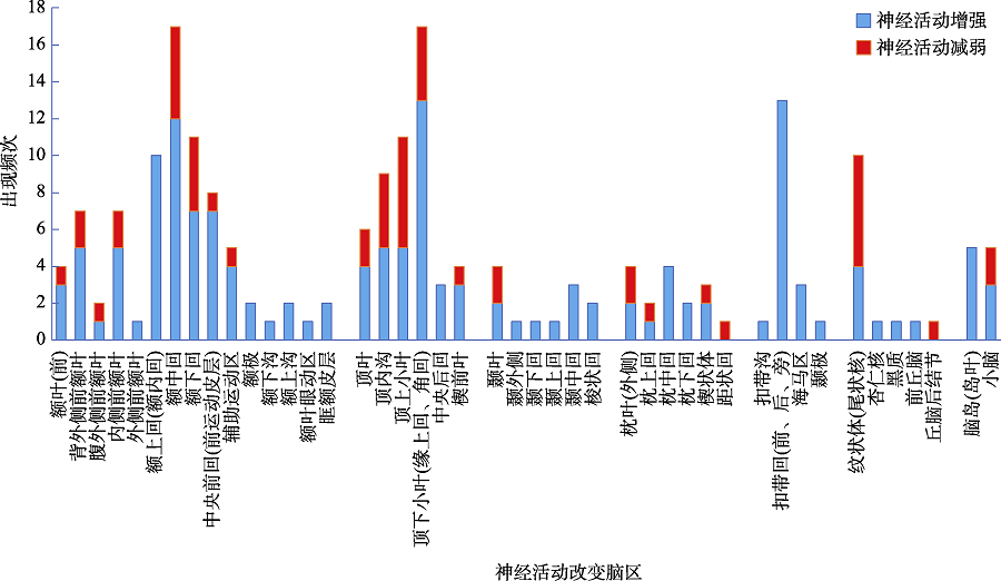
图1 WMT诱发神经活动减弱与增强的具体脑区频数分布(37篇文献, 42个实验)
| 1.额上回(额内回) | 2. 额中回 | 3. 额下回 | 4. 顶内沟 | 5. 顶上小叶 | 6. 顶下小叶 | 7. 扣带回 | 8. 纹状体 | |
|---|---|---|---|---|---|---|---|---|
| 减弱频次 | 10 | 12 | 7 | 5 | 5 | 13 | 13 | 4 |
| 增强频次 | 0 | 5 | 4 | 4 | 6 | 4 | 0 | 6 |
| df | 1 | 1 | 1 | 1 | 1 | 1 | 1 | 1 |
| χ2 | * | 2.88 | 0.82 | 0.11 | 0.09 | 4.77 | * | 0.40 |
| p | * | 0.143 | 0.549 | 1.000 | 1.000 | 0.049 | * | 0.754 |
表4 WMT诱发大脑子区改变的卡方检验(28篇文献, 30个实验)
| 1.额上回(额内回) | 2. 额中回 | 3. 额下回 | 4. 顶内沟 | 5. 顶上小叶 | 6. 顶下小叶 | 7. 扣带回 | 8. 纹状体 | |
|---|---|---|---|---|---|---|---|---|
| 减弱频次 | 10 | 12 | 7 | 5 | 5 | 13 | 13 | 4 |
| 增强频次 | 0 | 5 | 4 | 4 | 6 | 4 | 0 | 6 |
| df | 1 | 1 | 1 | 1 | 1 | 1 | 1 | 1 |
| χ2 | * | 2.88 | 0.82 | 0.11 | 0.09 | 4.77 | * | 0.40 |
| p | * | 0.143 | 0.549 | 1.000 | 1.000 | 0.049 | * | 0.754 |
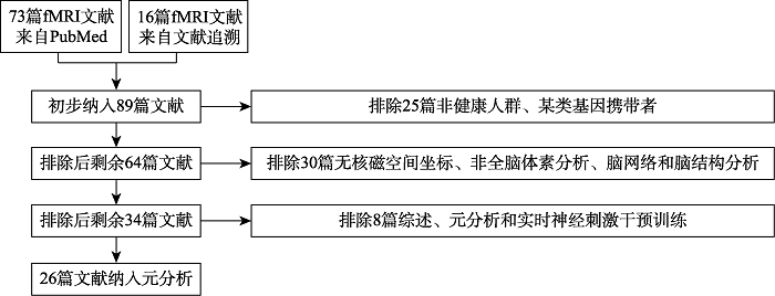
图2 逐步排除和纳入元分析的标准
| 文献 编号 | 激活减弱 | 文献 编号 | 激活增强 | ||||||
|---|---|---|---|---|---|---|---|---|---|
| 作者(年份) | 被试 数量 | 坐标 数量 | 坐标系 | 作者(年份) | 被试 数量 | 坐标 数量 | 坐标系 | ||
| 1 | Schneiders et al., | 32 | 7 | MNI | 22 | Jolles et al., | 15 | 18 | MNI |
| 2 | Schneiders et al., | 16 | 5 | MNI | 23 | Westerberg & Klingberg, | 3 | 15 | MNI |
| 3 | Milham et al., | 11 | 7 | MNI | 24 | Beatty et al., | 22 | 15 | MNI |
| 4 | Jansma et al., | 15 | 6 | MNI | 25 | Wang et al., | 27 | 7 | MNI |
| 5 | Miró-Padilla et al., | 25 | 3 | MNI | 26 | Li, Hu, et al., | 17 | 7 | MNI |
| 6 | Heinzel et al., | 15 | 6 | MNI | 14 | Dahlin et al., | 26 | 17 | MNI |
| 7 | Garavan et al., | 17 | 17 | MNI | 15 | Kühn et al., | 26 | 2 | MNI |
| 8 | Emch et al., | 30 | 39 | MNI | 18 | Olesen et al., | 11 | 8 | Talairach |
| 9 | Clark et al., | 25 | 7 | MNI | 19 | Salminen et al., | 18 | 1 | Talairach |
| 10 | Landau et al., | 10 | 7 | MNI | 20 | Nęcka et al., | 23 | 3 | MNI |
| 11 | Miró-Padilla et al., | 25 | 23 | MNI | |||||
| 12 | Schweizer et al., | 17 | 30 | MNI | |||||
| 13 | Flegal et al., | 26 | 9 | MNI | |||||
| 14 | Dahlin et al., | 15 | 3 | MNI | |||||
| 15 | Kühn et al., | 26 | 2 | MNI | |||||
| 16 | Sayala et al., | 10 | 4 | Talairach | |||||
| 17 | Kelly et al., | 18 | 5 | Talairach | |||||
| 18 | Olesen et al., | 11 | 3 | Talairach | |||||
| 19 | Salminen et al., | 18 | 10 | Talairach | |||||
| 20 | Nęcka et al., | 23 | 6 | MNI | |||||
| 21 | Takeuchi et al., | 41 | 2 | MNI | |||||
| 汇总 | 21篇 | 426 | 201 | 10篇 | 188 | 93 | |||
表5 纳入元分析文献的基本信息(共26篇文献, 31个实验)
| 文献 编号 | 激活减弱 | 文献 编号 | 激活增强 | ||||||
|---|---|---|---|---|---|---|---|---|---|
| 作者(年份) | 被试 数量 | 坐标 数量 | 坐标系 | 作者(年份) | 被试 数量 | 坐标 数量 | 坐标系 | ||
| 1 | Schneiders et al., | 32 | 7 | MNI | 22 | Jolles et al., | 15 | 18 | MNI |
| 2 | Schneiders et al., | 16 | 5 | MNI | 23 | Westerberg & Klingberg, | 3 | 15 | MNI |
| 3 | Milham et al., | 11 | 7 | MNI | 24 | Beatty et al., | 22 | 15 | MNI |
| 4 | Jansma et al., | 15 | 6 | MNI | 25 | Wang et al., | 27 | 7 | MNI |
| 5 | Miró-Padilla et al., | 25 | 3 | MNI | 26 | Li, Hu, et al., | 17 | 7 | MNI |
| 6 | Heinzel et al., | 15 | 6 | MNI | 14 | Dahlin et al., | 26 | 17 | MNI |
| 7 | Garavan et al., | 17 | 17 | MNI | 15 | Kühn et al., | 26 | 2 | MNI |
| 8 | Emch et al., | 30 | 39 | MNI | 18 | Olesen et al., | 11 | 8 | Talairach |
| 9 | Clark et al., | 25 | 7 | MNI | 19 | Salminen et al., | 18 | 1 | Talairach |
| 10 | Landau et al., | 10 | 7 | MNI | 20 | Nęcka et al., | 23 | 3 | MNI |
| 11 | Miró-Padilla et al., | 25 | 23 | MNI | |||||
| 12 | Schweizer et al., | 17 | 30 | MNI | |||||
| 13 | Flegal et al., | 26 | 9 | MNI | |||||
| 14 | Dahlin et al., | 15 | 3 | MNI | |||||
| 15 | Kühn et al., | 26 | 2 | MNI | |||||
| 16 | Sayala et al., | 10 | 4 | Talairach | |||||
| 17 | Kelly et al., | 18 | 5 | Talairach | |||||
| 18 | Olesen et al., | 11 | 3 | Talairach | |||||
| 19 | Salminen et al., | 18 | 10 | Talairach | |||||
| 20 | Nęcka et al., | 23 | 6 | MNI | |||||
| 21 | Takeuchi et al., | 41 | 2 | MNI | |||||
| 汇总 | 21篇 | 426 | 201 | 10篇 | 188 | 93 | |||
| 脑区 | 半球 | MNI坐标 | 体积(mm3) | ALE值 | p | BA | ||
|---|---|---|---|---|---|---|---|---|
| x | y | z | ||||||
| 额中回 | 右 | 36 | 20 | 40 | 352 | 0.021 | p < 0.001 | 6, 8 |
| 额上回/额内回/腹侧前扣带回 | 左 | -2 | 12 | 52 | 1024 | 0.028 | p < 0.001 | 6, 32, 24 |
表6 WMT减弱脑区激活的元分析结果(共26篇文献, 31个实验)
| 脑区 | 半球 | MNI坐标 | 体积(mm3) | ALE值 | p | BA | ||
|---|---|---|---|---|---|---|---|---|
| x | y | z | ||||||
| 额中回 | 右 | 36 | 20 | 40 | 352 | 0.021 | p < 0.001 | 6, 8 |
| 额上回/额内回/腹侧前扣带回 | 左 | -2 | 12 | 52 | 1024 | 0.028 | p < 0.001 | 6, 32, 24 |
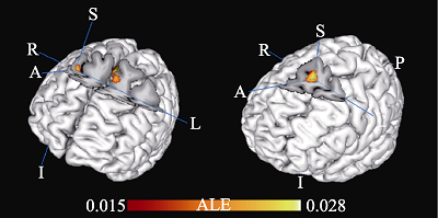
图3 WMT减弱脑区激活的2个激活簇区域(共26篇文献, 31个实验)

图4 WMT脑区分布递减时空模型(倒金字塔图) 注:左侧倒三角为频数分析结果(37篇文献, 42个实验), 右侧倒三角为元分析结果(26篇文献, 31个实验); 棕色框内的脑区为元分析差异具有统计学意义; 双黑线框内的脑区为只出现激活减弱, 单黑线框内的脑区为激活减弱或增强并存; 红线框内的脑区为激活减弱明显多于激活增强。SFG = Superior Frontal Gyrus (额上回), MeFG = Medial Frontal Gyrus (额内回), MFG = Middle Frontal Gyrus (额中回), IFG = Inferior Frontal Gyrus (额下回), SPL = Superior Parietal Lobule (顶上小叶), IPL = Inferior Parietal Lobule (顶下小叶), IPS = Intraparietal Sulcus (顶内沟), ACC = Anterior Cingulate Gyrus (前扣带回), PCC = Posterior Cingulate Gyrus (后扣带回)。图中间位置的4个脑区模型图片来自:A+医学百科网(www.a-hospital.com), 作者在其上新增了色彩和英文缩写。

图5 WMT效应调节因素的神经活动减弱与增强频数分布(31篇文献。33个实验)
| 序号 | 影响因素 | 减弱频次(秩次平均与总和) | 增强频次(秩次平均与总和) | df | χ2/Z | p |
|---|---|---|---|---|---|---|
| 1 | 人数(秩和检验) | 21(16.48, 346) | 12(17.92, 215) | -0.41 | 0.680 | |
| 2 | 神经技术 | 21 | 12 | 1 | 2.94 | 0.125 |
| BOLD | 20 | 1 | ||||
| FC | 9 | 3 | ||||
| 3 | 年龄(3个阶段) | 21 | 12 | 2 | 5.81 | 0.064 |
| 儿童 | 0 | 3 | ||||
| 青年 | 17 | 7 | ||||
| 中老年 | 4 | 2 | ||||
| 4 | 训练任务类型(6类) | 21 | 12 | 5 | 10.56 | 0.023 |
| 更新 | 14 | 3 | ||||
| 短时记忆 | 4 | 5 | ||||
| 抑制 | 0 | 3 | ||||
| 转换 | 1 | 0 | ||||
| 计算 | 1 | 0 | ||||
| 综合 | 1 | 1 | ||||
| 5 | 训练时间(3段) | 21 | 12 | 2 | 7.57 | 0.019 |
| 4周以下 | 15 | 4 | ||||
| 4~6周 | 6 | 5 | ||||
| 6周以上 | 0 | 3 |
表7 WMT效应的调节因素影响神经活动改变的非参数检验(31篇文献, 33个实验)
| 序号 | 影响因素 | 减弱频次(秩次平均与总和) | 增强频次(秩次平均与总和) | df | χ2/Z | p |
|---|---|---|---|---|---|---|
| 1 | 人数(秩和检验) | 21(16.48, 346) | 12(17.92, 215) | -0.41 | 0.680 | |
| 2 | 神经技术 | 21 | 12 | 1 | 2.94 | 0.125 |
| BOLD | 20 | 1 | ||||
| FC | 9 | 3 | ||||
| 3 | 年龄(3个阶段) | 21 | 12 | 2 | 5.81 | 0.064 |
| 儿童 | 0 | 3 | ||||
| 青年 | 17 | 7 | ||||
| 中老年 | 4 | 2 | ||||
| 4 | 训练任务类型(6类) | 21 | 12 | 5 | 10.56 | 0.023 |
| 更新 | 14 | 3 | ||||
| 短时记忆 | 4 | 5 | ||||
| 抑制 | 0 | 3 | ||||
| 转换 | 1 | 0 | ||||
| 计算 | 1 | 0 | ||||
| 综合 | 1 | 1 | ||||
| 5 | 训练时间(3段) | 21 | 12 | 2 | 7.57 | 0.019 |
| 4周以下 | 15 | 4 | ||||
| 4~6周 | 6 | 5 | ||||
| 6周以上 | 0 | 3 |
 |
附表1 WMT诱发神经活动改变的脑区次数分布(37篇文献,42个报告)
|
| *代表元分析用到的文献 | |
| [1] | 宋杰, 赵琬, 张秋梅, 李君. (2020). 大学生工作记忆广度训练迁移效应的行为学与影像学研究. 中华行为医学与脑科学杂志, 29(10), 909-914. |
| [2] |
Alagapan, S., Lustenberger, C., Hadar, E., Shin, H. W., & Frӧhlich, F. (2019). Low-frequency direct cortical stimulation of left superior frontal gyrus enhances working memory performance. NeuroImage, 184, 697-706.
doi: 10.1016/j.neuroimage.2018.09.064 URL |
| [3] |
Assem, M., Blank, I. A., Mineroff, Z., Ademoğlu, A., & Fedorenko, E. (2020). Activity in the fronto-parietal multiple-demand network is robustly associated with individual differences in working memory and fluid intelligence. Cortex, 131, 1-16.
doi: 10.1016/j.cortex.2020.06.013 URL |
| [4] |
Bäckman, L., Waris, O., Johansson, J., Andersson, M., Rinne, J. O., Alakurtti, K., … Nyberg, L. (2017). Increased dopamine release after working-memory updating training: Neurochemical correlates of transfer. Scientific Reports, 7(1), 7160.
doi: 10.1038/s41598-017-07577-y pmid: 28769095 |
| [5] |
* Beatty, E. L., Jobidon, M.-E., Bouak, F., Nakashima, A., Smith, I., Lam, Q., … Vartanian, O. (2015). Transfer of training from one working memory task to another: Behavioural and neural evidence. Frontiers in Systems Neuroscience, 9, 86.
doi: 10.3389/fnsys.2015.00086 pmid: 26082694 |
| [6] |
Briggs, R. G., Khan, A. B., Chakraborty, A. R., Abraham, C. J., Anderson, C. D., Karas, P. J., … Sughrue, M. E. (2020). Anatomy and white matter connections of the superior frontal gyrus. Clinical Anatomy, 33(6), 823-832.
doi: 10.1002/ca.v33.6 URL |
| [7] |
Brooks, S. J., Mackenzie-Phelan, R., Tully, J., & Schiöth, H. B. (2020). Review of the neural processes of working memory training: Controlling the impulse to throw the baby out with the bathwater. Frontiers in Psychiatry, 11, 512761.
doi: 10.3389/fpsyt.2020.512761 URL |
| [8] | Buschkuehl, M., Jaeggi, S. M., & Jonides, J. (2012). Neuronal effects following working memory training. Developmental Cognitive Neuroscience, 2, 167-179. |
| [9] |
* Clark, C. M., Lawlor-Savage, L., & Goghari, V. M. (2017). Functional brain activation associated with working memory training and transfer. Behavioural Brain Research, 334, 34-49.
doi: 10.1016/j.bbr.2017.07.030 URL |
| [10] |
Constantinidis, C., & Klingberg, T. (2016). The neuroscience of working memory capacity and training. Nature Reviews. Neuroscience, 17(7), 438-449.
doi: 10.1038/nrn.2016.43 pmid: 27225070 |
| [11] |
* Dahlin, E., Neely, A. S., Larsson, A., Backman, L., & Nyberg, L. (2008). Transfer of learning after updating training mediated by the striatum. Science, 320(5882), 1510-1512.
doi: 10.1126/science.1155466 pmid: 18556560 |
| [12] |
Dörrenbächer, S., Wu, C., Zimmer, H., & Kray, J. (2020). Plasticity in brain activity dynamics after task-shifting training in older adults. Neuropsychologia, 136, 107285.
doi: S0028-3932(19)30326-4 pmid: 31809779 |
| [13] |
Eickhoff, S. B., Laird, A. R., Grefkes, C., Wang, L. E., Zilles, K., & Fox, P. T. (2009). Coordinate-based activation likelihood estimation meta-analysis of neuroimaging data: A random-effects approach based on empirical estimates of spatial uncertainty. Human Brain Mapping, 30(9), 2907-2926.
doi: 10.1002/hbm.20718 pmid: 19172646 |
| [14] |
* Emch, M., Ripp, I., Wu, Q., Yakushev, I., & Koch, K. (2019). Neural and behavioral effects of an adaptive online verbal working memory training in healthy middle-aged adults. Frontiers in Aging Neuroscience, 11, 300.
doi: 10.3389/fnagi.2019.00300 URL |
| [15] |
Eschmann, K. C. J., Bader, R., & Mecklinger, A. (2020). Improving episodic memory: Frontal-midline theta neurofeedback training increases source memory performance. NeuroImage, 222, 117219.
doi: S1053-8119(20)30705-9 pmid: 32750499 |
| [16] |
* Flegal, K. E., Ragland, J. D., & Ranganath, C. (2019). Adaptive task difficulty influences neural plasticity and transfer of training. NeuroImage, 188, 111-121.
doi: 10.1016/j.neuroimage.2018.12.003 URL |
| [17] |
* Garavan, H., Kelley, D., Rosen, A., Rao, S. M., & Stein, E. A. (2000). Practice-related functional activation changes in a working memory task. Microscopy Research and Technique, 51(1), 54-63.
pmid: 11002353 |
| [18] |
Genç, E., Fraenz, C., Schlüter, C., Friedrich, P., Hossiep, R., Voelkle, M. C., … Jung, R. E. (2018). Diffusion markers of dendritic density and arborization in gray matter predict differences in intelligence. Nature Communications, 9(1):1905.
doi: 10.1038/s41467-018-04268-8 URL |
| [19] |
Gogulski, J., Zetter, R., Nyrhinen, M., Pertovaara, A., & Carlson, S. (2017). Neural substrate for metacognitive accuracy of tactile working memory. Cerebral Cortex, 27, 5343-5352.
doi: 10.1093/cercor/bhx219 URL |
| [20] |
Gur, R. C., Butler, E. R., Moore, T. M., Rosen, A. F. G., Ruparel, K., Satterthwaite, T. D., … Gur, R. E. (2020). Structural and functional brain parameters related to cognitive performance across development: Replication and extension of the Parieto-Frontal Integration Theory in a single sample. Cerebral Cortex, 31(3), 1444-1463.
doi: 10.1093/cercor/bhaa282 URL |
| [21] |
Han, Q., Zhang, Y., Liu, D. H., Wang, Y., Feng, Y. J., Yin, X. T., & Wang, J. (2018). Disrupted local neural activity and functional connectivity in subjective tinnitus patients: Evidence from resting-state fMRI study. Neuroradiology, 60(11), 1193-1201.
doi: 10.1007/s00234-018-2087-0 pmid: 30159629 |
| [22] | Harms, M. P., Wang, L., Csernansky, J. G., & Barch, D. M. (2013). Structure-function relationship of working memory activity with hippocampal and prefrontal cortex volumes. Brain Structure & Function, 218(1), 173-186. |
| [23] |
Heinzel, S., Lorenz, R. C., Brockhaus, W.-R., Wüstenberg, T., Kathmann, N., Heinz, A., & Rapp, M. A. (2014). Working memory load-dependent brain response predicts behavioral training gains in older adults. Journal of Neuroscience, 34(4), 1224-1233.
doi: 10.1523/JNEUROSCI.2463-13.2014 URL |
| [24] |
* Heinzel, S., Lorenz, R. C., Pelz, P., Heinz, A., Walter, H., Kathmann, N., … Stelzel, C. (2016). Neural correlates of training and transfer effects in working memory in older adults. NeuroImage, 134, 236-249.
doi: 10.1016/j.neuroimage.2016.03.068 URL |
| [25] |
Hempel, A., Giesel, F. L., Garcia Caraballo, N. M., Amann, M., Meyer, H., Wüstenberg, T., … Schröder, J. (2004). Plasticity of cortical activation related to working memory during training. The American Journal of Psychiatry, 161(4), 745-747.
doi: 10.1176/appi.ajp.161.4.745 URL |
| [26] |
Iordan, A. D., Cooke, K. A., Moored, K. D., Katz, B., Buschkuehl, M., Jaeggi, S. M., … Reuter-Lorenz, P. A. (2020). Neural correlates of working memory training: Evidence for plasticity in older adults. NeuroImage, 217, 116887.
doi: 10.1016/j.neuroimage.2020.116887 URL |
| [27] |
* Jansma, J. M., Ramsey, N. F., Slagter, H. A., & Kahn, R. S. (2001). Functional anatomical correlates of controlled and automatic processing. Journal of Cognitive Neuroscience, 13(6), 730-743.
pmid: 11564318 |
| [28] | Japee, S., Holiday, K., Satyshur, M. D., Mukai, I., & Ungerleider, L. G. (2015). A role of right middle frontal gyrus in reorienting of attention: A case study. Frontiers in Systems Neuroscience, 9, 23. |
| [29] |
* Jolles, D. D., Grol, M. J., van Buchem, M. A., Rombouts, S. A. R. B., & Crone, E. A. (2010). Practice effects in the brain: Changes in cerebral activation after working memory practice depend on task demands. NeuroImage, 52(2), 658-668.
doi: 10.1016/j.neuroimage.2010.04.028 URL |
| [30] |
Jolles, D. D., van Buchem, M. A., Crone, E. A., & Rombouts, S. A. R. B. (2013). Functional brain connectivity at rest changes after working memory training. Human Brain Mapping, 34(2), 396-406.
doi: 10.1002/hbm.21444 URL |
| [31] |
Jung, R. E., & Haier, R. J. (2007). The Parieto-Frontal Integration Theory (P-FIT) of intelligence: Converging neuroimaging evidence. The Behavioral and Brain Sciences, 30(2), 135-187.
doi: 10.1017/S0140525X07001185 URL |
| [32] |
Kanske, P., & Kotz, S. A. (2011). Emotion triggers executive attention: Anterior cingulate cortex and amygdala responses to emotional words in a conflict task. Human Brain Mapping, 32(2), 198-208.
doi: 10.1002/hbm.21012 URL |
| [33] |
* Kelly, A. M. C., Hester, R., Foxe, J. J., Shpaner, M., & Garavan, H. (2006). Flexible cognitive control: Effects of individual differences and brief practice on a complex cognitive task. NeuroImage, 31(2), 866-886.
pmid: 16520064 |
| [34] |
* Kühn, S., Schmiedek, F., Noack, H., Wenger, E., Bodammer, N. C., Lindenberger, U., & Lövden, M. (2013). The dynamics of change in striatal activity following updating training. Human Brain Mapping, 34(7), 1530-1541.
doi: 10.1002/hbm.v34.7 URL |
| [35] |
Lancaster, J. L., Tordesillas-Gutiérrez, D., Martinez, M., Salinas, F., Evans, A., Zilles, K., … Fox, P. T. (2007). Bias between MNI and Talairach coordinates analyzed using the ICBM-152 brain template. Human Brain Mapping, 28(11), 1194-1205.
pmid: 17266101 |
| [36] |
Landau, S. M., Garavan, H., Schumacher, E. H., & D'Esposito, M. (2007). Regional specificity and practice: Dynamic changes in object and spatial working memory. Brain Research, 1180, 78-89.
pmid: 17916334 |
| [37] |
* Landau, S. M., Schumacher, E. H., Garavan, H., Druzgal, T. J., & D'Esposito, M. (2004). A functional MRI study of the influence of practice on component processes of working memory. NeuroImage, 22(1), 211-221.
pmid: 15110011 |
| [38] |
Li, W., Qin, W., Liu, H. G., Fan, L. Z., Wang, J. J., Jiang, T. Z., & Yu, C. S. (2013). Subregions of the human superior frontal gyrus and their connections. NeuroImage, 78, 46-58.
doi: 10.1016/j.neuroimage.2013.04.011 URL |
| [39] |
* Li, Y. X., Hu, Y. Z., Zhao, M., Wang, Y. Q., Huang, J., & Chen, F. Y. (2013). The neural pathway underlying a numerical working memory task in abacus-trained children and associated functional connectivity in the resting brain. Brain Research, 1539, 24-33.
doi: 10.1016/j.brainres.2013.09.030 URL |
| [40] |
Lobier, M., Palva, J. M., & Palva, S. (2018). High-alpha band synchronization across frontal, parietal and visual cortex mediates behavioral and neuronal effects of visuospatial attention. NeuroImage, 165, 222-237.
doi: 10.1016/j.neuroimage.2017.10.044 URL |
| [41] |
* Milham, M. P., Banich, M. T., Claus, E. D., & Cohen, N. J. (2003). Practice-related effects demonstrate complementary roles of anterior cingulate and prefrontal cortices in attentional control. NeuroImage, 18(2), 483-493.
pmid: 12595201 |
| [42] |
* Miró-Padilla, A., Bueichekú, E., & Ávila, C. (2020). Locating neural transfer effects of n-back training on the central executive: A longitudinal fMRI study. Scientific Reports, 10(1), 5226.
doi: 10.1038/s41598-020-62067-y pmid: 32251354 |
| [43] |
* Miró-Padilla, A., Bueichekú, E., Ventura-Campos, N., Flores-Compañ, M.-J., Parcet, M. A., & Ávila, C. (2019). Long-term brain effects of N-back training: An fMRI study. Brain Imaging and Behavior, 13(4), 1115-1127.
doi: 10.1007/s11682-018-9925-x pmid: 30006860 |
| [44] |
Neale, C., Johnston, P., Hughes, M., & Scholey, A. (2015). Functional activation during the rapid visual information processing task in a middle aged cohort: An fMRI study. PloS One, 10(10), e0138994.
doi: 10.1371/journal.pone.0138994 URL |
| [45] |
* Nęcka, E., Gruszka, A., Hampshire, A., Sarzyńska-Wawer, J., Anicai, A.-E., … Soreq, E. (2021). The effects of working memory training on brain activity. Brain Sciences, 11(2), 155.
doi: 10.3390/brainsci11020155 URL |
| [46] |
* Olesen, P. J., Westerberg, H., & Klingberg, T. (2004). Increased prefrontal and parietal activity after training of working memory. Nature Neuroscience, 7(1), 75-79.
doi: 10.1038/nn1165 URL |
| [47] |
Owens, M. M., Duda, B., Sweet, L. H., & MacKillop, J. (2018). Distinct functional and structural neural underpinnings of working memory. NeuroImage, 174, 463-471.
doi: S1053-8119(18)30228-3 pmid: 29551458 |
| [48] |
Pozzi, E., Vijayakumar, N., Rakesh, D., & Whittle, S. (2020). Neural correlates of emotion regulation in adolescents and emerging adults: A meta-analytic study. Biological Psychiatry, 89(2), 194-204.
doi: 10.1016/j.biopsych.2020.08.006 URL |
| [49] |
Salmi, J., Nyberg, L., & Laine, M. (2018). Working memory training mostly engages general-purpose large-scale networks for learning. Neuroscience & Biobehavioral Reviews, 93, 108-122.
doi: 10.1016/j.neubiorev.2018.03.019 URL |
| [50] |
Salmi, J., Vilà-Balló, A., Soveri, A., Rostan, C., Rodríguez-Fornells, A., Lehtonen, M., & Laine, M. (2019). Working memory updating training modulates a cascade of event-related potentials depending on task load. Neurobiology of Learning and Memory, 166, 107085.
doi: S1074-7427(19)30152-2 pmid: 31513849 |
| [51] |
* Salminen, T., Kühn, S., Frensch, P. A., & Schubert, T. (2016). Transfer after dual n-back training depends on striatal activation change. Journal of Neuroscience, 36(39), 10198-10213.
doi: 10.1523/JNEUROSCI.2305-15.2016 pmid: 27683914 |
| [52] |
Sánchez-Pérez, N., Inuggi, A., Castillo, A., Campoy, G., García-Santos, J. M., González-Salinas, C., & Fuentes, L. J. (2019). Computer-based cognitive training improves brain functional connectivity in the attentional networks: A study with primary school-aged children. Frontiers in Behavioral Neuroscience, 13, 1-12.
doi: 10.3389/fnbeh.2019.00001 URL |
| [53] |
* Sayala, S., Sala, J. B., & Courtney, S. M. (2006). Increased neural efficiency with repeated performance of a working memory task is information-type dependent. Cerebral Cortex, 16(5), 609-617.
pmid: 16079245 |
| [54] |
* Schneiders, J. A., Opitz, B., Krick, C. M., & Mecklinger, A. (2011). Separating intra-modal and across-modal training effects in visual working memory: An fMRI investigation. Cerebral Cortex, 21(11), 2555-2564.
doi: 10.1093/cercor/bhr037 URL |
| [55] |
* Schneiders, J. A., Opitz, B., Tang, H., Deng, Y., Xie, C. X., Li, H., & Mecklinger, A. (2012). The impact of auditory working memory training on the fronto-parietal working memory network. Frontiers in Human Neuroscience, 6, 173.
doi: 10.3389/fnhum.2012.00173 pmid: 22701418 |
| [56] |
* Schweizer, S., Grahn, J., Hampshire, A., Mobbs, D., & Dalgleish, T. (2013). Training the emotional brain: Improving affective control through emotional working memory training. Journal of Neuroscience, 33(12), 5301-5311.
doi: 10.1523/JNEUROSCI.2593-12.2013 pmid: 23516294 |
| [57] |
Song, P., Lin, H., Liu, C., Jiang, Y., Lin, Y., Xue, Q., … Wang, Y. (2019). Transcranial magnetic stimulation to the middle frontal gyrus during attention modes induced dynamic module reconfiguration in brain networks. Frontiers in Neuroinformatics, 13, 1-9.
doi: 10.3389/fninf.2019.00001 URL |
| [58] |
Takeuchi, H., Taki, Y., Nouchi, R., Hashizume, H., Sekiguchi, A., Kotozaki, Y., … Kawashima, R. (2013). Effects of working memory training on functional connectivity and cerebral blood flow during rest. Cortex, 49(8), 2106-2125.
doi: 10.1016/j.cortex.2012.09.007 pmid: 23079491 |
| [59] |
* Takeuchi, H., Taki, Y., Nouchi, R., Hashizume, H., Sekiguchi, A., Kotozaki, Y., … Kawashima, R. (2014). Working memory training improves emotional states of healthy individuals. Frontiers in Systems Neuroscience, 8, 200.
doi: 10.3389/fnsys.2014.00200 pmid: 25360090 |
| [60] |
Thompson, T. W., Waskom, M. L., & Gabrieli, J. D. E. (2016). Intensive working memory training produces functional changes in large-scale frontoparietal networks. Journal of Cognitive Neuroscience, 28(4), 575-588.
doi: 10.1162/jocn_a_00916 pmid: 26741799 |
| [61] |
Turkeltaub, P. E., Eickhoff, S. B., Laird, A. R., Fox, M., Wiener, M., & Fox, P. (2012). Minimizing within-experiment and within-group effects in Activation Likelihood Estimation meta-analyses. Human Brain Mapping, 33(1), 1-13.
doi: 10.1002/hbm.21186 pmid: 21305667 |
| [62] |
* Wang, C., Xu, T. Y., Geng, F. J., Hu, Y. Z., Wang, Y. Q., Liu, H. F., & Chen, F. Y. (2019). Training on abacus-based mental calculation enhances visuospatial working memory in children. The Journal of Neuroscience, 39(33), 6439-6448.
doi: 10.1523/JNEUROSCI.3195-18.2019 URL |
| [63] |
* Westerberg, H., & Klingberg, T. (2007). Changes in cortical activity after training of working memory--A single-subject analysis. Physiology & Behavior, 92(1-2), 186-192.
doi: 10.1016/j.physbeh.2007.05.058 URL |
| [64] |
Zanto, T. P., & Gazzaley, A. (2013). Fronto-parietal network: Flexible hub of cognitive control. Trends in Cognitive Sciences, 17(12), 602-603.
doi: 10.1016/j.tics.2013.10.001 URL |
| [65] |
Zhou, H., Geng, F. J., Wang, Y. Q., Wang, C. J., Hu, Y. Z., & Chen, F. Y. (2019). Transfer effects of abacus training on transient and sustained brain activation in the frontal- parietal network. Neuroscience, 408, 135-146.
doi: S0306-4522(19)30239-8 pmid: 30981864 |
| [66] |
Zhu, J., Li, J. X., Li, X. W., Rao, J., Hao, Y. R., Ding, Z. J., & Wang, G. P. (2018). Neural basis of the emotional conflict processing in major depression: ERPs and source localization analysis on the N450 and P300 components. Frontiers in Human Neuroscience, 12, 214.
doi: 10.3389/fnhum.2018.00214 URL |
| [1] | 冉光明, 李睿, 张琪. 高社交焦虑者识别动态情绪面孔的神经机制[J]. 心理科学进展, 2020, 28(12): 1979-1988. |
| [2] | 李丹阳, 李鹏, 李红. 反馈负波及其近10年理论解释[J]. 心理科学进展, 2018, 26(9): 1642-1650. |
| [3] | 郭丽月, 严超, 邓赐平. 数学能力的改善:针对工作记忆训练的元分析[J]. 心理科学进展, 2018, 26(9): 1576-1589. |
| [4] | 杜玮玮, 宋婷, 李富洪. 双价效应及其认知机制[J]. 心理科学进展, 2018, 26(11): 1969-1975. |
| [5] | 王晨茜, 陈天勇, 韩布新. 前额叶在老年阶段的可塑性及相关机制[J]. 心理科学进展, 2018, 26(11): 2003-2012. |
| [6] | 师海宁, 王丽萍, 陈宏 . 基于Gabor的双重N-back工作记忆训练显著提高大学生视觉对比敏感度和流体智力[J]. 心理科学进展, 2017, 25(suppl.): 79-79. |
| [7] | 潘东旎, 李雪冰. 工作记忆训练在精神疾病中的应用[J]. 心理科学进展, 2017, 25(9): 1527-1543. |
| [8] | 杨紫嫣;罗宇;古若雷;刘云芝;蔡华俭. 自尊的认知神经机制[J]. 心理科学进展, 2017, 25(5): 788-798. |
| [9] | 周临舒, 赵怀阳, 蒋存梅. 音乐表演训练对神经可塑性的影响:元分析研究[J]. 心理科学进展, 2017, 25(11): 1877-1887. |
| [10] | 管西婷;林文娟. 抑郁症发病机理中的重要调节因子:吲哚胺2,3-双加氧酶[J]. 心理科学进展, 2013, 21(6): 951-957. |
| [11] | 刘春雷;周仁来. 工作记忆训练对认知功能和大脑神经系统的影响[J]. 心理科学进展, 2012, 20(7): 1003-1011. |
| [12] | 聂其阳;罗劲. “啊哈!”和“哈哈!”:顿悟与幽默的脑认知成分比较[J]. 心理科学进展, 2012, 20(2): 219-227. |
| [13] | 杜新;陈天勇. 老年执行功能的认知可塑性和神经可塑性[J]. 心理科学进展, 2010, 18(9): 1471-1480. |
| [14] | 李旭;胡金生. 脑神经回路预警系统:研究社会公正的新视角[J]. 心理科学进展, 2010, 18(7): 1175-1181. |
| [15] | 刘春雷;张庆林. 错误加工的神经机制[J]. 心理科学进展, 2009, 17(2): 341-348. |
| 阅读次数 | ||||||
|
全文 |
|
|||||
|
摘要 |
|
|||||