

1 引言
运动技能(motor skill)是指借助于神经、骨骼和肌肉系统来实现在身体活动中所需的各种行为操作, 通常是由多个动作单元构成的复杂系统, 需要在学习过程中通过反复练习逐步形成和发展(Shmuelof et al., 2012)。运动技能水平会随着学习进程的推进而逐步提高, 但受技能复杂程度、学习方法等因素的影响, 技能提高的速度会呈现出不同的趋势, 有的运动技能(如短跑等)在学习开始阶段提高迅速, 之后提升速度逐渐减缓; 有的运动技能(如游泳等)在学习开始阶段技能提高比较缓慢, 学习一段时间后技能提高逐渐加快; 而有一些运动技能(如网球等)在学习过程中还会出现技能提高暂时变缓甚至停顿的情况(陈琦, 刘儒德, 2019; 张力为, 毛志雄, 2018)。总的来说, 在运动技能学习过程中, 学习者的技能提高速度会表现出较为显著的阶段性的变化特点, 但在此过程中大脑功能发生了怎样的变化?却鲜有研究进行过探索。
大脑具有可塑性, 大脑的可塑性是指人脑具有的根据行为、学习等内外环境的变化而改变其结构和功能的能力, 是个体心理和行为适应性变化的物质基础(Kolb & Gibb, 2014; Sale et al., 2014)。近年来的脑成像研究发现, 运动技能学习可以塑造大脑静息态和任务态功能的变化(Yang, 2015; 娄虎, 刘萍, 2020; 任占兵 等, 2019)。然而既往研究多采用专家−新手的横断面设计, 探索某一运动技能的长期学习经验对大脑功能的影响, 发现不同项目的运动专家相比于新手, 具有特定的脑功能优势(Abreu et al., 2012; Bishop et al., 2013; Tomasino et al., 2013; Wei et al., 2014)。例如, 跳水运动员在表象跳水动作时海马旁回的激活更高(Wei & Luo, 2009); 羽毛球运动员左侧顶下小叶的静息态自发神经活动更低(Di et al., 2012); 乒乓球运动员在完成go/no-go视觉空间任务时, 额中回、眶额区会表现出更低的激活(Guo et al., 2017), 但左侧小脑和左侧颞中回的自发神经活动更高(张牧 等, 2020)。Wu等人(2018)认为脑区激活的降低可能与神经加工效率的提高有关, 而脑区激活的增强可能与专业化程度更高的神经加工能力有关, 两者都伴随着相同或更优的行为表现。但此类研究设计只能了解长期学习经验引起的最后变化, 无法了解学习过程中大脑功能的变化情况是否也如技能提高速度一样, 表现出阶段性特征?特别是运动技能学习早期大脑功能发生了怎样的变化无法得知。运动技能学习的早期阶段是观察大脑功能动态变化非常好的窗口期, 对此阶段进行观察比了解长期运动技能学习对脑功能的影响更有价值, 因为对于大多数普通人而言, 复杂运动技能的学习往往无法达到运动专家的水平。对运动技能学习早期大脑功能动态变化过程的考察, 能够帮助人们更好地理解运动与脑可塑性的关系, 一方面可以完善运动技能学习与控制的相关理论, 另一方面也能够为运动健脑提供更加坚实的实证支撑。
对手指序列动作技能学习过程的研究发现(Park et al., 2010), 在技能学习早期(前4分钟左右), 技能水平先显著提高, 后趋于稳定, 同时在感觉运动皮层、辅助运动区等脑区的激活也出现了先增加后降低的变化特点。从该研究可知, 简单动作技能学习早期, 大脑相应脑区的功能变化与技能水平的变化具有相关性, 那么, 复杂运动技能学习的早期过程是否也具有同样的表现?目前尚未得知。
磁共振成像(magnetic resonance imaging, MRI)是当前进行大脑结构和功能研究最为先进的技术, 但采集的过程需要头部固定、采用仰卧姿势, 因此无法在运动技能实际执行时采集脑数据。为了建立运动技能与脑的关联, 较多学者采用运动表象任务替代技能实际执行(Ross et al., 2003; Wei & Luo, 2009; Zhang et al., 2018)。运动表象(motor imagery)是指在无外部动作输出或者肌肉活动的情况下对简单或复杂动作进行心理模拟的过程(Feltz & Landers, 1983), 包括视觉表象和动觉表象(Hanakawa et al., 2003)。人们发现运动表象和运动实际执行(motor execution)存在着关联, 心理计时任务的研究发现两者耗费的时间通常比较接近(Guillot et al., 2012); 肌电(electromyogram, EMG)等外周生理测量技术的研究发现, 两者存在着生理水平上的功能一致性(Collet et al., 2011); 脑成像的研究也发现两者之间的神经基础具有重叠性(Guillot et al., 2008), 从而推测运动表象和运动实际执行之间具有“功能等同性” (functional equivalence) (Lafleur et al., 2002)。由于运动表象质量会受到运动技能水平的影响(Zhang et al., 2018), 因此认为对运动技能学习引发的大脑功能动态变化的观测, 能够用运动表象任务替代运动的实际执行(Holmes & Collins, 2001)。
综上所述, 一方面, 目前聚焦于大脑功能动态变化的研究较为缺乏, 尚未发现针对太极拳等复杂运动技能学习过程大脑功能动态变化的研究; 另一方面, 已有研究呈现的大多是长期太极拳技能学习的效果, 仍不清楚太极拳技能学习早期大脑功能的动态变化特点。基于上述分析, 本研究拟采用多时点纵向追踪设计, 借助于功能磁共振成像技术(fMRI), 采集太极拳技能学习早期不同时点个体完成运动表象任务的行为和脑功能数据, 同时结合太极拳技能水平和运动表象质量, 考察学习者大脑功能的动态变化特点。我们着重考察是否存在着一些特定脑区的激活, 可以伴随着技能水平的提高而发生变化, 且两者之间具有相关性。
2 方法
2.1 被试
被试招募自华东师范大学在读硕士研究生, 实验组20名, 其中1人因有幽闭恐惧症退出, 19名被试入组(10名女性, 平均年龄23.37岁)。同时为了排除表象任务熟悉性的影响, 本研究招募了10名在性别、年龄和受教育年限匹配的对照组被试(6名女性, 平均年龄23岁)。所有被试均为右利手, 视力或矫正视力正常, 无色盲或色弱, 无身体疾病或精神疾病, 太极拳技能均为零基础, 使用运动表象问卷评价动觉表象能力, 均符合研究要求, 两组被试的人口统计学特征见表1, 由表1可知, 两组被试在性别、年龄、身高、体重、受教育年限和动觉表象得分上均不存在显著差异(p > 0.05)。本实验按照最新版本赫尔辛基宣言所规定的伦理标准执行, 得到了华东师范大学人体受试者保护委员会的批准(批准号:HR 222-2018), 实验前所有被试签署了知情同意书, 完成任务后被试会获得一定报酬。
表1 实验组和对照组人口统计学特征
| 变量 | 实验组 | 对照组 | t(χ2) | p |
|---|---|---|---|---|
| 性别(男/女) | 9/10 | 4/6 | 0.14 | 0.705 |
| 年龄(年) | 23.37 ± 0.90 | 23.00 ± 0.82 | 1.08 | 0.288 |
| 身高(米) | 1.69 ± 0.08 | 1.66 ± 0.09 | 0.93 | 0.362 |
| 体重(公斤) | 62.69 ± 6.79 | 58.00 ± 8.40 | 1.63 | 0.114 |
| 受教育年限(年) | 16.32 ± 0.48 | 16.50 ± 0.71 | −0.84 | 0.411 |
| 动觉表象得分 | 6.20 ± 0.71 | 5.63 ± 0.76 | 2.03 | 0.053 |
注:性别采用χ2检验, 其余采用独立样本t检验。
2.2 太极拳技能学习
实验组被试接受系统的太极拳技能培养, 由一名获得国家武术二级运动员证书且有20多年太极拳教学经验的教师全程指导和监督。学习安排为:每次90分钟、每周5次, 其中包括3次教学指导课和2次自主练习课, 持续时间为14周。每次包括10分钟热身运动, 60分钟太极拳技能训练, 10分钟呼吸训练以及10分钟放松训练。训练内容以24式杨氏太极拳为主, 也学习8式、16式太极拳。在实验开始前, 与实验组和对照组被试签订了协议, 明确要求实验组被试在实验期间不进行其他运动技能的学习和有规律的锻炼(每周3次、每次30分钟以上、持续至少3个月) (Tao et al., 2016)。对照组被试保持正常的学习和生活, 也不进行任何运动技能的学习和有规律的锻炼。研究表明运动表象训练能够提高参与者的运动技能水平和静息态脑功能(Chiacchiero et al., 2015; Wang et al., 2019; 游茂林 等, 2020; 钟霞, 柯敏, 2011), 为了避免表象训练对太极拳运动技能提升和被试的脑功能产生影响, 要求两组被试在实验期间也不进行太极拳动作的表象训练, 并在每次数据采集前口头询问被试是否遵循了上述要求。
由于太极拳是复杂运动技能, 为保证表象任务更好地完成, 以便有利于脑数据的采集, 在征询太极拳教师意见后, 将第一次数据采集时间定在学习2周后。前2周先进行太极拳基本身法、手法和步法的学习。根据完整的学习计划和学习内容, 并考虑技能学习的阶段性特点, 确定了后两次数据采集的时间, 分别为学习8周和14周, 三次采集间隔时间均为6周。对照组被试共采集两次数据, 对应于实验组的第一次和第三次数据采集时间。
2.3 研究工具
(1)运动表象问卷
采用Williams等人(2012)修订的运动表象问卷(Movement Imagery Questionnaire-3, MIQ-3)对被试的运动表象能力进行评估, 该问卷具有良好的信效度。问卷共包含12道题, 要求被试对所描述的动作进行动觉或视觉表象, 随后要求被试对表象动作的难易程度进行7点评分, 1为“很难感受到或看到”, 7为“很容易感受到或看到”, 分别评估动觉(问卷1~4题)和视觉(问卷5~12题)表象能力。由于本研究重点关注动觉表象, 参照前人研究对被试的筛选方法(白学军 等, 2016; 沈诚 等, 2016), 以动觉表象(问卷1~4题)平均分超过4为入组标准, 本研究被试全部符合要求。
(2)运动表象任务
使用E-Prime 2.0软件编写, 采用组块和事件相关相结合的混合任务设计。整个任务共包含8个组块, 每个组块任务结束后, 会提示被试休息10秒, 有7段休息时间。每个组块包含4个试次, 每个试次表象其中一个示范动作, 动作呈现顺序随机。每个试次开始先呈现红色的“+”, 呈现时间为1秒。然后呈现1张示范动作的图片和文字, 如“起势”的图片和文字, 呈现时间为3秒, 接着呈现任务要求界面“请你立刻闭眼开始想象”, 被试按照要求采用动觉表象想象动作, 表象结束后按“1”键, 此时仍然保持闭眼状态, 直到听到“嘟嘟”提示音后, 被试睁开眼, 然后进入下一个试次(见图1)。
图1
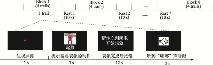
示范动作包含4个太极拳动作, 全部节选自24式杨氏太极拳, 分别是“起势” “野马分鬃” “白鹤亮翅”和“云手”。这4个动作是杨氏太极拳的基本动作, 四肢和躯干都参与运动, 难度适中, 具有一定的代表性。
2.4 实验流程
被试来到实验室后, 除第一次需要填写个人基本信息和运动表象问卷外, 其余流程如下(见图2):首先, 被试观看示范动作视频, 并在主试带领下进行练习。当被试对4个示范动作熟悉后, 独立完成动作, 每个动作3次, 主试用秒表记录完成时间, 并拍摄动作视频, 用于专家对其太极拳技能水平进行评分。
图2
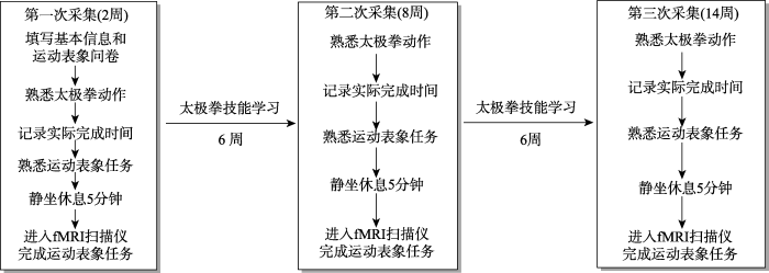
在完成上述任务后, 被试先在MRI扫描仪外进行一次运动表象任务练习, 练习程序和正式程序一致。在练习的过程中, 要求被试参照自己实际完成动作时的节奏, 采用动觉表象想象每一个动作从开始到结束的完整过程。待被试对实验流程和任务熟悉后, 静坐休息5分钟, 待心率恢复到安静状态后, 进入MRI扫描仪完成运动表象任务, 采集行为与脑数据。此外, 为了尽可能排除生物节律的影响, 安排同一名被试三次采集尽量在相同或相近的时间段。
2.5 数据采集与分析
2.5.1 行为数据采集
(1)太极拳技能水平
主试将拍摄的被试实际完成4个太极拳动作的视频进行匿名编号, 并对所有被试的脸部信息进行模糊化处理, 然后提供给太极拳专家(中国武术段位七段), 由专家对被试的太极拳技能水平进行评分(评分标准见网络版附表1)。
(2)运动表象质量
运动表象和运动实际执行的时间一致性是反映和评价运动表象质量的常用指标之一(Collet et al., 2011; Guillot et al., 2012; Malouin et al., 2008)。运动表象时间与运动实际执行时间越接近, 时间一致性数值越小, 说明运动表象质量就越好(Nakano et al., 2020)。参照沈诚等人(2016)的做法, 本研究采用运动表象时间与实际执行时间差值的绝对值。运动表象时间为被试进入MRI扫描仪完成表象任务的平均时间(4个动作, 每个动作重复8次), 通过E-Prime记录。实际执行时间为每名被试完成12次(4个动作, 每个动作重复3次)动作的平均时间。
2.5.2 脑数据采集和预处理
采用西门子3.0 T磁共振扫描仪(型号为Prismafit)进行脑功能像数据的采集。采用标准20通道头部线圈, 使用平面回波序列(Echo planar imaging, EPI)采集T2*功能像, 扫描参数为TR = 2000 ms, TE = 30 ms, 翻转角度为90度, 层厚3.5 mm, 视野为220 mm × 220 mm, 体素大小为3.4 mm × 3.4 mm × 3.5 mm, 共33层, 隔层扫描。
使用基于MATLAB R2013b 中的SPM 12工具包对功能像数据进行预处理。首先进行时间校正, 以去除同一时间图像因采集时间差异引起的误差; 接着进行头动校正, 排除头部平动大于3 mm、转动大于3度的被试, 本研究所有采集图像的头动参数均在许可范围内; 然后将校正后的图像进行空间标准化, 采用SPM自带EPI 模板, 归一化到蒙特利尔(Montreal Neurological Institute, MNI)坐标系统, 并将每个体素重新采样至3 mm × 3 mm × 3 mm; 最后使用高斯核函数进行平滑, 降低空间噪声, 全宽半高值(FWHM)为6 mm。
2.5.3 行为数据统计分析
行为数据基本服从正态分布, 使用SPSS 25.0对行为数据进行统计分析。首先进行2 (组别:实验组、对照组) × 2 (时间:2周、14周)两因素重复测量方差分析, 探究两组在太极拳技能水平和运动表象质量上的差异; 然后采用单因素重复测量方差分析, 考察实验组在不同时点太极拳技能水平和运动表象质量的变化, 事后多重比较采用Bonferroni校正, t检验和F检验分别采用Cohen’s d和η2p计算效应量。此外, 为了使研究结果更为可靠, 我们在已有的零假设检验(NHST)的基础上补充了贝叶斯因子BF10。
2.5.4 fMRI数据统计分析
首先使用SPM 12进行基于一般线性模型(general linear model, GLM)的个体分析, 感兴趣的回归量(regressor)为表象动作诱发的脑激活, 其开始时间(onset)锁定在提示开始表象界面呈现的时间, 时长(duration)为保持闭眼的12秒。不感兴趣的回归量为提示动作的图片和提醒睁眼的铃声, 开始时间为各自界面呈现的时间, 时长分别为3秒和2秒。此外, 在设计矩阵中还纳入了6个头动协变量, 以排除头动的潜在影响。将组块之间休息阶段设置为基线, 对比基线条件得到的每名被试的对比图像即为运动表象诱发的脑激活, 用于后续组分析。
然后分别对实验组被试三次时间的对比图像进行单样本t检验, 得到实验组在不同时间的激活结果。接着在SPM中选择flexible factorial设计构建单因素重复测量方差分析, 上述涉及到脑数据多体素检验的校正阈值设置为体素水平未校正, p < 0.001, 团块水平的FWE校正, p < 0.05。并以方差分析得到的差异脑区峰值点的坐标为球心, 以6 mm为半径重新作球形感兴趣区(region of interest, ROI), 使用RESTplus提取不同时间实验组被试在各ROI内的平均信号值, 并使用SPSS 25.0对提取出来的信号值进行方差分析和事后检验, 考察实验组在不同时点相关脑区激活的变化, ROI平均信号值基本服从正态分布, 事后多重比较采用Bonferroni校正, 采用Cohen’s d和η2p计算效应量, 并计算了贝叶斯因子BF10作为结果的补充。为考察实验组被试表象任务诱发的脑区激活和太极拳表象质量以及技能水平的关系, 首先对三次采集的ROI平均信号值、时间一致性和太极拳技能水平进行汇总, 然后采用皮尔逊相关检验分析三个变量之间的关系, 其次对不同采集时间ROI平均信号值、时间一致性和太极拳技能水平的变化值进行皮尔逊相关检验, 以探究三个变量的变化是否存在关联。为进一步排除表象任务熟悉性对脑功能变化的影响, 在SPM中构建配对样本t检验比较对照组前、后测运动表象诱发的脑激活是否存在显著差异。
3 结果
3.1 行为学结果
3.1.1 两因素重复测量方差分析结果
两组被试技能水平和运动表象质量的描述统计结果见表2。重复测量方差分析发现, 在太极拳技能水平上, 组别主效应显著, F(1, 27) = 304.54, p < 0.001, η2p = 0.92, 90% CI [0.87,0.95], BF10 > 100, 时间主效应显著, F(1, 27) = 25.17, p < 0.001, η2p = 0.48, 90% CI [0.25,0.64], BF10 > 100, 组别与时间的交互作用也显著, F(1, 27) = 28.29, p < 0.001, η2p = 0.51, 90% CI [0.28,0.66], BF10 > 100; 在运动表象质量上, 组别主效应显著, F(1, 27) = 4.60, p = 0.041, η2p = 0.15, 90% CI [0.00,0.35], BF10 > 1, 时间主效应以及组别与时间的交互作用均不显著(p > 0.05) (见网络版附表2)。事后检验发现, 实验组太极拳技能水平在两次测试时差异显著, t(27) = 8.80, p < 0.001, Cohen’s d = 2.06, 95% CI [1.58,2.54], BF10 > 100, 而时间一致性差异不显著(p > 0.05) (见网络版附图1); 对照组两次测试的太极拳技能水平和时间一致性均不存在显著差异(p > 0.05) (见网络版附表3), 说明在没有经过系统的太极拳学习的情况下, 对照组被试的太极拳技能水平和运动表象质量变化不大, 没有达到显著性水平。由于本研究重点考察的是学习过程中脑功能的动态变化, 因此, 后继统计分析将主要聚焦于实验组。
表2 两组行为学的描述性统计(M ± SD)
| 变量 | 组别 | 2周 | 14周 |
|---|---|---|---|
| 太极拳技能水平 | 实验组(n = 19) | 5.84 ± 0.50 | 7.55 ± 0.58 |
| 对照组(n = 10) | 3.25 ± 0.92 | 3.20 ± 0.79 | |
| 时间一致性(ms) | 实验组(n = 19) | 2045.81 ± 719.99 | 1641.70 ± 915.47 |
| 对照组(n = 10) | 2276.24 ± 1116.19 | 2799.13 ± 1601.13 |
3.1.2 实验组单因素重复测量方差结果
对实验组太极拳技能水平进行单因素重复测量方差分析发现, 时间主效应显著, F(2, 36) = 66.84, p < 0.001, η2p = 0.79, 90% CI [0.68,0.85], BF10 > 100; 事后检验发现, 14周的技能水平相比于2周(t(18) = 9.93, p < 0.001, Cohen’s d = 2.48, 95% CI [1.82,3.15], BF10 > 100)和8周(t(18) = 8.95, p < 0.001, Cohen’s d = 2.05, 95% CI [1.44,2.65], BF10 > 100)有显著提高, 但是2周和8周的技能水平差异不显著(p > 0.05)。说明从2周到8周太极拳技能水平变化不大, 而8周后技能水平显著提高, 从图3A可知, 技能水平表现出先缓慢后快速的变化特点。对时间一致性进行单因素重复测量方差分析发现, 虽然时间主效应不显著(p > 0.05, ηp2 = 0.11), 但随着学习进程的推进运动表象质量有逐渐提高的趋势(见图3B)。
图3
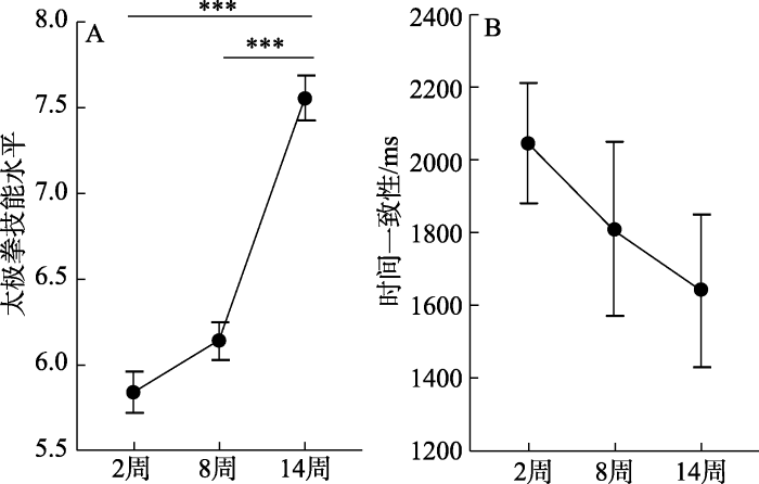
图3
不同时间(2周、8周和14周)技能水平(A)和时间一致性(B)的差异
注:**代表p < 0.01, ***代表p < 0.001, 误差线表示平均值的标准误差(standard error, SE), 事后多重比较采用Bonferroni校正。
3.2 实验组fMRI结果
3.2.1 单样本t检验
图4
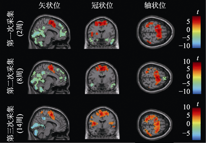
图4
不同时点运动表象激活的脑区
注:校正阈值为体素水平未校正, p < 0.001, 团块水平的FWE校正, p < 0.05; 第一次采集体素数量 > 147, 第二次采集体素数量 > 119, 第三次采集体素数量 > 55。
表3 不同时点运动表象激活的脑区
| 激活脑区 | 半球 | 体素数量 | 峰值点MNI坐标 | t值 | ||
|---|---|---|---|---|---|---|
| X | Y | Z | ||||
| 第一次采集(2周) | ||||||
| 辅助运动区/中央前回/顶下小叶/中央后回/颞上回 | 左 | 2542 | −9 | −3 | 63 | 8.92 |
| 额下回 | 右 | 203 | 39 | 15 | 6 | 6.76 |
| 额中回 | 左 | 148 | −33 | 36 | 27 | 7.80 |
| 中央后回/顶下小叶 | 右 | 231 | 42 | −36 | 45 | 7.75 |
| 距状裂/楔前叶 | 左/右 | 4360 | 27 | −27 | −6 | −11.07 |
| 颞中回 | 右 | 376 | 24 | 15 | −21 | −7.43 |
| 颞中回 | 左 | 329 | −48 | 6 | −33 | −8.01 |
| 内侧额上回 | 左/右 | 1579 | 3 | 63 | 3 | −8.43 |
| 第二次采集(8周) | ||||||
| 脑岛/中央前回/颞上回 | 左 | 120 | −45 | 0 | 3 | 6.17 |
| 顶下小叶/中央后回 | 左 | 398 | −57 | −27 | 39 | 7.07 |
| 辅助运动区/中央前回 | 左 | 1010 | −6 | −3 | 60 | 9.99 |
| 顶下小叶/中央后回 | 右 | 157 | 39 | −39 | 42 | 7.27 |
| 小脑9区 | 右 | 296 | 3 | −54 | −45 | −8.93 |
| 距状裂/楔前叶 | 左/右 | 5804 | 9 | −45 | 39 | −11.79 |
| 颞中回 | 右 | 222 | 51 | 3 | −39 | −7.11 |
| 颞中回 | 左 | 640 | −54 | 3 | −30 | −6.60 |
| 内侧额上回 | 左/右 | 1918 | 30 | 63 | 9 | −9.75 |
| 第三次采集(14周) | ||||||
| 颞上回 | 右 | 414 | 63 | −21 | 9 | 8.81 |
| 中央后回/中央前回/颞上回/辅助运动区 | 左 | 3352 | −51 | −21 | 0 | 10.28 |
| 额下回 | 右 | 144 | 57 | 9 | 24 | 7.94 |
| 中央后回 | 右 | 511 | 42 | −36 | 57 | 8.30 |
| 小脑6区 | 右 | 56 | 30 | −48 | −33 | 6.02 |
| 距状裂/楔前叶 | 左/右 | 2240 | −6 | −75 | 6 | −7.64 |
| 小脑9区 | 右 | 81 | 15 | −45 | −48 | −7.22 |
| 旁海马回 | 左/右 | 100 | 27 | −27 | −6 | −8.11 |
| 内侧扣带回 | 左 | 359 | −9 | −39 | 39 | −8.28 |
| 额中回 | 右 | 233 | 24 | 42 | 45 | −6.02 |
注:校正阈值为体素水平未校正, p < 0.001, 团块水平的FWE校正, p < 0.05; 第一次采集体素数量 > 147, 第二次采集体素数量 > 119, 第三次采集体素数量 > 55。
3.2.2 方差分析
为考察不同时点相关脑区激活的变化情况, 进行单因素重复测量方差分析, 结果显示, 左侧颞上回和左侧楔前叶在不同时点的激活存在显著差异(见表4)。
表4 不同时点运动表象激活差异的脑区
| 激活脑区 | 半球 | 体素数量 | 峰值点MNI坐标 | F值 | ||
|---|---|---|---|---|---|---|
| X | Y | Z | ||||
| 左侧颞上回 | 左 | 106 | −57 | −21 | 9 | 15.04 |
| 左侧楔前叶 | 左 | 61 | −3 | −54 | 33 | 11.33 |
注:校正阈值为体素水平未校正, p < 0.001, 团块水平的FWE校正, p < 0.05, 体素数量 > 60。
3.2.3 ROI方差分析和事后检验
以方差分析得到的脑区峰值点坐标为球心, 以6 mm为半径重新作球形ROI, 使用Restplus提取每个时点每名被试在两个ROI内的平均信号值进行方差分析和事后检验。结果显示(见图5), 在左侧颞上回上, 时间主效应显著, F(2, 36) = 13.45, p < 0.001, η2p = 0.43, 90% CI [0.21,0.58], BF10 > 100; 事后检验发现, 与2周(t(18) = 3.24, p = 0.014, Cohen’s d = 0.79, 95% CI [0.15,1.43], BF10 > 10)和8周(t(18) = 5.05, p < 0.001, Cohen’s d = 1.17, 95% CI [0.56,1.78], BF10 > 100)相比, 14周的激活显著增强, 但2周和8周之间不存在显著差异(p > 0.05)。在左侧楔前叶上, 时间主效应显著, F(2, 36) = 9.58, p < 0.001, η2p = 0.35, 90% CI [0.13,0.51], BF10 > 30; 事后检验发现, 14周的激活相比于2周 (t(18) = 3.89, p = 0.003, Cohen’s d = 0.90, 95% CI [0.29,1.51], BF10 > 30)和8周(t(18) = 3.85, p = 0.004, Cohen’s d = 0.84, 95% CI [0.26,1.41], BF10 > 30)出现了显著增强, 但2周和8周之间不存在显著差异(p > 0.05)。说明随着学习时间的延长, 左侧颞上回和左侧楔前叶的功能变化显著, 特别是从8周到14周两个脑区的功能发生了显著改变。
图5
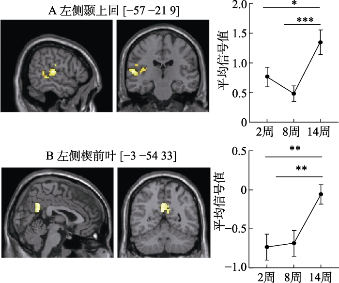
图5
不同时点左侧颞上回(A)和左侧楔前叶(B)的平均信号值差异
注:左列为交互脑区的矢状面和冠状面, 右列分别为以峰值点坐标为球心、6 mm为半径所作球形ROI 的平均信号值的事后检验, *代表p < 0.05, **代表p < 0.01, ***代表p < 0.001, 误差线表示平均值的标准误差(standard error, SE), 事后多重比较采用Bonferroni校正。
3.3 实验组ROI、时间一致性和技能水平的关系
首先, 为了解大脑功能、表象质量和技能水平三者的总体关系, 对三次采集的左侧颞上回和左侧楔前叶的平均信号值、时间一致性和太极拳技能水平汇总后进行皮尔逊相关分析; 其次, 为探究随着学习时间的延长, 大脑功能变化、运动表象质量变化和技能水平变化之间的关系, 对不同采集时点左侧颞上回和左侧楔前叶平均信号值的差值、时间一致性的差值和太极拳技能水平的差值进行皮尔逊相关分析。汇总的相关分析结果显示(见表5和图6), 被试ROI平均信号值和太极拳技能水平存在显著的中等程度的正相关(左侧颞上回, r = 0.41, p = 0.001; 左侧楔前叶, r = 0.50, p < 0.001), 表明太极拳技能水平越高, ROI激活越强。时间一致性和太极拳技能水平存在显著的较弱的负相关(r = −0.27, p = 0.042), 表明太极拳技能水平越高, 时间一致性数值越小, 运动表象质量就越好。ROI平均信号值和时间一致性的相关没有达到0.05的显著性水平, 但两者存在着负向关联的趋势。差值的相关分析结果显示(见图7), 从学习2周到14周, 被试左侧楔前叶平均信号值的变化和太极拳技能水平的变化呈显著的中等程度的正相关(r = 0.56, p = 0.013); 而从学习8周到14周, 被试在左侧楔前叶平均信号值的变化和时间一致性的变化呈显著的中等程度的负相关(r = −0.52, p = 0.022)。
表5 各变量的相关分析
| 变量 | M | SD | 1 | 2 | 3 |
|---|---|---|---|---|---|
| 1 技能水平 | 6.51 | 0.91 | — | ||
| 2 时间一致性(ms) | 1831.66 | 905.61 | −0.27* | — | |
| 3 左侧颞上回 | 0.86 | 0.81 | 0.41** | −0.16 | — |
| 4 左侧楔前叶 | −0.49 | 0.72 | 0.50*** | −0.18 | 0.41** |
注:*代表p < 0.05, **代表p < 0.01, ***代表p < 0.001。
图6
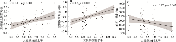
图7
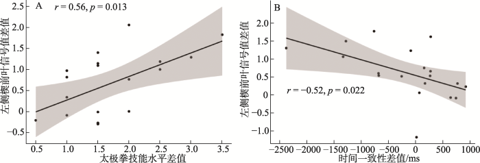
图7
左侧楔前叶平均信号值差值和太极拳技能水平差值(A)、时间一致性差值(B)的相关
注:(A)的差值为第三次采集(14周)减去第一次采集(2周), (B)的差值为第三次(14周)减去第二次(8周), 图中阴影部分为拟合线的置信区间。
总的来看, 表象任务诱发的脑区激活、太极拳表象质量和技能水平之间存在着较为紧密的关联, 随着太极拳技能水平的提高, 运动表象质量变好, 相关脑区的激活也得到了增强, 显示出太极拳技能学习对脑功能的积极影响。
3.4 对照组fMRI结果
在分析实验组脑数据的基础上, 同时对对照组进行了配对样本t检验。对照组全脑配对样本t检验结果显示, 即使在未校正的条件下(体素水平未校正, p < 0.001, 体素数量 > 10)也不存在显著差异的脑区。进一步的ROI配对样本t检验显示(见网络版附表4), 对照组在左侧颞上回和左侧楔前叶的激活均不存在显著差异(p > 0.05)。
4 讨论
本研究采用多时点纵向追踪设计, 从神经科学视角动态观测了复杂运动技能学习早期大脑功能发生的变化。发现14周的太极拳技能学习, 学习者的技能水平逐渐提高, 提高速度表现出先慢后快的变化特征, 时间一致性(运动表象质量)也具有变好的趋势; 脑数据结果发现, 随着学习时间的延长, 左侧颞上回和左侧楔前叶的功能发生了显著的变化, 特别是从8周到14周两个脑区的激活显著增强; 脑和行为的相关分析发现, 随着太极拳技能水平的提高, 时间一致性(运动表象质量)变好, 左侧颞上回和左侧楔前叶的激活也得到了增强。本研究结果为早期太极拳技能学习有助于大脑功能的积极变化提供了行为和脑激活层面的证据。值得注意的是, 我们并没有发现时间一致性(运动表象质量)在不同采集时点的显著性差异, 推测可能是本研究的样本量并不十分充足, 导致差异没能达到显著性水平; 此外, 由于磁共振扫描仪的特殊性, 被试在完成运动表象任务的时候处于仰卧位, 这与被试实际完成太极拳动作的站立位存在着较大的差异, 体位的不同可能会对时间一致性指标的精确性造成一定的影响。
本研究发现, 在太极拳技能学习的早期, 技能提高的速度并不是呈线性上升的, 而是显示出了明显的阶段性, 具体表现为从2周到8周技能水平变化不大, 而从8周到14周技能水平显著提高, 表现出先慢后快的变化特点。陈琦和刘儒德(2019)以及张力为和毛志雄(2018)认为, 技能学习过程中技能水平的提高呈现先慢后快的特点可能是因为个体新学习的动作技能和过去已有的动作经验关联不大, 可以利用的动作结构比较有限, 需要在一些基本动作上投入较多的精力, 因此刚开始学习时技能变化速度较为缓慢, 经过一段时间的学习, 一旦掌握了基本的动作结构, 技能水平的提高速度就会显著加快。本研究中的太极拳是一项包含了一系列手脚协同且精细、连贯的动作组成的复杂运动技能, 具有节奏缓慢、强度适中及身心合一的特点。在刚开始单个基本动作的学习阶段, 学习者可以利用的运动经验较少, 动作的协调和控制较差, 需要不断重复练习建立新的动作联结, 因此这一阶段技能水平变化不大。待掌握了单个基本动作后, 将这些单个动作组合成完整动作结构就相对容易, 学习者只需要思考动作之间的前后顺序和连接动作, 相对前一个阶段学习难度有所下降, 动作的协调性越来越好, 因此这一阶段技能提升速度会加快。
本研究发现, 运动表象任务在不同时点都激活了类似的额顶认知、运动控制和躯体感觉相关的脑区, 包括额下回、顶下小叶、中央前回、辅助运动区和中央后回等。额顶脑区与高阶认知控制过程相关(Jerde & Curtis, 2013), 中央前回参与运动控制与计划, 辅助运动区还和复杂动作序列的编排有关(Marchand et al., 2013), 而中央后回是参与躯体感觉加工的脑区(Tamè et al., 2016), 三次数据采集都发现了上述脑区的激活, 这可能与太极拳运动表象过程中认知加工和感觉运动加工的整合有关。本研究还发现, 从8周到14周左侧颞上回和左侧楔前叶由表象任务诱发的激活存在显著的增强, 这可能是8周到14周太极拳技能学习过程中, 伴随着技能水平的显著快速提高, 相关脑区激活也显著增强的结果。本研究发现脑区功能的变化和太极拳技能的提高表现出了类似的趋势, 8周到14周既是太极拳技能显著提高的阶段, 也是脑功能显著优化的阶段。而且, 脑和行为的相关分析也发现, 左侧颞上回和左侧楔前叶由表象任务诱发的激活和太极拳的技能水平显著相关, 这进一步说明了太极拳技能学习会对左侧颞上回和左侧楔前叶的功能产生显著的积极影响。值得注意的是, 本研究发现, 从2周到8周, 左侧颞上回的平均信号值虽然出现了暂时的下降, 但是差异并不显著, 推测可能是学习早期, 被试可以利用的已有运动经验较少, 需要不断重复练习单个基本动作, 导致技能的学习出现了一些反复, 表现为这一阶段技能水平变化不大, 因此, 相应的运动表象任务诱发的左侧颞上回的激活也变化不大。
颞上回被认为是镜像神经系统的一部分(Grèzes et al., 2004), 与动作的观察、执行、表象以及动作的理解有关(Fogassi et al., 2005; Tkach et al., 2007), 参与了序列动作的感知加工过程(Bachrach et al., 2016); 楔前叶属于默认网络的一部分(Utevsky et al., 2014), 与正念冥想以及高水平的身体表征相关(Ives- Deliperi et al., 2011), 在动作执行和动作表象过程中负责协调注意在空间目标之间的转换(Oshio et al., 2010)。已有研究也报告了运动技能学习或运动训练引发的颞上回和楔前叶脑区功能或结构的变化, 如赵琦等人(2017)的研究发现专业舞蹈运动员颞上回的局部一致性显著高于没有舞蹈经验的对照组; 张兰兰等人(2017)发现和新手相比, 篮球运动员表象篮球投篮动作时楔前叶的激活显著较强; Yamashita等人(2021)的研究结果显示, 运动员群体楔前叶的灰质体积显著大于对照组, 并且还与其参与运动训练的时长显著相关; Cui等人(2019)的研究也发现8周的太极拳锻炼显著增加了大学生颞上回、楔前叶等脑区的灰质体积。本研究中, 经过14周的系统学习, 特别是从8周到14周, 随着太极拳技能水平的显著提升, 个体完成太极拳动作越来越熟练, 完成动作的准确性也明显提高, 对涉及到与太极拳动作有关的感知、表象以及内部表征过程变得也越来越好, 而上述心理与行为表现的积极变化可能与颞上回、楔前叶神经加工能力的增强紧密相关, 从而表现出了相关脑区激活的增强, 上述结果为早期太极拳技能学习有助于优化与序列动作学习有关的脑区功能提供了证据。
当然本研究也存在着一些不足。首先, 采用运动表象任务反映实际的运动执行, 尽管该范式已得到广泛的认可和应用, 但是仍然无法替代被试实际执行动作, 未来希望能够利用更加便携的脑成像设备来探索运动技能学习大脑功能的动态变化特征; 其次, 由于客观实验条件的限制, 本研究仅采集了三次数据, 未来对此问题的后续研究会增加采集次数; 最后, 本研究对照组的样本量偏小, 虽然不会影响到本研究主要的研究发现, 但是可能无法充分揭示表象任务熟悉性潜在的影响, 后续将扩充样本量进一步深入分析。
5 结论
太极拳技能学习早期, 大脑左侧颞上回和左侧楔前叶的功能会发生积极变化, 表现为由运动表象任务诱发的神经激活显著增强, 脑区的功能变化与技能水平的提高存在显著的中等程度的正相关, 呈现出先慢后快的变化特征。研究结果提示随着太极拳技能水平的提高, 左侧颞上回和左侧楔前叶神经加工能力显著增强, 表明早期的太极拳等复杂运动技能的学习会对与序列动作学习有关的脑区功能产生积极影响, 表现为能够优化与序列动作有关的感知、表象以及内部表征过程。本研究的结果能够为运动健脑提供更加坚实的实证支撑, 从而帮助人们更好地理解运动与脑可塑性的关系。
附录:
太极拳训练计划
(1)训练内容:每次90分钟、每周5次, 其中包括3次教学指导课和2次自主练习课, 持续时间为14周, 老师全程指导和监督。每次包括10分钟热身运动, 60分钟太极拳技能训练, 10分钟呼吸训练以及10分钟放松训练。训练内容以24式杨氏太极拳为主, 也学习8式、16式太极拳。
(2)训练时间和频率:
第一个阶段为期2周。热身运动10分钟, 以拉伸练习和太极站桩为主; 技能训练60分钟, 主要包括太极拳基本的手法、步法练习以及8式太极拳的讲解和练习; 结束部分20分钟, 包括10分钟的呼吸练习和10分钟的放松练习。
第二个阶段为期6周。热身运动10分钟, 以8式太极拳练习为主; 技能训练60分钟, 主要包括16式和24式太极拳的讲解和练习; 结束部分20分钟, 包括10分钟的呼吸练习和10分钟的放松练习。
第三个阶段为期6周。热身运动10分钟, 以24式太极拳练习为主; 技能训练60分钟, 主要包括24式太极拳完整和连贯的练习, 强化动作规范, 提高动作质量; 结束部分20分钟, 包括10分钟的呼吸练习和10分钟的放松练习。
附表1 太极拳技能评分标准
| 评分分值 | 评分标准 |
|---|---|
| 0~2 | 动作不熟悉, 不能完成单式练习 |
| 2~4 | 动作不标准, 演练不连贯 |
| 4~6 | 动作基本标准, 演练基本连贯 |
| 6~8 | 动作标准, 演练流畅, 劲力柔和 |
| 8~10 | 节奏、呼吸、神态整体符合太极拳韵味 |
附表2 两因素重复测量方差分析(实验组19人, 对照组10人)
| 因变量 | F | p | ηp2 | |
|---|---|---|---|---|
| 太极拳技能水平 | 组别 | 304.54*** | < 0.001 | 0.92 |
| 时间 | 25.17*** | < 0.001 | 0.48 | |
| 组别 × 时间 | 28.29*** | < 0.001 | 0.51 | |
| 表象时间(ms) | 组别 | 0.71 | 0.406 | 0.03 |
| 时间 | 0.14 | 0.709 | 0.01 | |
| 组别 × 时间 | 3.05 | 0.092 | 0.10 | |
| 实际执行时间(ms) | 组别 | 66.69*** | < 0.001 | 0.71 |
| 时间 | 10.50** | 0.003 | 0.28 | |
| 组别 × 时间 | 5.10* | 0.032 | 0.16 | |
| 时间一致性(ms) | 组别 | 4.60* | 0.041 | 0.15 |
| 时间 | 0.06 | 0.812 | < 0.01 | |
| 组别 × 时间 | 3.51 | 0.072 | 0.12 |
注:*代表p < 0.05, **代表p < 0.01, ***代表p < 0.001。
附表3 组别和时间的事后检验(实验组19人, 对照组10人)
| 变量 | 事后检验 | t | p | Cohen’s d | |
|---|---|---|---|---|---|
| 太极拳技能水平 | 实验组 | 前测−后测 | −8.80*** | < 0.001 | −2.06 |
| 对照组 | 前测−后测 | 0.19 | 0.853 | −0.06 | |
| 前测 | 实验组−对照组 | 9.89*** | < 0.001 | 3.12 | |
| 后测 | 实验组−对照组 | 17.04*** | < 0.001 | 5.23 | |
| 表象时间(ms) | 实验组 | 前测−后测 | 1.81 | 0.082 | 0.42 |
| 对照组 | 前测−后测 | −0.85 | 0.405 | −0.27 | |
| 前测 | 实验组−对照组 | 1.54 | 0.135 | 0.62 | |
| 后测 | 实验组−对照组 | −0.20 | 0.845 | −0.07 | |
| 实际执行时间(ms) | 实验组 | 前测−后测 | 4.68*** | < 0.001 | 1.09 |
| 对照组 | 前测−后测 | 0.61 | 0.549 | 0.20 | |
| 前测 | 实验组−对照组 | 7.61*** | < 0.001 | 2.73 | |
| 后测 | 实验组−对照组 | 5.61*** | < 0.001 | 1.83 | |
| 时间一致性(ms) | 实验组 | 前测−后测 | 1.39 | 0.176 | 0.36 |
| 对照组 | 前测−后测 | −1.31 | 0.203 | −0.42 | |
| 前测 | 实验组−对照组 | −0.68 | 0.504 | −0.19 | |
| 后测 | 实验组−对照组 | −2.49* | 0.019 | −0.93 | |
注:*代表p < 0.05, **代表p < 0.01, ***代表p < 0.001。
附表4 对照组ROI的配对样本t检验(n = 10)
| 前测 | 后测 | t | p | Cohen’s d | |
|---|---|---|---|---|---|
| 左侧颞上回 | 0.25 ± 0.48 | 0.37 ± 0.52 | −0.57 | 0.580 | −0.18 |
| 左侧楔前叶 | −0.59 ± 0.65 | −0.53 ± 0.63 | −0.28 | 0.786 | −0.09 |
附图1
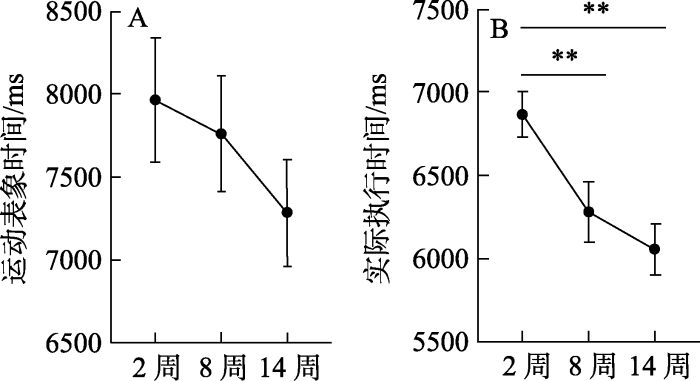
附图1
不同时间(2周、8周和14周)运动表象时间(A)和实际执行时间(B)的差异
注:**代表p < 0.01, 误差线表示平均值的标准误差(standard error, SE), 事后多重比较采用Bonferroni校正。
参考文献
Action anticipation beyond the action observation network: A functional magnetic resonance imaging study in expert basketball players
DOI:10.1111/ejn.2012.35.issue-10 URL [本文引用: 1]
Neuronal bases of structural coherence in contemporary dance observation
DOI:S1053-8119(15)00793-4
PMID:26348557
[本文引用: 1]

The neuronal processes underlying dance observation have been the focus of an increasing number of brain imaging studies over the past decade. However, the existing literature mainly dealt with effects of motor and visual expertise, whereas the neural and cognitive mechanisms that underlie the interpretation of dance choreographies remained unexplored. Hence, much attention has been given to the action observation network (AON) whereas the role of other potentially relevant neuro-cognitive mechanisms such as mentalizing (theory of mind) or language (narrative comprehension) in dance understanding is yet to be elucidated. We report the results of an fMRI study where the structural coherence of short contemporary dance choreographies was manipulated parametrically using the same taped movement material. Our participants were all trained dancers. The whole-brain analysis argues that the interpretation of structurally coherent dance phrases involves a subpart (superior parietal) of the AON as well as mentalizing regions in the dorsomedial prefrontal cortex. An ROI analysis based on a similar study using linguistic materials (Pallier et al., 2011) suggests that structural processing in language and dance might share certain neural mechanisms.Copyright © 2015 Elsevier Inc. All rights reserved.
Comparison of motor execution and motor imagery brain activation patterns: A fNIRS Study
DOI:10.3724/SP.J.1041.2016.00495
[本文引用: 1]
<p>It is widely believed that activity in the primary motor cortex relates only to motor execution. However, the extent to which similar activity occurs when imagining motor movements remains to be determined and, while some researchers report activity in the primary motor cortex during both motor execution and motor imagery tasks (e.g.Solodkin et al., 2004; Sharma et al., 2008), others report no effects of motor imagery (e.g., Binkofski et al., 2000; Hanakawa et al., 2003; Hétu et al., 2013). It is still unknown whether brain activation patterns of motor execution and motor imagery are similar, and whether both tasks activate the primary motor cortex. In addition, it is also unclear about the effect of imagination intensity on the primary motor cortex (this effect has been well established in motor execution tasks). Accordingly, the present research investigated the relationship between the intensity of real and imagined exercise on cortical activity using functional near-infrared spectroscopy (fNIRS), especially in the primary motor cortex. A preliminary assessment used 10 participants (5 male, 5 female), who did not take part in the main experiment, to establish an appropriate level of exercise intensity. For the main experiment, 30 participants (15 male, 15 female) with high imagination ability were selected using the Motor Imagery Questionnaire (Revised). These participants performed a motor execution task in which they actually lifted dumbbells under two levels of exercise intensity (males, 4 pounds and 8 pounds; females, 2 pounds and 4 pounds) and an imagery version of this task in which they imagined lifting dumbbells of these weights. The fNIRS was used to measure cortical changes in oxygen level during the performance of the two tasks. Finally, on completion of the imagery task, the “motor imagery self-assessment questionnaire” was administered to assess the quality of the participants’ imagination. All participants reported that they could imagine dumbbell movement under different levels of exercise intensity. The fNIRS showed that although both motor imagery and motor execution produced increases in cortical oxygen levels, this was greater, and lateralized to the left hemisphere, during motor execution. In addition, effects of exercise intensity were observed during motor execution but not during motor imagery, such that oxygen levels were higher with increased exercise intensity. The indication from the present findings is that both motor execution and motor imagery activate the primary motor cortex. This challenges the conventional view that activity in the primary motor cortex relates only to motor execution and shows that this activity is also connected with motor imagery, plan, and control. The present findings therefore provide an important theoretical basis for the use of motor imagery therapy in the field of neural prosthetics. However, the present findings also reveal important differences in the effects of motor execution and motor imagery, and in particular that effects of exercise intensity on cortical activation may be observed only during motor execution and not motor imagery. However, further research will be required to more fully understand the nature of these differences in cortical activation.</p>
基于fNIRS的运动执行与运动想象脑激活模式比较
Neural bases for anticipation skill in soccer: An fMRI study
Motor imagery improves balance in older adults
DOI:10.1097/TGR.0000000000000063 URL [本文引用: 1]
Measuring motor imagery using psychometric, behavioral, and psychophysiological tools
DOI:10.1097/JES.0b013e31820ac5e0
PMID:21206282
[本文引用: 2]
This review examines the measurement of motor imagery (MI) processes. First, self-report measures of MI are evaluated. Next, mental chronometry measures are considered. Then, we explain how physiological indices of the autonomic nervous system can measure MI. Finally, we show how these indices may be combined to produce a measure of MI quality called the Motor Imagery Index.
Tai Chi Chuan vs general aerobic exercise in brain plasticity: A multimodal MRI study
DOI:10.1038/s41598-019-53731-z
PMID:31754170
[本文引用: 1]
This study contrasted the impact of Tai Chi Chuan and general aerobic exercise on brain plasticity in terms of an increased grey matter volume and functional connectivity during structural magnetic resonance imaging (sMRI) and resting-state functional magnetic resonance imaging (rs-fMRI), explored the advantages of Tai Chi Chuan in improving brain structure and function. Thirty-six college students were grouped into Tai Chi Chuan (Bafa Wubu of Tai Chi), general aerobic exercise (brisk walking) and control groups. Individuals were assessed with a sMRI and rs-fMRI scan before and after an 8-week training period. The VBM toolbox was used to conduct grey matter volume analyses. The CONN toolbox was used to conduct several seed-to-voxel functional connectivity analyses. We can conclude that compared with general aerobic exercise, eight weeks of Tai Chi Chuan exercise has a stronger effect on brain plasticity, which is embodied in the increase of grey matter volume in left middle occipital gyrus, left superior temporal gyrus and right middle temporal gyrus and the enhancement of functional connectivity between the left middle frontal gyrus and left superior parietal lobule. These findings demonstrate the potential and advantages of Tai Chi Chuan exercises in eliciting brain plasticity.
Altered resting brain function and structure in professional badminton players
DOI:10.1089/brain.2011.0050
PMID:22840241
[本文引用: 1]
Neuroimaging studies of professional athletic or musical training have demonstrated considerable practice-dependent plasticity in various brain structures, which may reflect distinct training demands. In the present study, structural and functional brain alterations were examined in professional badminton players and compared with healthy controls using magnetic resonance imaging (MRI) and resting-state functional MRI. Gray matter concentration (GMC) was assessed using voxel-based morphometry (VBM), and resting-brain functions were measured by amplitude of low-frequency fluctuation (ALFF) and seed-based functional connectivity. Results showed that the athlete group had greater GMC and ALFF in the right and medial cerebellar regions, respectively. The athlete group also demonstrated smaller ALFF in the left superior parietal lobule and altered functional connectivity between the left superior parietal and frontal regions. These findings indicate that badminton expertise is associated with not only plastic structural changes in terms of enlarged gray matter density in the cerebellum, but also functional alterations in fronto-parietal connectivity. Such structural and functional alterations may reflect specific experiences of badminton training and practice, including high-capacity visuo-spatial processing and hand-eye coordination in addition to refined motor skills.
Statistical power analyses using G*Power 3.1: Tests for correlation and regression analyses
DOI:10.3758/BRM.41.4.1149
PMID:19897823
[本文引用: 1]
G*Power is a free power analysis program for a variety of statistical tests. We present extensions and improvements of the version introduced by Faul, Erdfelder, Lang, and Buchner (2007) in the domain of correlation and regression analyses. In the new version, we have added procedures to analyze the power of tests based on (1) single-sample tetrachoric correlations, (2) comparisons of dependent correlations, (3) bivariate linear regression, (4) multiple linear regression based on the random predictor model, (5) logistic regression, and (6) Poisson regression. We describe these new features and provide a brief introduction to their scope and handling.
The effects of mental practice on motor skill learning and performance: A meta-analysis
DOI:10.1123/jsp.5.1.25
URL
[本文引用: 1]
A longstanding research question in the sport psychology literature has been whether a given amount of mental practice prior to performing a motor skill will enhance one's subsequent performance. The research literature, however, has not provided any clear-cut answers to this question and this has prompted the present, more comprehensive review of existing research using the meta-analytic strategy proposed by Glass (1977). From the 60 studies yielding 146 effect sizes the overall average effect size was.48, which suggests, as did Richardson (1967a), that mentally practicing a motor skill influences performance somewhat better than no practice at all. Effect sizes were also compared on a number of variables thought to moderate the effects of mental practice. Results from these comparisons indicated that studies employing cognitive tasks had larger average effect sizes than motor or strength tasks and that published studies had larger average effect sizes than unpublished studies. These findings are discussed in relation to several existing explanations for mental practice and four theoretical propositions are advanced.
Parietal lobe: From action organization to intention understanding
DOI:10.1126/science.1106138
PMID:15860620
[本文引用: 1]
Inferior parietal lobule (IPL) neurons were studied when monkeys performed motor acts embedded in different actions and when they observed similar acts done by an experimenter. Most motor IPL neurons coding a specific act (e.g., grasping) showed markedly different activations when this act was part of different actions (e.g., for eating or for placing). Many motor IPL neurons also discharged during the observation of acts done by others. Most responded differentially when the same observed act was embedded in a specific action. These neurons fired during the observation of an act, before the beginning of the subsequent acts specifying the action. Thus, these neurons not only code the observed motor act but also allow the observer to understand the agent's intentions.
Brain mechanisms for inferring deceit in the actions of others
DOI:10.1523/JNEUROSCI.0219-04.2004
URL
[本文引用: 1]
During social interactions, it is important to judge accurately whether a person is honest or deceitful. We often use nonverbal cues to infer whether others are trying to deceive us. Using functional magnetic resonance imaging, we studied subjects watching videos of actors lifting a box and judged whether or not the actors were trying to deceive them concerning the real weight of the box. When the subjects judged the actions as reflecting deceptive intention, there was activation of the amygdala and rostral anterior cingulate cortex. These areas were not activated when subjects made judgements about the beliefs rather than the intentions of others. We suggest that these activations reflect the observers' judgements of social intentions toward themselves and might reflect an emotional response to being deceived.
Functional neuroanatomical networks associated with expertise in motor imagery
DOI:10.1016/j.neuroimage.2008.03.042
PMID:18479943
[本文引用: 1]
Although numerous behavioural studies provide evidence that there exist wide differences within individual motor imagery (MI) abilities, little is known with regards to the functional neuroanatomical networks that dissociate someone with good versus poor MI capacities. For the first time, we thus compared, through functional magnetic resonance imaging (fMRI), the pattern of cerebral activations in 13 skilled and 15 unskilled imagers during both physical execution and MI of a sequence of finger movements. Differences in MI abilities were assessed using well-established questionnaire and chronometric measures, as well as a new index based upon the subject's peripheral responses from the autonomic nervous system. As expected, both good and poor imagers activated the inferior and superior parietal lobules, as well as motor-related regions including the lateral and medial premotor cortex, the cerebellum and putamen. Inter-group comparisons revealed that good imagers activated more the parietal and ventrolateral premotor regions, which are known to play a critical role in the generation of mental images. By contrast, poor imagers recruited the cerebellum, orbito-frontal and posterior cingulate cortices. Consistent with findings from the motor sequence learning literature and Doyon and Ungerleider's model of motor learning [Doyon, J., Ungerleider, L.G., 2002. Functional anatomy of motor skill learning. In: Squire, L.R., Schacter, D.L. (Eds.), Neuropsychology of memory, Guilford Press, pp. 225-238], our results demonstrate that compared to skilled imagers, poor imagers not only need to recruit the cortico-striatal system, but to compensate with the cortico-cerebellar system during MI of sequential movements.
Understanding the timing of motor imagery: Recent findings and future directions
DOI:10.1080/1750984X.2011.623787 URL [本文引用: 2]
"Neural efficiency" of athletes’ brain during visuo-spatial task: An fMRI study on table tennis players
DOI:10.3389/fnbeh.2017.00072 URL [本文引用: 1]
Functional properties of brain areas associated with motor execution and imagery
DOI:10.1152/jn.00132.2002
PMID:12574475
[本文引用: 1]
Imagining motor acts is a cognitive task that engages parts of the executive motor system. While motor imagery has been intensively studied using neuroimaging techniques, most studies lack behavioral observations. Here, we used functional MRI to compare the functional neuroanatomy of motor execution and imagery using a task that objectively assesses imagery performance. With surface electromyographic monitoring within a scanner, 10 healthy subjects performed sequential finger-tapping movements according to visually presented number stimuli in either a movement or an imagery mode of performance. We also examined effects of varied and fixed stimulus types that differ in stimulus dependency of the task. Statistical parametric mapping revealed movement-predominant activity, imagery-predominant activity, and activity common to both movement and imagery modes of performance (movement-and-imagery activity). The movement-predominant activity included the primary sensory and motor areas, parietal operculum, and anterior cerebellum that had little imagery-related activity (-0.1 ~ 0.1%), and the caudal premotor areas and area 5 that had mild-to-moderate imagery-related activity (0.2 ~ 0.7%). Many frontoparietal areas and posterior cerebellum demonstrated movement-and-imagery activity. Imagery-predominant areas included the precentral sulcus at the level of middle frontal gyrus and the posterior superior parietal cortex/precuneus. Moreover, activity of the superior precentral sulcus and intraparietal sulcus areas, predominantly on the left, was associated with accuracy of the imagery task performance. Activity of the inferior precentral sulcus (area 6/44) showed stimulus-type effect particularly for the imagery mode. A time-course analysis of activity suggested a functional gradient, which was characterized by a more "executive" or more "imaginative" property in many areas related to movement and/or imagery. The results from the present study provide new insights into the functional neuroanatomy of motor imagery, including the effects of imagery performance and stimulus-dependency on brain activity.
The PETTLEP approach to motor imagery: A functional equivalence model for sport psychologists
DOI:10.1080/10413200109339004 URL [本文引用: 1]
The neural substrates of mindfulness: An fMRI investigation
DOI:10.1080/17470919.2010.513495
PMID:20835972
[本文引用: 1]
"Mindfulness" is a capacity for heightened present-moment awareness that we all possess to a greater or lesser extent. Enhancing this capacity through training has been shown to alleviate stress and promote physical and mental well-being. As a consequence, interest in mindfulness is growing and so is the need to better understand it. This study employed functional magnetic resonance imaging (fMRI) to identify the brain regions involved in state mindfulness and to shed light on its mechanisms of action. Significant signal decreases were observed during mindfulness meditation in midline cortical structures associated with interoception, including bilateral anterior insula, left ventral anterior cingulate cortex, right medial prefrontal cortex, and bilateral precuneus. Significant signal increase was noted in the right posterior cingulate cortex. These findings lend support to the theory that mindfulness achieves its positive outcomes through a process of disidentification.
Maps of space in human frontoparietal cortex
Searching for the principles of brain plasticity and behavior
DOI:10.1016/j.cortex.2013.11.012
PMID:24457097
[本文引用: 1]
An important development in behavioral neuroscience in the past 25 years has been the demonstration that the brain is far more flexible in structure and function than was previously believed. Studies of laboratory animals have provided an important tool for understanding the nature of brain plasticity and behavior at many levels ranging from detailed behavioral paradigms, electrophysiology, neuronal morphology, protein chemistry, and epigenetics. Here we seek a synthesis of the multidisciplinary work on brain plasticity and behavior to identify some general principles on how the brain changes in response to a wide range of experiences over the lifetime. Copyright © 2013 Elsevier Ltd. All rights reserved.
Motor learning produces parallel dynamic functional changes during the execution and imagination of sequential foot movements
DOI:10.1006/nimg.2001.1048
PMID:11969325
[本文引用: 1]
The aim of the present positron emission tomography study was to measure the dynamic changes in cerebral activity before and after practice of an explicitly known sequence of foot movements when executed physically and to compare them to those elicited during motor imagery of the same movements. Nine healthy volunteers were scanned while performing both types of movement at an early phase of learning and after a 1-h training period of a sequence of dorsiflexions and plantarflexions with the left foot. These experimental conditions were compared directly, as well as to a perceptual control condition. Changes in regional cerebral blood flow associated with physical execution of the sequence early in the learning process were observed bilaterally in the dorsal premotor cortex and cerebellum, as well as in the left inferior parietal lobule. After training, however, most of these brain regions were no longer significantly activated, suggesting that they are critical for establishing the cognitive strategies and motor routines involved in executing sequential foot movements. By contrast, after practice, an increased level of activity was seen bilaterally in the medial orbitofrontal cortex and striatum, as well as in the left rostral portion of the anterior cingulate and a different region of the inferior parietal lobule, suggesting that these structures play an important role in the development of a long lasting representation of the sequence. Finally, as predicted, a similar pattern of dynamic changes was observed in both phases of learning during the motor imagery conditions. This last finding suggests that the cerebral plasticity occurring during the incremental acquisition of a motor sequence executed physically is reflected by the covert production of this skilled behavior using motor imagery.2002 Elsevier Science (USA).
The influence of sports training on the brain plasticity of athletes: An ALE analysis of fMRI studies
运动训练对运动员大脑功能可塑性变化的影响——基于fMRI研究的ALE分析
Reliability of mental chronometry for assessing motor imagery ability after stroke
DOI:10.1016/j.apmr.2007.11.006
PMID:18226656
[本文引用: 1]
To examine the reproducibility of 2 chronometric tests: time-dependent motor imagery (TDMI) screening test and temporal congruence test.Test-retest 10 to 14 days apart.Laboratory of a university-affiliated center for research in rehabilitation.Twenty persons post cerebrovascular accident (CVA) and 46 healthy persons (controls).The reproducibility of the TDMI screening test, wherein the number of stepping movements (performed in sitting) imagined over 15, 25, and 45 seconds is recorded, and of the temporal congruence test wherein the duration of physically executed (E) and imagined (I) stepping movements is recorded, was evaluated.The test-retest reliability of the number of imagined movements (TDMI screening test), movement duration and I/E time ratios (temporal congruence test), and intrasession reliability of the temporal congruence test were assessed by using intraclass correlation coefficients (ICCs).For the TDMI screening test, the ICCs ranged from.88 to.93 (CVA, n=20) and from.87 to.92 (controls, n=9). For the temporal congruence test, when the total duration of 2 series of 5 stepping movements was averaged, ICCs ranged from.76 to.97 (CVA, n=20) and from.77 to.93 (controls, n=46), whereas for 1 series the ICCs ranged from.71 to.95 and from.63 to.95 in the CVA and control groups, respectively. The ICCs for intrasession reliability for the CVA (n=20) and control (n=46) groups, respectively, ranged from.90 to.98 and.95 to.97.The present findings support the reproducibility of both tests in both groups. Mental chronometry can be used reliably for the screening of patients capable of motor imagery or for measuring temporal congruence between real and imagined movements poststroke.
Functional architecture of the cortico-basal ganglia circuitry during motor task execution: Correlations of strength of functional connectivity with neuropsychological task performance among female subjects
DOI:10.1002/hbm.21505
PMID:22287185
[本文引用: 1]
The primary aim of this study was to enhance our understanding of the functional architecture of the cortico-basal ganglia circuitry during motor task execution. Twenty right-handed female subjects without any history of neuropsychiatric illness underwent fMRI at 3 T. The activation paradigm was a complex motor task completed with the nondominant hand. Analyses of functional connectivity strength were conducted for pairs of structures in input, intrinsic, and output segments of the circuitry. Next, connectivity strengths were correlated with results of neurocognitive testing conducted outside of the scanner, which provided information about both motor and cognitive processes. For input pathways, results indicate that SMA-striatum interactions are particularly relevant for motor behavior and disruptions may impact both motor and cognitive functions. For intrinsic pathways, results indicate that thalamus (VA nucleus) to striatum feedback pathway appears to have an important role during task execution and carries information relevant for motor planning. Together, these findings add to accumulating evidence that the GPe may play a role in higher order basal ganglia processing. A potentially controversial finding was that strong functional connectivity appears to occur across intrinsic inhibitory pathways. Finally, output (thalamus to cortex) feedback was only correlated with motor planning. This result suggests circuit processes may be more relevant for future behaviors than the execution of the current task.Copyright © 2012 Wiley Periodicals, Inc.
Increased time difference between imagined and physical walking in older adults at a high risk of falling
DOI:10.3390/brainsci10060332
URL
[本文引用: 1]
Walking motor imagery ability is thought to be associated with a fear of falling; however, no studies have compared fall risk and motor imagery ability. This study aimed to ascertain the time difference between imagined and physical walking in older adults at low and high risks of falling. Motor imagery ability was assessed using mental chronometry, which measures the imagined time required for movement. Participants included 31 older adults classified as having a high (n = 15) or low (n = 16) risk of falling based on single leg stance time. The time required for imagined and physical walking was measured using 5 m long walkways with three different widths (15, 25, and 50 cm), and the temporal errors (absolute and constant error) were compared. Physical walking time was significantly longer in the high-risk group than in the low-risk group for the 15 and 25 cm wide walkways. The absolute error between the imagined and physical walking times was significantly larger in the high-risk group than in the low-risk group for the 15 and 25 cm wide walkways. There was also a significant difference in the constant error between the high- and low-risk groups between the imagined and physical walking times for all three walkways. Older adults who may be at a higher risk of falling showed longer walking times during action execution but overestimated their performance (i.e., they believe they would be faster) during motor imagery. Therefore, the time difference between imagined and physical walking could, in part, be useful as a tool for assessing fall risk based on motor imagery.
Differential effect of double-pulse TMS applied to dorsal premotor cortex and precuneus during internal operation of visuospatial information
DOI:10.1016/j.neuroimage.2009.07.034
PMID:19632337
[本文引用: 1]
Human neuro-imaging studies have often reported co-activation of the dorsal premotor cortex (PMd) and the posterior parietal cortex (PPC) during internal operation of visuospatial information, referred to here as "visuospatial mental operation". However, the functions assigned to the PMd and PPC during these tasks are still unclear. Here, we examined the significance of these two areas for a visuospatial mental operation using the transcranial magnetic stimulation (TMS) technique. Subjects performed a task in which a visuospatial mental operation was required. A localization study conducted prior to the TMS experiment using functional magnetic resonance imaging (fMRI) revealed that the PMd and the medial part of the PPC, precuneus (PCu), were specifically activated during the visuospatial mental operation. Then, we impeded the activities of the PMd and the PCu in the right hemisphere during the same task using double-pulse TMS to determine whether these activities were necessary for the task. The TMS was applied at different times in relation to the visuospatial mental operation cue. Consequently, only the TMS applied at 300 ms after the cue affected the task performance. Furthermore, we found that the TMS at this time to each area differentially affected the performance: TMS to the PMd hindered the performance of the task whereas TMS to the PCu facilitated it without a speed/accuracy trade-off. These effects were not found in the control condition that lacked a visuospatial mental operation. These findings suggest that the PMd and the PCu are involved in differential aspects of visuospatial mental operations.
Dynamic changes in the cortico-subcortical network during early motor learning
DOI:10.3233/NRE-2010-0540 URL [本文引用: 1]
Cognition and brain function in elderly Tai Chi practitioners: A case-control study
DOI:10.1016/j.explore.2018.04.007 URL [本文引用: 1]
A review of brain plasticity of motor skill experts: Evidence from magnetic resonance imaging
运动技能专家脑可塑性研究进展: 来自磁共振成像的证据
The mind's eye: Functional MR imaging evaluation of golf motor imagery
Mental imagery involves rehearsing or practicing a task in the mind with no physical movement. The technique is commonly used, but the actual physical foundation of imagery has not been evaluated for the fast, complex, automatic motor movement of the golf swing. This study evaluated motor imagery of the golf swing, of golfers of various handicaps, by using functional MR imaging to assess whether areas of brain activation could be defined by this technique and to define any association between activated brain areas and golf skill.Six golfers of various handicap levels were evaluated with functional MR imaging during a control condition and during mental imagery of their golf swing. Two control conditions were evaluated--"rest" and "wall"--and were then subtracted from the experimental condition to give the functional activation map. These control conditions were then tested against the golf imagery; the participants were told to mentally rehearse their golf swings from a first person perspective. The percentages of activated pixels in 137 defined regions of interest were calculated.The "rest-versus-golf" paradigm showed activation in motor cortex, parietal cortex, frontal lobe, cerebellum, vermis, and action planning areas (frontal and parietal cortices, supplementary motor area, cerebellum) and areas involved with error detection (cerebellum). Vermis, supplementary motor area, cerebellum, and motor regions generally showed the greatest activation. Little activation was seen in the cingulate gyrus, right temporal lobe, deep gray matter, and brain stem. A correlation existed between increased number of areas of activation and increased handicap.This study showed the feasibility of defining areas of brain activation during imagery of a complex, coordinated motor task. Decreased brain activation occurred with increased golf skill level for the supplementary motor area and cerebellum with little activation of basal ganglia.
Environment and brain plasticity: Towards an endogenous pharmacotherapy
DOI:10.1152/physrev.00036.2012
PMID:24382886
[本文引用: 1]
Brain plasticity refers to the remarkable property of cerebral neurons to change their structure and function in response to experience, a fundamental theoretical theme in the field of basic research and a major focus for neural rehabilitation following brain disease. While much of the early work on this topic was based on deprivation approaches relying on sensory experience reduction procedures, major advances have been recently obtained using the conceptually opposite paradigm of environmental enrichment, whereby an enhanced stimulation is provided at multiple cognitive, sensory, social, and motor levels. In this survey, we aim to review past and recent work concerning the influence exerted by the environment on brain plasticity processes, with special emphasis on the underlying cellular and molecular mechanisms and starting from experimental work on animal models to move to highly relevant work performed in humans. We will initiate introducing the concept of brain plasticity and describing classic paradigmatic examples to illustrate how changes at the level of neuronal properties can ultimately affect and direct key perceptual and behavioral outputs. Then, we describe the remarkable effects elicited by early stressful conditions, maternal care, and preweaning enrichment on central nervous system development, with a separate section focusing on neurodevelopmental disorders. A specific section is dedicated to the striking ability of environmental enrichment and physical exercise to empower adult brain plasticity. Finally, we analyze in the last section the ever-increasing available knowledge on the effects elicited by enriched living conditions on physiological and pathological aging brain processes.
The effect of different proprioception input on motor imagery of complex movement: A fMRI study
不同本体感觉输入对复杂运动动作表象的影响: fMRI研究
How is a motor skill learned? Change and invariance at the levels of task success and trajectory control
DOI:10.1152/jn.00856.2011
PMID:22514286
[本文引用: 1]
The public pays large sums of money to watch skilled motor performance. Notably, however, in recent decades motor skill learning (performance improvement beyond baseline levels) has received less experimental attention than motor adaptation (return to baseline performance in the setting of an external perturbation). Motor skill can be assessed at the levels of task success and movement quality, but the link between these levels remains poorly understood. We devised a motor skill task that required visually guided curved movements of the wrist without a perturbation, and we defined skill learning at the task level as a change in the speed-accuracy trade-off function (SAF). Practice in restricted speed ranges led to a global shift of the SAF. We asked how the SAF shift maps onto changes in trajectory kinematics, to establish a link between task-level performance and fine motor control. Although there were small changes in mean trajectory, improved performance largely consisted of reduction in trial-to-trial variability and increase in movement smoothness. We found evidence for improved feedback control, which could explain the reduction in variability but does not preclude other explanations such as an increased signal-to-noise ratio in cortical representations. Interestingly, submovement structure remained learning invariant. The global generalization of the SAF across a wide range of difficulty suggests that skill for this task is represented in a temporally scalable network. We propose that motor skill acquisition can be characterized as a slow reduction in movement variability, which is distinct from faster model-based learning that reduces systematic error in adaptation paradigms.
Bilateral representations of touch in the primary somatosensory cortex
DOI:10.1080/02643294.2016.1159547
PMID:27314449
[本文引用: 1]
According to current textbook knowledge, the primary somatosensory cortex (SI) supports unilateral tactile representations, whereas structures beyond SI, in particular the secondary somatosensory cortex (SII), support bilateral tactile representations. However, dexterous and well-coordinated bimanual motor tasks require early integration of bilateral tactile information. Sequential processing, first of unilateral and subsequently of bilateral sensory information, might not be sufficient to accomplish these tasks. This view of sequential processing in the somatosensory system might therefore be questioned, at least for demanding bimanual tasks. Evidence from the last 15 years is forcing a revision of this textbook notion. Studies in animals and humans indicate that SI is more than a simple relay for unilateral sensory information and, together with SII, contributes to the integration of somatosensory inputs from both sides of the body. Here, we review a series of recent works from our own and other laboratories in favour of interactions between tactile stimuli on the two sides of the body at early stages of processing. We focus on tactile processing, although a similar logic may also apply to other aspects of somatosensation. We begin by describing the basic anatomy and physiology of interhemispheric transfer, drawing on neurophysiological studies in animals and behavioural studies in humans that showed tactile interactions between body sides, both in healthy and in brain-damaged individuals. Then we describe the neural substrates of bilateral interactions in somatosensation as revealed by neurophysiological work in animals and neuroimaging studies in humans (i.e., functional magnetic resonance imaging, magnetoencephalography, and transcranial magnetic stimulation). Finally, we conclude with considerations on the dilemma of how efficiently integrating bilateral sensory information at early processing stages can coexist with more lateralized representations of somatosensory input, in the context of motor control.
Increased hippocampus-medial prefrontal cortex resting-state functional connectivity and memory function after Tai Chi Chuan practice in elder adults
DOI:10.3389/fnagi.2016.00025
PMID:26909038
[本文引用: 1]
Previous studies provide evidence that aging is associated with the decline of memory function and alterations in the hippocampal (HPC) function, including functional connectivity to the medial prefrontal cortex (mPFC). In this study, we investigated if longitudinal (12-week) Tai Chi Chuan and Baduanjin practice can improve memory function and modulate HPC resting-state functional connectivity (rs-FC). Memory function measurements and resting-state functional magnetic resonance imaging (rs-fMRI) were applied at the beginning and the end of the experiment. The results showed that (1) the memory quotient (MQ) measured by the Wechsler Memory Scale-Chinese Revision significantly increased after Tai Chi Chuan and Baduanjin practice as compared with the control group, and no significant difference was observed in MQ between the Tai Chi Chuan and Baduanjin groups; (2) rs-FC between the bilateral hippocampus and mPFC significantly increased in the Tai Chi Chuan group compared to the control group (also in the Baduanjin group compared to the control group, albeit at a lower threshold), and no significant difference between the Tai Chi Chuan and Baduanjin groups was observed; (3) rs-FC increases between the bilateral hippocampus and mPFC were significantly associated with corresponding memory function improvement across all subjects. Similar results were observed using the left or right hippocampus as seeds. Our results suggest that both Tai Chi Chuan and Baduanjin may be effective exercises to prevent memory decline during aging.
Congruent activity during action and action observation in motor cortex
DOI:10.1523/JNEUROSCI.2895-07.2007
URL
[本文引用: 1]
A variety of studies have shown that motor cortical areas can be activated by observation of familiar actions. Here, we describe single-neuron responses in monkey primary motor (MI) and dorsal premotor (PMd) cortices during passive observation and execution of a familiar task. We show that the spiking modulation, preferred directions, and encoded information of cells in MI and PMd remain consistent during both observation and movement. Furthermore, we find that the presence of a visual target is necessary to elicit this congruent neural activity during observation. These findings along with results from our analysis of the oscillatory power in the beta frequency of the local field potential are consistent with previous imaging and EEG studies that have suggested that congruence between observation and action is a general feature of the motor system, even outside of canonical “mirror” areas. Such congruent activity has proposed relevance to motor learning, mimicry, and communication and has practical applications for the development of motor-cortical neuroprostheses in paralyzed patients.
How are the motor system activity and functional connectivity between the cognitive and sensorimotor systems modulated by athletic expertise?
DOI:10.1016/j.brainres.2013.09.048
PMID:24099840
[本文引用: 1]
Expertise offers a unique insight into how our brain functions. The purpose of this experiment was to determine if motor system activity and functional connectivity between the cognitive system and sensorimotor system is differentially modulated by an individual's level of expertise. This goal was achieved through the acquisition of functional neuroimaging data in 10 expert volleyball players and 10 novice individuals who were presented with a series of sentences describing possible technical volleyball-specific motor acts and acts that cannot be performed as positive ("Do …!") or negative ("Don't …") commands, while they were silently reading them and deciding whether the actions were technically feasible or not. Compared with novices, experts' activity in the left primary motor cortex hand area (M1) and in the left premotor cortex (Pm) was decreased by impossible actions presented as positive commands. Sensorimotor activation in response to action-related stimuli is not that automatic as held since we found that these areas were deactivated during the task, and their functional connectivity to the primary visual cortex was strengthened for possible actions presented as positive commands, reflecting the neural processes underlying the interaction between motor and visual imagery. These results suggest that the neural activity within the key areas implicitly triggered by motor simulation is a function of the expertise, action feasibility, and context. © 2013 Elsevier B.V. All rights reserved.
Precuneus is a functional core of the default-mode network
DOI:10.1523/JNEUROSCI.4227-13.2014
URL
[本文引用: 1]
Efforts to understand the functional architecture of the brain have consistently identified multiple overlapping large-scale neural networks that are observable across multiple states. Despite the ubiquity of these networks, it remains unclear how regions within these large-scale neural networks interact to orchestrate behavior. Here, we collected functional magnetic resonance imaging data from 188 human subjects who engaged in three cognitive tasks and a resting-state scan. Using multiple tasks and a large sample allowed us to use split-sample validations to test for replication of results. We parceled the task-rest pairs into functional networks using a probabilistic spatial independent components analysis. We examined changes in connectivity between task and rest states using dual-regression analysis, which quantifies voxelwise connectivity estimates for each network of interest while controlling for the influence of signals arising from other networks and artifacts. Our analyses revealed systematic state-dependent functional connectivity in one brain region: the precuneus. Specifically, task performance led to increased connectivity (compared to rest) between the precuneus and the left frontoparietal network (lFPN), whereas rest increased connectivity between the precuneus and the default-mode network (DMN). The absolute magnitude of this effect was greater for DMN, suggesting a heightened specialization for resting-state cognition. All results replicated within the two independent samples. Our results indicate that the precuneus plays a core role not only in DMN, but also more broadly through its engagement under a variety of processing states.
The reorganization of resting-state brain networks associated with motor imagery training in chronic stroke patients
DOI:10.1109/TNSRE.2019.2940980
PMID:31536007
[本文引用: 1]
A number of studies have suggested that motor imagery training (MIT) has a positive influence on the upper extremity motor recovery in stroke patients, but little is known about its neural basis. To investigate the cortical motor network plasticity after MIT, 34 chronic hemiplegic subjects with subcortical stroke were recruited and randomly allocated to either the conventional rehabilitation therapy (CRT) or the CRT + MIT. The patients were assessed with the upper limb section of Fugl-Meyer assessment Scale (FM-UL) and resting-state fMRI before and after the 4 weeks of treatment. Seed-based functional connectivity (FC) of the ipsilesional primary motor cortex (M1) and graph-theory based analysis were used to explore the relationships between the motor recovery and reorganization of motor networks. We found that the patients in the MIT group showed more improvement in the FM-UL scores compared with the CRT group. Both groups presented increased inter-hemispheric and decreased intra-hemispheric FC of the ipsilesional M1 after intervention. However, the MIT group showed increased FC of the ipsilesional M1 with the ipsilesional precentral and postcentral gyri, middle cingulate gyrus and supramarginal gyrus after intervention, while the CRT group showed decreased FC in these regions. In addition, the clustering coefficient was significantly increased in the MIT group but not in the CRT group, and the increment of clustering coefficient was significantly positively correlated with improvement of FM-UL scores. Therefore, MIT might contribute to the motor recovery in stroke patients through the following network reorganization, i.e., promoting the efficiency of regional neuronal communication and the reorganization of intrinsic FC of the ipsilesional M1 involving widely distributed motor network in both hemispheres.
Sport expert's motor imagery: Functional imaging of professional motor skills and simple motor skills
DOI:10.1016/j.brainres.2009.08.014 URL [本文引用: 2]
Tai Chi Chuan optimizes the functional organization of the intrinsic human brain architecture in older adults
Further validation and development of the movement imagery questionnaire
Task-switching performance improvements after Tai Chi Chuan training are associated with greater prefrontal activation in older adults
DOI:10.3389/fnagi.2018.00280 URL [本文引用: 1]
Impact of early- commenced and continued sports training on the precuneus in older athletes
DOI:10.3389/fnhum.2021.766935
URL
[本文引用: 1]
Intervention studies on sedentary older adults have demonstrated that commencing physical exercise at an older age has a positive effect on brain structure. Although this suggests that older athletes with lifelong sports training have larger gray matter volume (GMV) in some brain regions compared to age-matched non-athletes, evidence in the literature is scarce. Moreover, it remains unclear whether a larger GMV is associated with training intensity or period of training in life. To address these gaps in the literature, we compared regional brain GMV between 24 older athletes (mean age, 71.4 years; age at the commencement of sports training, 31.2 years, continuous sports training, 40.0 years; current training time, 7.9 h/week) and 24 age-matched non-athletes (mean age, 71.0 years). The period of sports training and the current training time of the athletes were assessed. Both groups were evaluated for physical activity intensity as well as cognitive and motor performance. Although no group differences were noted in cognitive and motor performance, athletes reported higher physical activity intensity than non-athletes. Whole-brain structural analysis revealed a significantly larger GMV in several brain regions in athletes. Notably, the GMV of the precuneus in athletes was positively correlated with earlier commencement of sports training and training duration but was negatively correlated with current training time. Our findings demonstrate that early-commenced and continued sports training predicts structural maintenance of the precuneus in old age. Our results also suggest that excessive training time in old age may have a negative impact on the GMV of the precuneus; thereby delineating how the precuneus is associated with lifelong sports training in older athletes.
The influence of motor expertise on the brain activity of motor task performance: A meta-analysis of functional magnetic resonance imaging studies
Design and effect evaluation of the performance of specific imagery training program: A case study of free throw
专项表象训练方案设计与效果测评——以篮球罚球训练为例
Regular Tai Chi practice is associated with improved memory as well as structural and functional alterations of the hippocampus in the elderly
DOI:10.3389/fnagi.2020.586770 URL [本文引用: 1]
The effects of motor skill level and somatosensory input on motor imagery: An fMRI study on basketball free shot
DOI:10.3724/SP.J.1041.2017.00307
[本文引用: 2]
The behavioral performance and neural responses of imagining a motor task may be different in individuals with different motor skill levels. Utilization of special instrument induces plastic changes in the brain in the way that tool users combine the tool in their body schema. However, the neural correlates underlying these effects are still not clear. Using functional magnetic resonance imaging (fMRI), we investigated the effects of motor skill level and somatosensory input on motor imagery. We tested 12 basketball players and 12 novices when they performed kinesthetic motor imagery of basketball free shot under with-ball and without-ball conditions. Motor imagery duration was calculated. Motor imagery strategy and quality were measured with self-evaluation questionnaires. Field training was implemented and the physical performance of the task was recorded before the main experiment. The time difference between the duration for motor imagery and physical performance was smaller in basketball players than that in novices. The motor imagery strategy scores with self-evaluation questionnaire were same in two groups, indicating that all participants performed kinesthetic imagery successfully. The general kinesthetic and visual imagery abilities between groups showed no significant differences whereas basketball players showed better kinesthetic imagery quality for free shot movements. The scores for the quality of both kinesthetic and visual imagery were higher under with-ball than that under without-ball condition in both groups, suggesting that participants in both groups performed better motor imagery under with-ball condition. fMRI analysis revealed (1) basketball players showed greater mirror neuron system activation relative to novices; (2) the activation of mirror neuron system in basketball players were lower in with-ball compared to without-ball condition. Moreover, the activity of right inferior frontal gyrus was negatively correlated with the self-evaluated kinesthetic imagery scores only for the basketball players. We conclude that better motor imagery is related to higher level motor skill. The application of task- specific instrument may be required for this facilitation effect. Utilization of special instrument induces plastic changes in mirror neuron system. The findings may provide evidence for the implication of motor imagery with utilization of specific instrument in the promotion of motor skill.
运动技能水平与躯体感觉输入对运动表象的影响
Expertise-level-dependent functionally plastic changes during motor imagery in basketball players
DOI:10.1016/j.neuroscience.2018.03.050 URL [本文引用: 2]
Brain plasticity of table tennis athletes based on dynamic amplitude of low-frequency fluctuation method
基于动态低频振荡振幅方法的乒乓球运动员脑可塑性变化
The optimization in sensorimotor system in dancers: Evidences from resting state fMRI
舞蹈运动员大脑感知运动系统的功能特征——一项静息态功能磁共振研究
