

1 问题提出
在熟悉的环境中穿行, 在陌生的环境中寻路, 空间导航活动时刻在我们的生活中发生。对空间的认知被认为是人类认知和智能的“核心知识” (Core Knowledge) (Spelke & Kinzler, 2007)。因此, 研究空间导航的发展、认知神经基础和遗传因素, 对我们理解智能的本质和起源, 有着重要的科学意义。英国伦敦大学学院教授John O'Keefe、挪威科技大学教授May-Britt Moser、Edvard Moser通过对老鼠的电生理实验研究(Fyhn et al., 2004; O'Keefe & Nadel, 1978), 首次发现了构成大脑空间定位系统的功能特异神经元细胞, 开启了理解人类空间导航神经基础的大门。凭借这一发现, 他们获得了2014年诺贝尔生理学或医学奖。此外, 青少年时期的空间能力与其将来能否在科学、技术、工程和数学等理工科领域(STEM)取得更好的学业及职业成就, 有着直接关联(Wai et al., 2009); 而且, 空间导航能力的退化, 又是阿尔兹海默症(Alzheimer's disease, AD)等与记忆有关的神经退行性疾病的核心临床表现(张家鑫 等, 2019; Coughlan et al., 2018)。因此, 对人类空间导航的认知神经和遗传基础的研究具有重要的教育意义和临床价值。
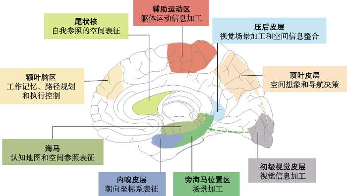
近几十年来, 空间导航研究领域取得了重要进展, 其研究对象涵盖不同动物模型、儿童和成年人, 研究方法涉及行为、电生理记录、功能和结构脑成像等。这些研究成果, 主要集中在对单个脑区特异性功能的刻画, 比如海马(hippocampus)中的位置细胞(place cell)和内嗅皮层(entorhinal cortex)的网格细胞(grid cell)等(Fyhn et al., 2004; O'Keefe & Nadel, 1978); 此外就是利用脑影像的方法, 找到不同脑区的特异性功能, 分别对应空间导航的不同认知成分, 比如位于内嗅皮层的类网格细胞功能和位于旁海马的场景加工脑区(Horner et al., 2016; Zhen et al., 2017)。这些研究, 极大地推进了我们对人类空间导航的认知加工和神经机制的理解(图1) (王欣 等, 2018; 许琴 等, 2010; Coughlan et al., 2018; Epstein et al., 2017)。
图1
然而, 空间导航是一个复杂的认知过程, 灵活的导航需要多感知模态整合、视空间编码、记忆和决策等多个认知成分的共同参与(Wolbers & Hegarty, 2010; Baumann & Mattingley, 2021)。而且, 研究者认为, 这些认知过程并非不同表征模块独立运作(modular processing), 而是一个高度动态、需要整合不同模态信息的加工过程(heuristic processing) (Ekstrom et al., 2017; Kong et al., 2017)。此外, 研究表明, 对导航至关重要的海马, 其功能不仅仅是对位置的表征, 它似乎更像是普遍的粘合剂(“the hippocampus as a binding device”) (Pu et al., 2020; Cornwell et al., 2008; Kessels et al., 2001; Mitchell et al., 2000), 联结了来自其他脑区的不同模态信息, 以支持人类的行为和决策。因此, 研究者指出, 研究空间导航的神经机制不应局限于某个特定脑区(如海马), 而应该从神经环路或网络的角度, 研究不同脑区如何交互以支撑复杂导航行为(Ekstrom et al., 2017; Baumann & Mattingley, 2021; Kong et al., 2017)。目前, 我们对人类大脑空间导航脑网络的架构和运作机制还处于起步阶段, 很多关键问题尚不清楚。例如, 导航脑网络的复杂网络属性(核心节点和模块化)是怎样的?通常被认为对空间导航至关重要的海马结构, 是导航脑网络的核心节点吗?一些与导航和记忆有关的神经退行性疾病, 如阿尔兹海默症, 是否表现出导航脑网络异常?如何实现对导航脑网络的精准调控, 从而改善导航行为表现?
鉴于此, 本项目拟采用跨学科研究手段, 对空间导航的脑网络基础和调控机制进行研究。本项目有望为我们理解空间导航在脑网络水平的神经机制提供新的科学证据, 同时对空间导航脑网络和调控机制的研究, 为研究AD等认知障碍脑疾病提供了新的重要视角, 为早期诊断和精准治疗新方案的提出提供参考。
2 研究现状
2.1 脑网络研究方法
随着神经成像技术的不断成熟和普及, 弥散加权成像(diffusion weighted imaging, DWI)、功能磁共振成像(functional magnetic resonance imaging, fMRI)和T1加权成像(T1-weighted MRI)等多种脑影像方法, 在考察多个脑区之间的内在交互研究中得到广泛应用。采用这些成像技术和不同的图像处理算法, 我们可以从白质纤维连接、功能连接、形态学协变关联等不同角度, 考察感兴趣脑区之间的连接特征和脑网络拓扑属性。
白质纤维连接主要采用DWI数据和纤维追踪方法, 可以量化不同脑区之间的白质纤维连接的路径和强度信息(Maier-Hein et al., 2017; Tournier et al., 2019)。功能连接主要采用静息态或任务态fMRI数据, 通过量化不同脑区功能活动相关的时间序列的同步性, 可以用于研究不同脑区之间的功能交互的方向性和强度信息(Kelly et al., 2012; Smith et al., 2013)。结构形态学的协变关联也是一种常用的结构连接建模方法, 其计算过程往往基于大脑结构形态学测量, 比如通过计算两个脑区的形态学测量跨个体或跨时间点的相关性来量化脑区间的共变性(He et al., 2007; Evans, 2013); 也有研究者提出, 通过计算不同脑区形态学测量分布的相似性来量化单被试水平的脑区间结构关联(Kong et al., 2015; Kong et al., 2014)。
2.2 空间导航的脑网络研究
利用皮层脑电图(electrocortigography, ECoG), Watrous等人研究了颞叶内侧、前额叶和部分顶叶脑区如何实现交互以支持空间和时间记忆(Watrous et al., 2015)。他们发现, 相比较不正确记忆的试次, 正确记忆的试次所展现出的脑区之间的功能连接更强。虽然, 由于ECoG的电极覆盖有限, 他们没能记录到其他一些与导航和记忆有关的重要脑区, 但他们的研究结果极大地表明脑区之间的协同对导航和记忆至关重要。
为了克服上述ECoG电极覆盖有限的缺点, 作者采用大尺度脑影像元分析(meta-analysis) (Yarkoni et al., 2011)对导航相关的脑区进行了全面定位, 并基于此导航脑功能区图谱, 采用功能连接方法考察了空间导航脑网络在无任务状态(静息态)时的网络拓扑属性(如核心节点和模块化等)及其行为学相关(Kong et al., 2017)。与以往脑网络研究中所采用的全脑网络方法不同, 该研究首先利用几十年关于空间导航的神经影像学研究成果(截止到2014年11月), 基于大尺度元分析定位导航相关的功能脑区, 并以此为节点, 结合静息态fMRI对空间导航脑网络进行建模和分析。研究结果显示, 基于静息态fMRI的空间导航脑网络, 表现出小世界和模块化的组织特征。此外, 近年来, 研究者不断认识到压后皮层(retrosplenial cortex, RSC)在空间导航中的关键作用, 提出了RSC作为导航网络核心节点(而非海马)的理论预测(Ekstrom et al., 2017; Weisberg & Ekstrom 2021)。该理论对于理解人类空间导航脑网络的认知和计算机制具有重要的意义, 但还尚待进一步验证。在前期的初步研究中, 我们发现, RSC在基于静息态fMRI构建的导航脑网络中表现出最高的中介度(betweenness), 并且RSC的核心程度在个体间的差异与自我报告的导航能力显著正相关(Kong et al., 2017)。这些研究结果为以RSC作为导航网络核心节点的理论预测提供了重要实证数据。该研究为我们理解空间导航的脑网络基础迈出了关键一步。
近十年来, 有更多关于空间导航的脑影像研究发表, 这些最新的研究为我们定位导航相关脑功能区提供了新的重要数据。在本项目中, 将最新的脑影像研究成果整合到元分析中, 从而对导航相关脑功能区进行更新。这些更新后的脑区定位可以帮助我们对导航脑网络进行更精准地建模和分析。
2.3 直接电刺激调控大脑活动
直接电刺激通过植入的电极将电流施加在大脑的特定区域, 是调控神经活动的新的有效方法(Hescham et al., 2020)。直接电刺激是治疗帕金森患者运动失能(Benabid et al., 1991; Bronstein et al., 2011)和确定癫痫发作部位(Fisher et al., 2010; McIntyre & Hahn, 2010)的标准方法, 也被用于探索强迫症等一些精神障碍疾病的诊疗方案(Greenberg et al., 2006)。直接电刺激作用效果的底层机制可能与其通过电流刺激打破了脑网络的失能状态有关(Ezzyat et al., 2018)。在关于脑功能的基础研究方面, 研究者利用癫痫病人术前植入的深度电极, 采用iEEG和直接电刺激方法调控海马和内嗅皮层的活动, 考察对导航行为或记忆能力的影响。例如, Suthana等人在导航学习阶段, 给5名术前癫痫病人内嗅皮层和海马连续施加50 Hz的微电流, 他们发现, 相较于没有电流刺激的条件, 刺激病人的内嗅皮层后导航行为有明显提高; 然而, 刺激海马的条件下病人的导航能力却没有明显变化(Suthana et al., 2012)。但在随后一个更大规模的实验里, 研究者发现, 不管刺激内嗅皮层还是海马, 均会使导航行为表现显著降低(Jacobs et al., 2016)。
在不同研究中, 直接电刺激对行为产生了不同的影响, 其原因也尚不明确(Mohan et al., 2020)。可能的因素包括不同研究中采用的刺激靶点脑区、刺激强度和频率、行为范式等方面的异质性。因此, 目前我们对如何精准地调控大脑以改善认知功能还知之甚少。这些亟待解决的关键核心问题包括:如何选择直接电刺激的靶点脑区以达到最佳的调控效果、刺激特定脑区对脑认知网络的动态影响以及刺激引起的脑网络的变化和行为改变的对应关系等。为此, 本项目将以得到的空间导航脑网络模型作为指导, 选取导航脑网络中的核心节点脑区和与之连接的白质纤维作为靶点进行微电流刺激, 结合iEEG脑活动记录, 探索核心节点脑区对导航网络动态和导航行为的调控机制。
3 研究构想
本项目将采用行为学测试、多模态磁共振脑成像技术、计算建模与机器学习、脑网络组学和神经调控技术等跨学科研究方法, 结合大样本开放数据库, 研究空间导航的脑网络基础及其调控机制。
3.1 研究1:空间导航脑网络的定位和建模
本部分我们将重点关注空间导航脑网络的定位、网络建模和复杂网络分析, 以揭示空间导航脑网络的空间分布和核心节点、模块化等复杂网络拓扑属性, 以及不同模态网络之间的交互关系。具体包括:1) 调研空间导航相关的神经影像研究, 采用大尺度脑影像元分析方法, 对研究文献中报告的功能激活坐标进行元分析, 形成空间导航的脑功能区分布图。进而, 参照大脑解剖先验知识和大脑分割图谱(atlas), 完成相关脑区的分割和命名, 形成空间导航脑功能区图谱。2) 基于得到的导航脑功能区图谱, 结合不同模态磁共振图像, 建立多模态脑网络:结合弥散磁共振成像数据和纤维追踪技术, 量化脑区间白质纤维连接, 构建白质连接网络; 结合静息态功能磁共振成像数据和功能连接方法, 量化脑区间功能连接, 构建功能连接网络; 结合结构磁共振脑成像数据和脑形态学分析方法, 采用作者在已有研究中提出的个体形态学相似性脑网络算法, 构建空间导航脑网络的形态学网络。3).采用复杂网络研究方法, 考察不同模态空间导航脑网络的核心节点、模块化等拓扑特征。采用多元统计方法考察不同模态导航脑网络的相似性, 考察功能网络与结构网络之间的交互关系。
3.2 研究2:空间导航脑网络的影响因素
本部分我们将采用大样本公开数据库(UK Biobank), 通过关联导航脑网络的拓扑属性指标与早期生活经历、全基因组数据, 探索该功能网络的早期生活经历因素和遗传基础。具体包括:1) 考察导航脑网络的拓扑属性指标与性别和出生地(城市或乡村)、家庭经济状况、饮食和酒精摄入等早期生活经历之间的关系; 2) 基于全基因组关联分析和遗传学功能分析, 考察导航脑网络的遗传基础, 探索导航脑网络的候选基因和通路。
3.3 研究3:空间导航脑网络的调控机制
本部分我们将结合前沿的iEEG和直接电刺激技术, 以本项目得到的空间导航脑网络模型为指导, 探索导航脑网络核心节点对网络状态和导航行为表现的调控作用。具体包括:1) 基于癫痫病人术前植入的深度电极(depth electrode), 记录导航脑网络相关脑区的颅内脑电活动, 并采用直接电刺激技术, 考察不同靶点脑区对网络状态的调控作用; 2) 基于癫痫病人术前植入的深度电极, 采用直接电刺激技术, 考察不同靶点脑区对导航行为表现的调控作用。在本部分研究中, 刺激靶点脑区的选择是关键。考虑到网络模型中核心节点的关键作用, 该研究中靶点脑区选择将主要参考上述研究建立的导航脑网络模型中识别出的核心节点。根据已有实验数据和相关理论假设, 海马(“the hippocampus as a binding device”) (Kessels et al., 2001; O’Reilly et al., 2022)和RSC (负责不同子系统的信息整合) (Ekstrom et al., 2017)是目前我们最感兴趣的靶点脑区。研究实施过程中, 靶点脑区和其他相关参数将结合最新的研究进展最终确定。
4 理论构建与创新
空间认知是人类认知和智能的“核心知识”, 空间导航能力缺陷被认为是AD的关键认知生物学标志物。揭示人类空间导航的认知神经机制和遗传基础, 不仅在心理学、认知神经科学、脑科学和医学等领域具有重要的科学意义, 同时也具有重要的教育意义和应用价值。
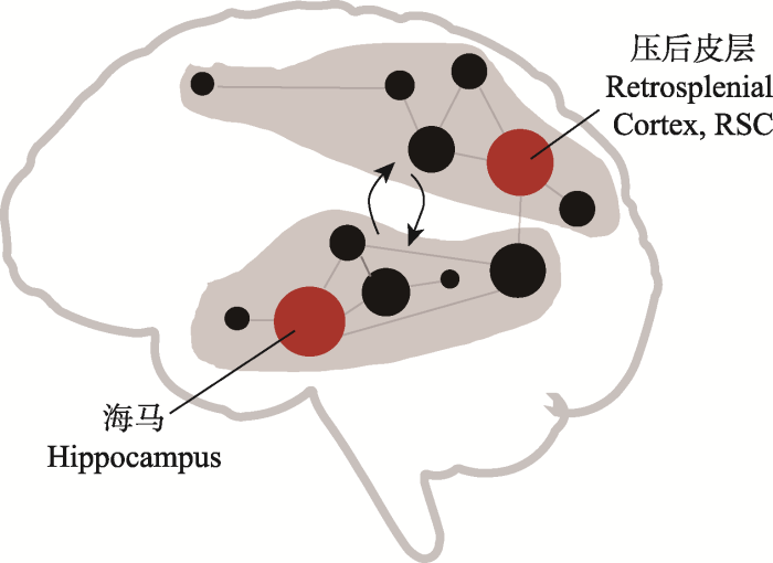
已有研究通常关注海马、内嗅皮质等单个脑区的功能特异性, 因此在传统理论中多局限于单个脑区在空间导航过程中的功能。空间导航是一个复杂的认知过程, 近年来, 研究者指出, 研究空间导航的神经机制不应局限于某个特定脑区(如海马), 而应该从神经环路或网络的角度, 研究不同脑区如何交互以支撑复杂导航行为(Ekstrom et al., 2017; Baumann & Mattingley, 2021; Kong et al., 2017; Weisberg & Ekstrom, 2021)。本项目打破传统的局限于单个脑区功能特异性的研究视角, 采用跨学科的研究手段和计算方法, 对空间导航的脑网络基础进行研究, 有望识别出空间导航脑网络的关键拓扑属性(如模块化和核心节点脑区等), 揭示导航网络中不同模块的内部结构特征和功能意义, 以及关键核心节点的分布特点, 这些结果对于构建空间导航的脑网络理论模型可以提供重要数据(图2)。以导航脑网络核心节点脑区为例, 近年来, 研究者基于RSC在空间导航中的关键作用, 提出了RSC作为导航脑网络核心脑区的理论假设(Ekstrom et al., 2017), 我们预期, 本研究的成果将为进一步检验和完善相关理论提供多个方面的实证数据。同时, 在此基础上, 我们将以导航脑网络的核心节点脑区为直接电刺激靶点, 结合基于磁共振技术的功能连接和白质纤维追踪技术, 探索靶点对导航脑网络的网络动态和导航行为的调控机制。完善的空间导航脑网络理论模型的建立, 可以催生大量重要的研究工作, 以解决新的关键科学问题。例如, 导航脑网络中不同功能模块的具体功能如何?不同模块之间的交互在不同导航任务下是如何动态变化的?交互动态的异常是否会导致脑疾病的产生?
图2
基于诸多细胞水平的研究发现, 研究者提出了整合位于不同脑区的不同类型神经细胞加工的向量编码理论模型, 试图揭示空间信息不同要素神经表征的统一机制(Bicanski & Burgess, 2020)。这些模型为我们更好地理解空间导航脑网络中不同节点脑区、不同功能模块之间的交互以及这些交互的功能意义, 提供了重要视角; 同时, 导航脑网络模型的建立, 又可以进一步帮助我们在神经环路和网络水平验证和理解这些计算理论。
此外, 大量研究表明, 空间导航能力衰退是AD等认知障碍的重要早期行为表现; 同时, 参与空间导航的重要脑区与早期AD影响的脑区大量重叠。随着社会老龄化, AD等老年痴呆症已经成为严重的健康、经济和社会问题。由北京宣武医院主导的一项最新大规模调查显示, 在我国60岁以上人群中有约1000万AD患者, 占比约为4% (Jia et al., 2020)。关于空间导航的脑网络的研究成果, 将帮助我们从脑认知网络的角度, 理解老龄化与相关脑疾病的底层神经机制, 帮助我们更好地开发新的诊断和干预策略, 以阻断或减缓疾病的发展, 推迟并降低发病率。目前, 针对AD的神经调控研究还相对比较初步, 且多采用TMS、tDCS等无创调控技术。这些技术在研究脑网络的调控机制中, 尤其是以深部脑结构为靶点脑区时, 具有很大的局限性。因此, 本项目选用更为直接、精准的直接电刺激技术, 探索特定脑结构对导航脑网络的调控机制。虽然, 该神经调控研究在癫痫患者中进行, 相关研究成果一方面将帮助我们理解导航脑网络的调控机制, 同时将为AD等空间导航损伤相关的脑疾病的干预和治疗提供重要参考。
综上所述, 本项目采用跨学科研究方法, 对空间导航的脑网络基础和调控机制进行研究。研究成果将为我们理解空间导航的神经环路机制、影响因素, 以及脑认知网络与行为之间的因果关系提供重要科学证据; 同时, 导航脑网络模型及其调控机制为研究AD等认知障碍脑疾病提供了新的重要的视角, 为早期诊断和精准治疗新方案的提出提供参考, 符合国家重大战略需求。
参考文献
Extrahippocampal contributions to spatial navigation in humans: A review of the neuroimaging evidence
DOI:10.1002/hipo.23313
PMID:33595156
[本文引用: 4]

Spatial navigation is a crucial everyday skill, which when impaired leads to a significant decrease in quality of life. In humans, functional magnetic resonance imaging (fMRI) has provided extensive insights into the neural underpinnings of navigation skills. Whereas the hippocampus has been recognized as the prime region underpinning navigation abilities, by providing a cognitive map of the environment, imaging studies have also implicated a range of other brain regions. In this review, we provide an overview of the fMRI evidence for extrahippocampal contributions to spatial navigation. We show that the parahippocampal cortex, retrosplenial cortex, dorsal striatum, and the posterior parietal cortex provide important complementary functions, and ultimately form part of a functional network that regulates successful way-finding behavior.© 2021 Wiley Periodicals LLC.
Long-term suppression of tremor by chronic stimulation of the ventral intermediate thalamic nucleus
DOI:10.1016/0140-6736(91)91175-t
PMID:1671433
[本文引用: 1]
The usefulness of high-frequency stimulation of the ventral intermediate nucleus (Vim) as the first neurosurgical procedure in disabling tremor was assessed in 26 patients with Parkinson's disease and 6 with essential tremor. 7 of these patients had already undergone thalamotomy contralateral to the stimulated side, and 11 others had bilateral Vim stimulation at the same time. Chronic stimulating electrodes connected to a pulse generator were implanted in the Vim. Tremor amplitude at rest, during posture holding, and during action and intention manoeuvres was assessed by means of accelerometry. Of the 43 thalami stimulated, 27 showed complete relief from tremor and 11 major improvement (88%). The improvement was maintained for up to 29 months (mean follow-up 13 [SD 9] months). Adverse effects were mild and could be eradicated by reduction or cessation of stimulation. This reversibility and adaptability, allowing control of side-effects, make thalamic stimulation preferable to thalamotomy, especially when treatment of both sides of the brain is needed.
Neuronal vector coding in spatial cognition
DOI:10.1038/s41583-020-0336-9
PMID:32764728
[本文引用: 1]
Several types of neurons involved in spatial navigation and memory encode the distance and direction (that is, the vector) between an agent and items in its environment. Such vectorial information provides a powerful basis for spatial cognition by representing the geometric relationships between the self and the external world. Here, we review the explicit encoding of vectorial information by neurons in and around the hippocampal formation, far from the sensory periphery. The parahippocampal, retrosplenial and parietal cortices, as well as the hippocampal formation and striatum, provide a plethora of examples of vector coding at the single neuron level. We provide a functional taxonomy of cells with vectorial receptive fields as reported in experiments and proposed in theoretical work. The responses of these neurons may provide the fundamental neural basis for the (bottom-up) representation of environmental layout and (top-down) memory-guided generation of visuospatial imagery and navigational planning.
Deep brain stimulation for Parkinson disease: An expert consensus and review of key issues
DOI:10.1001/archneurol.2010.260
URL
PMID:20937936
[本文引用: 1]
To provide recommendations to patients, physicians, and other health care providers on several issues involving deep brain stimulation (DBS) for Parkinson disease (PD).An international consortium of experts organized, reviewed the literature, and attended the workshop. Topics were introduced at the workshop, followed by group discussion.A draft of a consensus statement was presented and further edited after plenary debate. The final statements were agreed on by all members.(1) Patients with PD without significant active cognitive or psychiatric problems who have medically intractable motor fluctuations, intractable tremor, or intolerance of medication adverse effects are good candidates for DBS. (2) Deep brain stimulation surgery is best performed by an experienced neurosurgeon with expertise in stereotactic neurosurgery who is working as part of a interprofessional team. (3) Surgical complication rates are extremely variable, with infection being the most commonly reported complication of DBS. (4) Deep brain stimulation programming is best accomplished by a highly trained clinician and can take 3 to 6 months to obtain optimal results. (5) Deep brain stimulation improves levodopa-responsive symptoms, dyskinesia, and tremor; benefits seem to be long-lasting in many motor domains. (6) Subthalamic nuclei DBS may be complicated by increased depression, apathy, impulsivity, worsened verbal fluency, and executive dysfunction in a subset of patients. (7) Both globus pallidus pars interna and subthalamic nuclei DBS have been shown to be effective in addressing the motor symptoms of PD. (8) Ablative therapy is still an effective alternative and should be considered in a select group of appropriate patients.
Human hippocampal and parahippocampal theta during goal-directed spatial navigation predicts performance on a virtual Morris water maze
DOI:10.1523/JNEUROSCI.5001-07.2008
PMID:18524903
[本文引用: 1]
The hippocampus and parahippocampal cortices exhibit theta oscillations during spatial navigation in animals and humans, and in the former are thought to mediate spatial memory formation. Functional specificity of human hippocampal theta, however, is unclear. Neuromagnetic activity was recorded with a whole-head 275-channel magnetoencephalographic (MEG) system as healthy participants navigated to a hidden platform in a virtual reality Morris water maze. MEG data were analyzed for underlying oscillatory sources in the 4-8 Hz band using a spatial filtering technique (i.e., synthetic aperture magnetometry). Source analyses revealed greater theta activity in the left anterior hippocampus and parahippocampal cortices during goal-directed navigation relative to aimless movements in a sensorimotor control condition. Additional analyses showed that left anterior hippocampal activity was predominantly observed during the first one-half of training, pointing to a role for this region in early learning. Moreover, posterior hippocampal theta was highly correlated with navigation performance, with the former accounting for 76% of the variance of the latter. Our findings suggest human spatial learning is dependent on hippocampal and parahippocampal theta oscillations, extending to humans a significant body of research demonstrating such a pivotal role for hippocampal theta in animal navigation.
Spatial navigation deficits - overlooked cognitive marker for preclinical Alzheimer disease?
DOI:10.1038/s41582-018-0031-x
URL
PMID:29980763
[本文引用: 2]
Detection of incipient Alzheimer disease (AD) pathophysiology is critical to identify preclinical individuals and target potentially disease-modifying therapies towards them. Current neuroimaging and biomarker research is strongly focused in this direction, with the aim of establishing AD fingerprints to identify individuals at high risk of developing this disease. By contrast, cognitive fingerprints for incipient AD are virtually non-existent as diagnostics and outcomes measures are still focused on episodic memory deficits as the gold standard for AD, despite their low sensitivity and specificity for identifying at-risk individuals. This Review highlights a novel feature of cognitive evaluation for incipient AD by focusing on spatial navigation and orientation deficits, which are increasingly shown to be present in at-risk individuals. Importantly, the navigation system in the brain overlaps substantially with the regions affected by AD in both animal models and humans. Notably, spatial navigation has fewer verbal, cultural and educational biases than current cognitive tests and could enable a more uniform, global approach towards cognitive fingerprints of AD and better cognitive treatment outcome measures in future multicentre trials. The current Review appraises the available evidence for spatial navigation and/or orientation deficits in preclinical, prodromal and confirmed AD and identifies research gaps and future research priorities.
Interacting networks of brain regions underlie human spatial navigation: A review and novel synthesis of the literature
DOI:10.1152/jn.00531.2017
URL
PMID:28931613
[本文引用: 7]
Navigation is an inherently dynamic and multimodal process, making isolation of the unique cognitive components underlying it challenging. The assumptions of much of the literature on human spatial navigation are that 1) spatial navigation involves modality independent, discrete metric representations (i.e., egocentric vs. allocentric), 2) such representations can be further distilled to elemental cognitive processes, and 3) these cognitive processes can be ascribed to unique brain regions. We argue that modality-independent spatial representations, instead of providing exact metrics about our surrounding environment, more often involve heuristics for estimating spatial topology useful to the current task at hand. We also argue that egocentric (body centered) and allocentric (world centered) representations are better conceptualized as involving a continuum rather than as discrete. We propose a neural model to accommodate these ideas, arguing that such representations also involve a continuum of network interactions centered on retrosplenial and posterior parietal cortex, respectively. Our model thus helps explain both behavioral and neural findings otherwise difficult to account for with classic models of spatial navigation and memory, providing a testable framework for novel experiments.
The cognitive map in humans: Spatial navigation and beyond
DOI:10.1038/nn.4656
URL
PMID:29073650
[本文引用: 2]
The 'cognitive map' hypothesis proposes that brain builds a unified representation of the spatial environment to support memory and guide future action. Forty years of electrophysiological research in rodents suggest that cognitive maps are neurally instantiated by place, grid, border and head direction cells in the hippocampal formation and related structures. Here we review recent work that suggests a similar functional organization in the human brain and yields insights into how cognitive maps are used during spatial navigation. Specifically, these studies indicate that (i) the human hippocampus and entorhinal cortex support map-like spatial codes, (ii) posterior brain regions such as parahippocampal and retrosplenial cortices provide critical inputs that allow cognitive maps to be anchored to fixed environmental landmarks, and (iii) hippocampal and entorhinal spatial codes are used in conjunction with frontal lobe mechanisms to plan routes during navigation. We also discuss how these three basic elements of cognitive map based navigation-spatial coding, landmark anchoring and route planning-might be applied to nonspatial domains to provide the building blocks for many core elements of human thought.
Networks of anatomical covariance
DOI:10.1016/j.neuroimage.2013.05.054
URL
PMID:23711536
[本文引用: 1]
Functional imaging or diffusion-weighted imaging techniques are widely used to understand brain connectivity at the systems level and its relation to normal neurodevelopment, cognition or brain disorders. It is also possible to extract information about brain connectivity from the covariance of morphological metrics derived from anatomical MRI. These covariance patterns may arise from genetic influences on normal development and aging, from mutual trophic reinforcement as well as from experience-related plasticity. This review describes the basic methodological strategies, the biological basis of the observed covariance as well as applications in normal brain and brain disease before a final review of future prospects for the technique. Copyright © 2013 Elsevier Inc. All rights reserved.
Closed-loop stimulation of temporal cortex rescues functional networks and improves memory
DOI:10.1038/s41467-017-02753-0
URL
PMID:29410414
[本文引用: 1]
Memory failures are frustrating and often the result of ineffective encoding. One approach to improving memory outcomes is through direct modulation of brain activity with electrical stimulation. Previous efforts, however, have reported inconsistent effects when using open-loop stimulation and often target the hippocampus and medial temporal lobes. Here we use a closed-loop system to monitor and decode neural activity from direct brain recordings in humans. We apply targeted stimulation to lateral temporal cortex and report that this stimulation rescues periods of poor memory encoding. This system also improves later recall, revealing that the lateral temporal cortex is a reliable target for memory enhancement. Taken together, our results suggest that such systems may provide a therapeutic approach for treating memory dysfunction.
Electrical stimulation of the anterior nucleus of thalamus for treatment of refractory epilepsy
DOI:10.1111/j.1528-1167.2010.02536.x
URL
PMID:20331461
[本文引用: 1]
We report a multicenter, double-blind, randomized trial of bilateral stimulation of the anterior nuclei of the thalamus for localization-related epilepsy.Participants were adults with medically refractory partial seizures, including secondarily generalized seizures. Half received stimulation and half no stimulation during a 3-month blinded phase; then all received unblinded stimulation.One hundred ten participants were randomized. Baseline monthly median seizure frequency was 19.5. In the last month of the blinded phase the stimulated group had a 29% greater reduction in seizures compared with the control group, as estimated by a generalized estimating equations (GEE) model (p = 0.002). Unadjusted median declines at the end of the blinded phase were 14.5% in the control group and 40.4% in the stimulated group. Complex partial and "most severe" seizures were significantly reduced by stimulation. By 2 years, there was a 56% median percent reduction in seizure frequency; 54% of patients had a seizure reduction of at least 50%, and 14 patients were seizure-free for at least 6 months. Five deaths occurred and none were from implantation or stimulation. No participant had symptomatic hemorrhage or brain infection. Two participants had acute, transient stimulation-associated seizures. Cognition and mood showed no group differences, but participants in the stimulated group were more likely to report depression or memory problems as adverse events.Bilateral stimulation of the anterior nuclei of the thalamus reduces seizures. Benefit persisted for 2 years of study. Complication rates were modest. Deep brain stimulation of the anterior thalamus is useful for some people with medically refractory partial and secondarily generalized seizures.
Spatial representation in the entorhinal cortex
DOI:10.1126/science.1099901
PMID:15333832
[本文引用: 2]
As the interface between hippocampus and neocortex, the entorhinal cortex is likely to play a pivotal role in memory. To determine how information is represented in this area, we measured spatial modulation of neural activity in layers of medial entorhinal cortex projecting to the hippocampus. Close to the postrhinal-entorhinal border, entorhinal neurons had stable and discrete multipeaked place fields, predicting the rat's location as accurately as place cells in the hippocampus. Precise positional modulation was not observed more ventromedially in the entorhinal cortex or upstream in the postrhinal cortex, suggesting that sensory input is transformed into durable allocentric spatial representations internally in the dorsocaudal medial entorhinal cortex.
Three-year outcomes in deep brain stimulation for highly resistant obsessive-compulsive disorder
DOI:10.1038/sj.npp.1301165
URL
PMID:16855529
[本文引用: 1]
Deep brain stimulation (DBS) of the anterior limb of the internal capsule has been shown to be beneficial in the short term for obsessive-compulsive disorder (OCD) patients who exhaust conventional therapies. Nuttin et al, who published the first DBS for OCD series, found promising results using a capsule target immediately rostral to the anterior commissure extending into adjacent ventral capsule/ventral striatum (VC/VS). Published long-term outcome data are limited to four patients. In this collaborative study, 10 adult OCD patients meeting stringent criteria for severity and treatment resistance had quadripolar stimulating leads implanted bilaterally in the VC/VS. DBS was activated openly 3 weeks later. Eight patients have been followed for at least 36 months. Group Yale-Brown Obsessive Compulsive Scale (YBOCS) scores decreased from 34.6+/-0.6 (mean+/-SEM) at baseline (severe) to 22.3+/-2.1 (moderate) at 36 months (p < 0.001). Four of eight patients had a > or =35% decrease in YBOCS severity at 36 months; in two patients, scores declined between 25 and 35%. Global Assessment of Functioning scores improved from 36.6+/-1.5 at baseline to 53.8+/-2.5 at 36 months (p < 0.001). Depression and anxiety also improved, as did self-care, independent living, and work, school, and social functioning. Surgical adverse effects included an asymptomatic hemorrhage, a single seizure, and a superficial infection. Psychiatric adverse effects included transient hypomanic symptoms, and worsened depression and OCD when DBS was interrupted by stimulator battery depletion. This open study found promising long-term effects of DBS in highly treatment-resistant OCD.
Small-world anatomical networks in the human brain revealed by cortical thickness from MRI.
DOI:10.1093/cercor/bhl149
PMID:17204824
[本文引用: 1]
An important issue in neuroscience is the characterization for the underlying architectures of complex brain networks. However, little is known about the network of anatomical connections in the human brain. Here, we investigated large-scale anatomical connection patterns of the human cerebral cortex using cortical thickness measurements from magnetic resonance images. Two areas were considered anatomically connected if they showed statistically significant correlations in cortical thickness and we constructed the network of such connections using 124 brains from the International Consortium for Brain Mapping database. Significant short- and long-range connections were found in both intra- and interhemispheric regions, many of which were consistent with known neuroanatomical pathways measured by human diffusion imaging. More importantly, we showed that the human brain anatomical network had robust small-world properties with cohesive neighborhoods and short mean distances between regions that were insensitive to the selection of correlation thresholds. Additionally, we also found that this network and the probability of finding a connection between 2 regions for a given anatomical distance had both exponentially truncated power-law distributions. Our results demonstrated the basic organizational principles for the anatomical network in the human brain compatible with previous functional networks studies, which provides important implications of how functional brain states originate from their structural underpinnings. To our knowledge, this study provides the first report of small-world properties and degree distribution of anatomical networks in the human brain using cortical thickness measurements.
Graph theoretical modeling of brain connectivity
DOI:10.1097/WCO.0b013e32833aa567
URL
PMID:20581686
[本文引用: 1]
In recent years, there has been an explosion of studies on network modeling of brain connectivity. This review will focus mainly on recent findings concerning graph theoretical analysis of human brain networks with a variety of imaging modalities, including structural MRI, diffusion MRI, functional MRI, and EEG/MEG.Recent studies have utilized graph theoretical approaches to investigate the organizational principles of brain networks. These studies have consistently shown many important statistical properties underlying the topological organization of the human brain, including modularity, small-worldness, and the existence of highly connected network hubs. Importantly, these quantifiable network properties were found to change during normal development, aging, and various neurological and neuropsychiatric diseases such as Alzheimer's disease and schizophrenia. Moreover, several studies have also suggested that these network properties correlate with behavioral and genetic factors.The exciting research regarding graph theoretical analysis of brain connectivity yields truly integrative and comprehensive descriptions of the structural and functional organization of the human brain, which provides important implications for health and disease. Future research will most likely involve integrative models of brain structural and functional connectivity with multimodal neuroimaging data, exploring whether graph-based brain network analysis could yield reliable biomarkers for disease diagnosis and treatment.
Deep brain stimulation and cognition: Translational aspects
DOI:10.1016/j.nlm.2020.107283 URL [本文引用: 1]
Grid-like processing of imagined navigation
DOI:10.1016/j.cub.2016.01.042
PMID:26972318
[本文引用: 1]
Grid cells in the entorhinal cortex (EC) of rodents [1] and humans [2] fire in a hexagonally distributed spatially periodic manner. In concert with other spatial cells in the medial temporal lobe (MTL) [3-6], they provide a representation of our location within an environment [7, 8] and are specifically thought to allow the represented location to be updated by self-motion [9]. Grid-like signals have been seen throughout the autobiographical memory system [10], suggesting a much more general role in memory [11, 12]. Grid cells may allow us to move our viewpoint in imagination [13], a useful function for goal-directed navigation and planning [12, 14-16], and episodic future thinking more generally [17, 18]. We used fMRI to provide evidence for similar grid-like signals in human entorhinal cortex during both virtual navigation and imagined navigation of the same paths. We show that this signal is present in periods of active navigation and imagination, with a similar orientation in both and with the specifically 6-fold rotational symmetry characteristic of grid cell firing. We therefore provide the first evidence suggesting that grid cells are utilized during movement of viewpoint within imagery, potentially underpinning our more general ability to mentally traverse possible routes in the service of planning and episodic future thinking. Copyright © 2016 The Authors. Published by Elsevier Ltd.. All rights reserved.
Direct electrical stimulation of the human entorhinal region and hippocampus impairs memory
DOI:S0896-6273(16)30836-4
URL
PMID:27930911
[本文引用: 1]
Deep brain stimulation (DBS) has shown promise for treating a range of brain disorders and neurological conditions. One recent study showed that DBS in the entorhinal region improved the accuracy of human spatial memory. Based on this line of work, we performed a series of experiments to more fully characterize the effects of DBS in the medial temporal lobe on human memory. Neurosurgical patients with implanted electrodes performed spatial and verbal-episodic memory tasks. During the encoding periods of both tasks, subjects received electrical stimulation at 50 Hz. In contrast to earlier work, electrical stimulation impaired memory performance significantly in both spatial and verbal tasks. Stimulation in both the entorhinal region and hippocampus caused decreased memory performance. These findings indicate that the entorhinal region and hippocampus are causally involved in human memory and suggest that refined methods are needed to use DBS in these regions to improve memory.Copyright © 2016 Elsevier Inc. All rights reserved.
Prevalence, risk factors, and management of dementia and mild cognitive impairment in adults aged 60 years or older in China: A cross-sectional study
DOI:10.1016/S2468-2667(20)30185-7 URL [本文引用: 1]
Characterizing variation in the functional connectome: Promise and pitfalls
DOI:10.1016/j.tics.2012.02.001
PMID:22341211
[本文引用: 1]
The functional MRI (fMRI) community has zealously embraced resting state or intrinsic functional connectivity approaches to mapping brain organization. Having demonstrated their utility for charting the large-scale functional architecture of the brain, the field is now leveraging task-independent methods for the investigation of phenotypic variation and the identification of biomarkers for clinical conditions. Enthusiasm aside, questions regarding the significance and validity of intrinsic brain phenomena remain. Here, we discuss these challenges and outline current developments that, in moving the field toward discovery science, permit a shift from cartography toward a mechanistic understanding of the neural bases of variation in cognition, emotion and behavior.Copyright © 2012. Published by Elsevier Ltd.
Varieties of human spatial memory: A meta-analysis on the effects of hippocampal lesions
DOI:10.1016/S0165-0173(01)00058-3 URL [本文引用: 2]
Mapping individual brain networks using statistical similarity in regional morphology from MRI.
DOI:10.1371/journal.pone.0141840 URL [本文引用: 1]
Measuring individual morphological relationship of cortical regions
DOI:10.1016/j.jneumeth.2014.09.003 URL [本文引用: 1]
Human navigation network: The intrinsic functional organization and behavioral relevance
DOI:10.1007/s00429-016-1243-8 URL [本文引用: 5]
The challenge of mapping the human connectome based on diffusion tractography
DOI:10.1038/s41467-017-01285-x
URL
PMID:29116093
[本文引用: 1]
Tractography based on non-invasive diffusion imaging is central to the study of human brain connectivity. To date, the approach has not been systematically validated in ground truth studies. Based on a simulated human brain data set with ground truth tracts, we organized an open international tractography challenge, which resulted in 96 distinct submissions from 20 research groups. Here, we report the encouraging finding that most state-of-the-art algorithms produce tractograms containing 90% of the ground truth bundles (to at least some extent). However, the same tractograms contain many more invalid than valid bundles, and half of these invalid bundles occur systematically across research groups. Taken together, our results demonstrate and confirm fundamental ambiguities inherent in tract reconstruction based on orientation information alone, which need to be considered when interpreting tractography and connectivity results. Our approach provides a novel framework for estimating reliability of tractography and encourages innovation to address its current limitations.
Network perspectives on the mechanisms of deep brain stimulation
DOI:10.1016/j.nbd.2009.09.022
URL
PMID:19804831
[本文引用: 1]
Deep brain stimulation (DBS) is an established medical therapy for the treatment of movement disorders and shows great promise for several other neurological disorders. However, after decades of clinical utility the underlying therapeutic mechanisms remain undefined. Early attempts to explain the mechanisms of DBS focused on hypotheses that mimicked an ablative lesion to the stimulated brain region. More recent scientific efforts have explored the wide-spread changes in neural activity generated throughout the stimulated brain network. In turn, new theories on the mechanisms of DBS have taken a systems-level approach to begin to decipher the network activity. This review provides an introduction to some of the network based theories on the function and pathophysiology of the cortico-basal-ganglia-thalamo-cortical loops commonly targeted by DBS. We then analyze some recent results on the effects of DBS on these networks, with a focus on subthalamic DBS for the treatment of Parkinson's disease. Finally we attempt to summarize how DBS could be achieving its therapeutic effects by overriding pathological network activity.
fMRI evidence of age-related hippocampal dysfunction in feature binding in working memory
Richly detailed memories for particular events depend on processes that bind individual features of experience together. Previous cognitive behavioral research indicates that older adults have more difficulty than young adults in conditions requiring feature binding. We used functional magnetic resonance imaging (fMRI) during a working memory task to identify neural substrates of this age-related deficit in feature binding. For young, but not older, adults there was greater activation in left anterior hippocampus on combination trials (remember objects together with their locations) than on trials in which participants were told to remember only which objects or only which locations occurred. The results provide neuroimaging evidence for an age-related hippocampal dysfunction in feature binding in working memory.
The effects of direct brain stimulation in humans depend on frequency, amplitude, and white-matter proximity
DOI:S1935-861X(20)30108-X
URL
PMID:32446925
[本文引用: 1]
Researchers have used direct electrical brain stimulation to treat a range of neurological and psychiatric disorders. However, for brain stimulation to be maximally effective, clinicians and researchers should optimize stimulation parameters according to desired outcomes.The goal of our large-scale study was to comprehensively evaluate the effects of stimulation at different parameters and locations on neuronal activity across the human brain.To examine how different kinds of stimulation affect human brain activity, we compared the changes in neuronal activity that resulted from stimulation at a range of frequencies, amplitudes, and locations with direct human brain recordings. We recorded human brain activity directly with electrodes that were implanted in widespread regions across 106 neurosurgical epilepsy patients while systematically stimulating across a range of parameters and locations.Overall, stimulation most often had an inhibitory effect on neuronal activity, consistent with earlier work. When stimulation excited neuronal activity, it most often occurred from high-frequency stimulation. These effects were modulated by the location of the stimulating electrode, with stimulation sites near white matter more likely to cause excitation and sites near gray matter more likely to inhibit neuronal activity.By characterizing how different stimulation parameters produced specific neuronal activity patterns on a large scale, our results provide an electrophysiological framework that clinicians and researchers may consider when designing stimulation protocols to cause precisely targeted changes in human brain activity.Copyright © 2020 The Author(s). Published by Elsevier Inc. All rights reserved.
The structure of systematicity in the brain
DOI:10.1177/09637214211049233
PMID:35785023
[本文引用: 1]
A hallmark of human intelligence is the ability to adapt to new situations, by applying learned rules to new content (systematicity) and thereby enabling an open-ended number of inferences and actions (generativity). Here, we propose that the human brain accomplishes these feats through pathways in the parietal cortex that encode the abstract structure of space, events, and tasks, and pathways in the temporal cortex that encode information about specific people, places, and things (content). Recent neural network models show how the separation of structure and content might emerge through a combination of architectural biases and learning, and these networks show dramatic improvements in the ability to capture systematic, generative behavior. We close by considering how the hippocampal formation may form integrative memories that enable rapid learning of new structure and content representations.
Theta oscillations support the interface between language and memory
Functional connectomics from resting-state fMRI
DOI:10.1016/j.tics.2013.09.016
URL
PMID:24238796
[本文引用: 1]
Spontaneous fluctuations in activity in different parts of the brain can be used to study functional brain networks. We review the use of resting-state functional MRI (rfMRI) for the purpose of mapping the macroscopic functional connectome. After describing MRI acquisition and image-processing methods commonly used to generate data in a form amenable to connectomics network analysis, we discuss different approaches for estimating network structure from that data. Finally, we describe new possibilities resulting from the high-quality rfMRI data being generated by the Human Connectome Project and highlight some upcoming challenges in functional connectomics. Copyright © 2013 Elsevier Ltd. All rights reserved.
Core knowledge
Human cognition is founded, in part, on four systems for representing objects, actions, number, and space. It may be based, as well, on a fifth system for representing social partners. Each system has deep roots in human phylogeny and ontogeny, and it guides and shapes the mental lives of adults. Converging research on human infants, non-human primates, children and adults in diverse cultures can aid both understanding of these systems and attempts to overcome their limits.
Memory enhancement and deep-brain stimulation of the entorhinal area
DOI:10.1056/NEJMoa1107212
URL
PMID:22316444
[本文引用: 1]
The medial temporal structures, including the hippocampus and the entorhinal cortex, are critical for the ability to transform daily experience into lasting memories. We tested the hypothesis that deep-brain stimulation of the hippocampus or entorhinal cortex alters memory performance.We implanted intracranial depth electrodes in seven subjects to identify seizure-onset zones for subsequent epilepsy surgery. The subjects completed a spatial learning task during which they learned destinations within virtual environments. During half the learning trials, focal electrical stimulation was given below the threshold that elicits an afterdischarge (i.e., a neuronal discharge that occurs after termination of the stimulus).Entorhinal stimulation applied while the subjects learned locations of landmarks enhanced their subsequent memory of these locations: the subjects reached these landmarks more quickly and by shorter routes, as compared with locations learned without stimulation. Entorhinal stimulation also resulted in a resetting of the phase of the theta rhythm, as shown on the hippocampal electroencephalogram. Direct hippocampal stimulation was not effective. In this small series, no adverse events associated with the procedure were observed.Stimulation of the entorhinal region enhanced memory of spatial information when applied during learning. (Funded by the National Institutes of Health and the Dana Foundation.).
MRtrix3: A fast, flexible and open software framework for medical image processing and visualisation
DOI:10.1016/j.neuroimage.2019.116137 URL [本文引用: 1]
Spatial ability for STEM domains: Aligning over 50 years of cumulative psychological knowledge solidifies its importance
DOI:10.1037/a0016127 URL [本文引用: 1]
Phase-amplitude coupling supports phase coding in human ECoG
DOI:10.7554/eLife.07886 URL [本文引用: 1]
Hippocampal volume and navigational ability: The map (ping) is not to scale
DOI:10.1016/j.neubiorev.2021.03.012
URL
PMID:33722618
[本文引用: 2]
A critical question regards the neural basis of complex cognitive skill acquisition. One extensively studied skill is navigation, with evidence suggesting that humans vary widely in navigation abilities. Yet, data supporting the neural underpinning of these individual differences are mixed. Some evidence suggests robust structure-behavior relations between hippocampal volume and navigation ability, whereas other experiments show no such correlation. We focus on several possibilities for these discrepancies: 1) volumetric hippocampal changes are relevant only at the extreme ranges of navigational abilities; 2) hippocampal volume correlates across individuals but only for specific measures of navigation skill; 3) hippocampal volume itself does not correlate with navigation skill acquisition; connectivity patterns are more relevant. To explore this third possibility, we present a model emphasizing functional connectivity changes, particularly to extra-hippocampal structures. This class of models arises from the premise that navigation is dynamic and that good navigators flexibly solve spatial challenges. These models pave the way for research on other skills and provide more precise predictions for the neural basis of skill acquisition.Copyright © 2021. Published by Elsevier Ltd.
What determines our navigational abilities?
DOI:10.1016/j.tics.2010.01.001
PMID:20138795
[本文引用: 1]
The ability to find one's way in our complex environments represents one of the most fundamental cognitive functions. Although involving basic perceptual and memory related processes, spatial navigation is particularly complex because it is a multisensory process in which information needs to be integrated and manipulated over time and space. Not surprisingly, humans differ widely in this ability, and recent animal and human work has begun to unveil the underlying mechanisms. Here, we consider three interdependent domains that have been related to navigational abilities: cognitive and perceptual factors, neural information processing and variability in brain microstructure. Together, the findings converge into an emerging model of how different factors interact to produce individual patterns of navigational performance.Copyright 2010 Elsevier Ltd. All rights reserved.
Large-scale automated synthesis of human functional neuroimaging data
DOI:10.1038/nmeth.1635
PMID:21706013
[本文引用: 1]
The rapid growth of the literature on neuroimaging in humans has led to major advances in our understanding of human brain function but has also made it increasingly difficult to aggregate and synthesize neuroimaging findings. Here we describe and validate an automated brain-mapping framework that uses text-mining, meta-analysis and machine-learning techniques to generate a large database of mappings between neural and cognitive states. We show that our approach can be used to automatically conduct large-scale, high-quality neuroimaging meta-analyses, address long-standing inferential problems in the neuroimaging literature and support accurate 'decoding' of broad cognitive states from brain activity in both entire studies and individual human subjects. Collectively, our results have validated a powerful and generative framework for synthesizing human neuroimaging data on an unprecedented scale.
Quantifying the variability of scene-selective regions: Interindividual, interhemispheric, and sex differences
DOI:10.1002/hbm.23519
URL
PMID:28117508
[本文引用: 1]
Scene-selective regions (SSRs), including the parahippocampal place area (PPA), retrosplenial cortex (RSC), and transverse occipital sulcus (TOS), are among the most widely characterized functional regions in the human brain. However, previous studies have mostly focused on the commonality within each SSR, providing little information on different aspects of their variability. In a large group of healthy adults (N = 202), we used functional magnetic resonance imaging to investigate different aspects of topographical and functional variability within SSRs, including interindividual, interhemispheric, and sex differences. First, the PPA, RSC, and TOS were delineated manually for each individual. We then demonstrated that SSRs showed substantial interindividual variability in both spatial topography and functional selectivity. We further identified consistent interhemispheric differences in the spatial topography of all three SSRs, but distinct interhemispheric differences in scene selectivity. Moreover, we found that all three SSRs showed stronger scene selectivity in men than in women. In summary, our work thoroughly characterized the interindividual, interhemispheric, and sex variability of the SSRs and invites future work on the origin and functional significance of these variabilities. Additionally, we constructed the first probabilistic atlases for the SSRs, which provide the detailed anatomical reference for further investigations of the scene network. Hum Brain Mapp 38:2260-2275, 2017. © 2017 Wiley Periodicals, Inc.© 2017 Wiley Periodicals, Inc.
Network centrality in the human functional connectome
DOI:10.1093/cercor/bhr269 URL [本文引用: 1]
