

1 引言
情绪调节是人们保持情绪平衡的重要心理机制。以往有关情绪调节的研究较多关注个体如何有意识地采用策略改变情绪。近来有研究试图证明情绪调节在无意识(自动化)层面也能发挥作用(Bargh & Williams, 2007; Raio, Orederu, Palazzolo, Shurick, & Phelps, 2013)。自动情绪调节(Automatic Emotion Regulation, AER)是由无意识目标驱动的、不需要有意控制的情绪调节过程(Mauss, Bunge, & Gross, 2010)。已有研究证实自动情绪调节可以影响个体对情绪信息的认知加工、主观情绪体验以及相关生理反应, 如交感神经兴奋性(Mauss, Evers, Wilhelm, & Gross, 2006; Schwager, & Rothermund, 2013; Vogt, Lozo, Koster, & de Houwer, 2011; Zhang & Lu, 2012)。具体在对情绪信息的加工过程方面, 有研究已发现自动情绪调节可以改变个体对负性刺激的注意偏向, 促进对负性刺激的注意回避(Pourtois, Schwartz, Seghier, Lazeyras, & Vuilleumier, 2006; Waugh, Wager, Fredrickson, Noll, & Taylor, 2008)。然而, 当前该领域对与自动情绪调节下注意选择相关的脑活动尚不清楚。
已有研究表明, 眶额皮层(orbitofrontal cortex, OFC)参与在自动情绪调节与认知相整合的脑网络中(Rolls, 2000)。首先, OFC是与自动情绪调节相关的重要脑区, 它与外侧前额叶和腹内侧前额叶一同负责自动情绪调节(Mauss, Cook, & Gross, 2007)。一方面, OFC下行和杏仁核、纹状体、脑岛以及扣带回等与情绪密切相关的脑结构相连, 对情绪效价和唤醒度进行编码与调节(Rolls & Grabenhorst, 2008; Guillory & Bujarski, 2014), 并负责对未来的负性结果进行预期与评价, 以及促进奖赏驱动的行为习得等(Cooch et al., 2015; Rich & Wallis, 2016; Nogueira et al., 2017; Li, Sescousse, Amiez, & Dreher, 2017)。另一方面, OFC上行参与到控制情绪的脑网络中, 调节了背外侧前额叶和腹内侧前额叶与杏仁核的连接, 影响着情绪性行为与生理反应(Stein et al., 2007; Phillips, Ladouceur, & Drevets, 2008)。相关研究发现, 抑郁症病人往往伴随OFC活动异常增高, 而恢复期病人OFC激活强度会减弱至接近正常水平(Brody et al., 2001; Mayberg et al., 2005)。de Almeida等人(2009)发现与正常人相比, 抑郁症患者评价愉快面孔时, 内侧OFC与杏仁核的功能连接更弱。另外, 有关健康被试的研究发现, 当左侧OFC与杏仁核的心理生理交互(psychophysiological interactions, PPI)更强时, 被试体验到的负性情绪更少, 而这一相关模式在右侧OFC和背内侧前额叶上并未被发现(Banks, Eddy, Angstadt, Nathan, & Phan, 2007)。以上研究提示OFC可能在自动情绪调节相关脑网络中负责情绪信息的上下传递。
其次, 研究表明OFC也参与在自动情绪调节下负性注意偏向相关的脑活动中。例如, 高特质心理弹性个体的前部外侧OFC在非威胁线索条件下的激活强于威胁线索条件, 而低特质心理弹性个体则呈现相反的激活模式(Waugh et al., 2008)。类似的, 研究发现左侧OFC的激活与负性情绪刺激的注意选择相关(Pourtois et al., 2006)。Pourtois等人(2006)采用线索目标范式, 将左右呈现的面孔(快乐-中性, 恐惧-中性)作为线索刺激, 将随后立刻呈现在表情同侧位置(有效线索条件)或中性面孔所在位置(无效线索条件)的灰色条形作为目标刺激, 并请被试判断灰色条呈现在屏幕的左侧还是右侧。结果发现在恐惧-中性的线索下, 左侧OFC在无效线索条件下的激活强于有效线索条件。这提示当目标刺激出现在与恐惧表情相反的位置时, 恐惧面孔线索刺激获取的大量注意资源与目标刺激获取任务相关的注意资源产生了冲突(Pourtois et al., 2006)。该研究提示, 左侧OFC的激活与负性情绪刺激的注意分配过程相关。更进一步的, 临床研究发现OFC损伤病人比正常人对情绪性分心刺激投入更少的注意、对目标刺激投入更多的注意(Hartikainen, Ogawa, & Knight, 2012; Mäki-Marttunen et al., 2017)。例如, 在Hartikainen等人(2012)的研究中, 被试观看屏幕中央呈现的情绪图片或中性图片(150 ms), 在图片消失200 ms之后判断屏幕左侧或右侧呈现的三角形的上下朝向。结果发现OFC损伤病人在情绪刺激分心物下FCz电极点的N2-P3a的波幅减小, 对目标刺激的N2-P3b的波幅更大, 而正常人呈现相反的脑电模式(Hartikainen et al., 2012)。这一结果表明OFC受损会损伤对情绪刺激自动的注意朝向和对目标刺激的注意分配(Hartikainen et al., 2012)。
尽管以往通过fMRI的研究手段提示了左侧OFC在自动情绪调节下注意选择中的作用, 然而左侧OFC和自动情绪调节下注意选择的关系并未获得必要性证据。而经颅直流电刺激(transcranial direct current stimulation, tDCS)是一种非侵入性的脑刺激技术, 它采用微弱电流刺激(1~2 mA)调节大脑皮层的神经活动。tDCS能够对目标区域施加极其微弱的直流电以刺激大脑皮层, 通过阳极刺激和阴极刺激导致神经元细胞膜电位去极化和超极化, 来改变某些脑区的皮层兴奋程度, 进而促进或抑制与该脑区有密切相关的认知、情绪与行为活动 (Utz, Dimova, Oppenländer, & Kerkhoff, 2010; Nitsche & Paulus, 2000; Stagg & Nitsche, 2011; Civai, Miniussi, & Rumiati, 2015)。并且这种脑区兴奋性的持续时间为1小时以上(Nitsche & Paulus, 2001), 能够满足大多心理实验的时间需求, 因而tDCS越来越多地被用于检验认知行为与可能的神经回路之间的关系(Coffman, Clark, & Parasuraman, 2014; Jacobson, Koslowsky, & Lavidor, 2012)。本研究将通过tDCS的阴极刺激和假刺激操纵左侧OFC的激活程度, 检验自动情绪调节下恐惧刺激注意偏向是否能够随之改变。
前人研究大多通过无意识启动情绪调节目标的方法来诱发自动情绪调节。以往有研究使用双义词的句子整理任务, 发现启动情绪控制词可以降低被试自我报告的愤怒水平(Mauss et al., 2007), 启动认知重评相关词可以令被试体验到更少压力(Williams, Bargh, Nocera & Gray, 2009)。近来, 采用阈下启动的研究发现, 阈下呈现概念词会改变随后相关联的认知加工(Brooks et al., 2012; Brasel & Gips, 2011; Karremans, Stroebe, & Claus, 2006; Moskowitz, Li, & Kirk, 2004)。例如, Légal, Chappé, Coiffard和Villard-Forrest (2012)的研究发现, 阈下启动“信任”目标词会使个体对广告内容产生更为积极的评价。而Tong等人(2013)也发现阈下启动“不公平”的概念可以增强被试的愤怒表情并加快了被试对愤怒词汇的反应。与情绪性注意选择有关的研究发现, 阈下启动情绪控制目标词能够促进被试对负性刺激的注意回避, 使其在点探测任务中不一致线索条件下的反应时更快(刘珂, 张晶, 赵怡佳, 2016; Zhang, Lipp, & Hu, 2017)。与句子整理任务相比, 在阈下启动任务中, 被试于无意识觉察的情况下接受启动信息, 避免了对完成目标词义进行有意识加工而产生的认知需求(Blakemore, Neveu, & Vuilleumier, 2017)。因而, 本研究将使用阈下启动情绪控制词来激活相关目标表征, 从而引发相应的自动情绪调节。
目前尚未有研究通过tDCS来操纵OFC激活以检验其在自动情绪调节下负性注意选择中的作用。当前, 比较具有借鉴意义的发现是, Rao等人(2018)的研究表明, 使用植入电极刺激外侧OFC能够改善抑郁症病人的情绪体验、增强被试的快乐体验。也有研究证明, 正常人接受OFC的阳极电刺激会增强人们对情绪面孔(愤怒、厌恶、恐惧、高兴、悲伤和中性)的识别(Willis et al., 2015)。Schutter和van-Honk (2006)也发现, 对健康被试的左侧OFC进行重复经颅磁刺激增强能够促进被试对快乐面孔的记忆。同时, fMRI研究证明, 左侧OFC与杏仁核的连接增强与负性情绪体验降低有关, 并且左侧OFC激活强度与对负性刺激的注意选择相关(Pourtois et al., 2006)。因而, 本研究将使用tDCS技术考察阴极刺激左侧OFC激活和自动情绪调节下注意偏向的关系。在前测中, 改进的点探测范式首先阈下启动情绪控制目标词, 然后呈现线索刺激, 随后即刻出现探测点, 从而模拟自动情绪控制下对恐惧刺激的注意回避。在对左侧OFC施加tDCS阴极刺激或者假刺激之后, 被试进行后测, 完成与前测相同的流程。即, 再次接受阈下情绪控制目标的启动并完成注意选择任务。结果分析了前后测和tDCS刺激条件这两个自变量, 以阈下启动目标引发的注意回避为因变量, 来揭示左侧OFC是否参与在自动情绪调节影响注意选择的过程中。鉴于以往的研究, 本研究提出如下假设:在阴极刺激之后, 阈下启动情绪控制词引发的对恐惧性刺激的注意回避会消失。具体表现为阴极刺激前被试表现出对负性刺激的注意回避, 阴极刺激后被试对负性刺激的注意回避消失, 表现为注意警觉。在假刺激条件下, 被试在前后测中都表现为对负性刺激的注意回避。
2 方法
2.1 被试
40名在校大学生(非心理学专业或学习过心理学相关课程)参与本实验。其中男性20名, 女性20名。被试的平均年龄为21.42 ± 2.09岁。被试被随机分配成两组:实验组被试(10名女生, 10名男生, 年龄21.30 ± 1.81岁)接受阴极刺激, 对照组(10名女生, 10名男生, 年龄21.95 ± 2.50岁)接受假刺激。所有被试报告身体健康, 均为右利手, 无精神疾病及脑部损伤史, 无癫痫或癫痫家族史, 裸眼视力或矫正视力正常。所有被试在实验之前签署知情同意书, 实验后获取一定的报酬。
2.2 实验材料
实验材料包括刘珂等人(2016)评定的点探测任务图片材料共64张。点探测任务分为前测与后测两个阶段, 前测与后测的图片材料各32张。前测与后测点探测任务均包含恐惧刺激材料图片(蛇)16张、中性刺激材料图片(蘑菇)16张。前后测所用刺激图片不重复。在实验前对前后测恐惧刺激材料图片进行效价平衡。
经过前期评定的情绪控制目标启动词10个(如:控制、隐藏、冷静)和无关中性动作词10个(如:坐下、站立、弯腰)作为实验材料。词语均为中性效价、高频词语。在评定中, 请20名心理学专业的学生对40个双字词进行打分。首先是对其情绪控制的类别归属进行评价, 1 = 不属于, 2 = 属于, 3 = 不确定。其次是对词语的效价进行1~7等级的评定, 1 = 非常负性, 4 = 中性, 7 = 非常正性。最终选择其中属性明确、效价中性的两组词共20个作为实验材料。实验组词语的平均得分为3.98, 对照组的平均得分为4.18。两组的效价没有显著差异, t(19) = -1.26, p = 0.22。然后对两组启动词的使用频率进行配对样本t检验, 两组启动词使用频率没有显著差异, t(9) = -0.82, p = 0.44。
2.3 实验程序
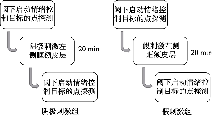
本实验为2(前后测:tDCS刺激前, tDCS刺激后) × 2(tDCS刺激:阴极刺激, 假刺激)×2(恐惧刺激与探测点位置匹配一致性:左右位置一致, 左右位置不一致)的混合实验设计。其中前后测、刺激与探测点位置匹配一致性为被试内变量, tDCS刺激条件为被试间变量。被试被随机分成2组, 每组被试接受不同的tDCS刺激。实验组被试接受tDCS阴极刺激, 对照组被试接受tDCS假刺激。
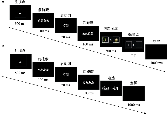
被试进入实验室之后先完成一份状态-特质焦虑问卷(李文利, 钱铭怡, 1995), 随后完成阈下启动情绪控制目标与点探测任务作为前测。该任务结束之后被试休息15分钟, 随后被试接受tDCS刺激。刺激结束后被试立刻完成与前测相同的任务(见图1)。实验任务共64个试次, 包括4个练习试次和60个实验试次。依据前人的研究, 阈下启动点探测任务中, 注视点首先呈现500 ms, 然后是100 ms的前掩蔽刺激, 紧接着启动词快速呈现20 ms, 然后又是100 ms的后掩蔽刺激(王佳莹, 缴润凯, 张明, 2016)。之后蛇和蘑菇的图片一左一右呈现, 刺激呈现时间为500 ms。蛇图片和探测点出现的位置在被试内平衡。图片消失后, 其中一个图片所在的位置会出现一个白色圆点。被试需要对其位置进行判断, 并进行按键操作(F键-左, J键-右)。被试做出按键反应后, 空屏出现, 1000 ms后进入下一个试次。
图1
任务结束之后再完成一份状态焦虑量表和一份蛇恐惧量表(Klorman, Weerts, Hastings, Melamed, & Lang, 1974)。最后完成启动词辨别任务, 以探察被试对启动刺激的觉察。辨别任务流程见图2。这些被试在辨别任务的正确率(36.26%~50.20%)与随机猜测水平没有显著差异。这表明被试没有觉察到与情绪控制相关的阈下启动词。本实验中的情绪控制目标阈下启动任务和启动词辨别任务均采用Eprime 2.0软件呈现并记录被试的正确率与反应时。
图2
主机型号为DELL OPTIPLEX 9020MT, 显示器采用19寸CRT纯平显示器, 屏幕分辨率为1024×768像素, 刷新率为60 Hz。本实验使用E-prime 2.0软件编程和呈现。所有数据用EXCEL 2007和SPSS 22.0整理与分析。
2.4 经颅直流电刺激参数及方法
本实验采用DC-STIMULATOR PLUS刺激仪器进行tDCS刺激, 该仪器由德国NeuroConn公司研发。将一对海绵表面电极片(面积5 cm × 7 cm)浸泡在生理盐水(0.90%的氯化钠) 4~5分钟, 然后将其应用到目标区域刺激皮层。在本实验中, 按照国际EEG 10-20系统的标准, FP1是针对左侧OFC的tDCS研究常用的刺激位置(Homan, Herman, & Purdy, 1987; Nejati, Salehinejad, Nitsche, Najian, & Javadi, 2017; Yang, Gao, Shi, Ye, & Chen, 2017), 因此在FP1位置进行阴极刺激或假刺激。为了降低通过头皮的电流分流和深度增加电流密度, 通过计算选择P4作为距离阴极电极最远的参考电极。基于前人的研究可知, tDCS研究的刺激时间一般为5~20分钟(Boggio, Zaghi, & Fregni, 2009)。为确保左侧OFC能够得到充分激活, 本实验设置刺激时间为20 min。阴极刺激电流为1.5 mA。假刺激条件下, 使用1.5 mA的直流电刺激被试15 s, 然后仪器自动停止刺激(电极片仍会戴在被试头上20 min)。刺激条件的fade in时间和fade out时间均为15 s (Keeser et al., 2011; 甘甜 等, 2013)。本实验的所有被试均不知道自己接受的是哪种刺激条件。
3 结果
3.1 问卷数据
对状态-特质焦虑问卷的得分进行2(刺激条件:阴极刺激, 假刺激) × 2(前后测:刺激前, 刺激后)的混合设计方差分析。结果显示, 状态焦虑与特质焦虑的刺激条件的组间效应均不显著, F(1, 38) = 2.85, p = 0.10; F(1, 38) = 0.69, p = 0.41; 前后测的主效应均不显著, F(1, 38) = 0.65, p = 0.43; F(1, 38) = 0.23, p = 0.68; 刺激条件与前后测的交互作用均不显著, F(1, 38) = 0.30, p = 0.59; F(1, 38) = 0.07, p = 0.80。对蛇恐惧问卷得分进行单因素方差分析, 结果表明, 蛇恐惧得分在刺激条件上没有显著差异, F(1, 38) = 0.01, p = 0.94。
3.2 反应时
采用2(前后测:tDCS刺激前, tDCS刺激后) × 2(tDCS刺激条件:阴极刺激, 假刺激) × 2(恐惧刺激与探测点位置匹配一致性:一致, 不一致)的混合设计方差分析各条件下的反应时。结果发现, 探测点位置匹配一致性的主效应显著, F(1, 38) = 8.26, p = 0.01, ηp2 = 0.18, 一致条件下的反应时显著大于不一致条件下的反应时。探测点位置匹配一致性与刺激条件的交互作用显著, F(1, 38) = 7.55, p = 0.01, ηp2 = 0.17。经过简单效应分析, 在假刺激的条件下, 一致条件的反应时大于不一致条件(p = 0.01)。阴极刺激条件下, 一致条件的反应时与不一致条件无显著差异(p = 0.06)。
表1 各实验条件下的反应时(ms, M ± SD)
| 刺激类型 | 前测反应时 | 后测反应时 | ||
|---|---|---|---|---|
| 一致条件 | 不一致条件 | 一致条件 | 不一致条件 | |
| 假刺激 | 429.52 ± 49.27 | 422.58 ± 49.49 | 412.30 ± 46.69 | 402.30 ± 38.08 |
| 阴极刺激 | 436.11 ± 44.71 | 428.87 ± 42.63 | 405.07 ± 30.43 | 411.93 ± 36.85 |
图3
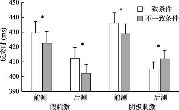
图3
tDCS刺激(阴极刺激, 假刺激) × 前后测(刺激前, 刺激后) × 刺激与探测点位置一致性(一致, 不一致)各实验处理水平下, 被试判断探测点位置的反应时(* ps < 0.05)
4 讨论
本研究使用tDCS操纵左侧眶额皮层(orbitofrontal cortex, OFC)的激活, 检验了其在自动情绪调节下负性注意偏向加工中的作用。结果表明, 在阴极刺激抑制左侧OFC激活的条件下, 前测中自动情绪调节对负性刺激注意偏向的作用在后测中消失。具体地, 当抑制了左侧OFC激活后, 被试在接受阈下情绪控制目标词后不再对负性刺激产生注意回避, 而是表现出对与负性刺激位置一致的探测点反应更快。这一结果提示了左侧OFC在阈下情绪调节目标影响情绪性注意的过程中扮演着重要角色。
本研究发现, 被试接受阴极刺激后, 后测中对负性刺激的注意选择模式与前测相反, 而接受假刺激的被试其注意偏向在前后测并未发生反转。具体表现为, 前测中阈下情绪控制目标引发被试对负性刺激的注意回避, 在阴极刺激抑制左侧OFC后, 转变为了注意警觉。而在假刺激组, 被试在前测与后测都维持着对负性刺激的注意回避。这说明, 左侧OFC可能参与在阈下情绪调节目标影响情绪性注意选择的脑活动中。这与前人操纵脑激活的相关研究有相同指向的发现。例如, Schutter和Van-Honk (2006)研究发现, 对健康被试的左侧OFC进行重复经颅磁刺激增强能够促进被试对开心面孔的记忆, 然而这种促进效应并未出现在增强左侧背外侧前额叶激活的条件中。Rao等人(2018)使用植入电极来刺激抑郁症病人外侧OFC, 发现被试的快乐体验获得增强。这些研究提示OFC激活的增强能够减少被试的负性情绪记忆, 提升被试的正性情绪。然而, 这些研究并未从抑制左侧OFC兴奋性的角度检验其在情绪调节中的作用。本研究则使用tDCS抑制了左侧OFC的激活, 消退了在前测中自动情绪调节下对负性刺激的注意回避, 使被试对负性刺激表现为注意警觉。
结合Mauss等人(2007)构建的自动情绪调节模型可以对结果做进一步解释。Mauss等人(2007)认为OFC与外侧前额叶、腹内侧前额叶负责了自上而下自动地改变对负性情绪注意偏向这一过程。本研究结果提示, 对左侧OFC活动的抑制可能减弱了外侧前额叶和腹内侧前额叶对负性注意偏向自上而下的控制, 使得行为表现从阈下目标调节下的注意回避转为了注意警觉。该结果一方面支持了左侧OFC是自动情绪调节的注意加工机制中的关键脑区; 另一方面也说明阈下启动情绪调节目标这一方法, 能够在较短时间内引发个体对负性刺激的注意回避, 印证了前人的研究结果。总之, 本研究的结果证实了左侧OFC在自动情绪调节下对恐惧刺激的注意选择中具有重要作用, 为进一步验证左侧OFC在情绪调节环路中的重要作用提供了前期支持。
另外, 本研究的结果可能在受到注意回避影响的同时也受到了返回抑制的作用。已有研究表明, 情绪注意偏向和返回抑制两种注意机制相互影响(关荐 等, 2018)。对负性情绪刺激的注意回避是情绪注意偏向的重要机制之一(Cisler & Koster, 2010)。在反应模式上, 注意回避和返回抑制体现的都是注意不再投入线索刺激位置的机制, 并共同表现为在一致条件下的反应时更长。而且, 已有研究者同时从注意回避和返回抑制角度解释了线索目标任务中一致条件下反应时更长的表现。例如, Waters等人(2007)在研究中使用的实验流程与本研究相同, 均为在500 ms的线索之后立刻呈现探测点, 要求被试对探测点的位置进行判断。结果发现, 低焦虑女性被试在厌恶刺激线索一致条件下反应时更长。Waters等人认为这一结果符合了返回抑制的模式, 反映的是被试对厌恶刺激的注意回避。基于此, 返回抑制的机制可能和注意回避共同作用了本研究的结果。
本研究仍然存在一些不足。首先, 研究仅关注了情绪加工过程中的注意偏向, 所选择的实验范式未涉及情绪体验层面的感受。以往研究已经表明OFC在改善情绪体验中具有重要作用(Mayberg et. al., 2005; Brody et al., 2001; Waugh et al., 2008)。在未来的研究中, 需要考虑检验阈下启动调节目标对情绪体验的影响, 从而回答情绪的意识感受是否受到阈下操纵的自动情绪调节的影响并进一步揭示OFC在其中的作用。其次, 由于OFC是面积较大的脑区, 已有研究也已经提示外侧OFC和内侧OFC在情绪加工中可能具有不同的功能(Rudebeck & Murray, 2011; Fettes et al., 2016)。因而, 旨在通过电刺激或者磁刺激操纵OFC激活的研究, 需要聚焦在不同的OFC分区, 进而为揭示OFC在情绪与情绪调节中的作用提供更深入的证据。再有, 实验未对左侧OFC的神经活动情况进行监测。所以, 本研究尚不能够确定实验操作能够精确有效作用于左侧眶额皮层。故而在未来的研究中, 将尽可能提升技术条件来精准确定tDCS的定位和检验皮层获得刺激的有效性。另外, 近来有研究发现, 同时增强左侧背外侧前额叶与抑制右侧OFC能够使个体更好地适应负性刺激和应对压力事件(Nejati, Salehinejad, & Nitsche, 2017)。未来的研究应该继续探讨OFC和左侧背外侧前额叶的激活, 以及彼此间的协同机制与自动情绪调节下注意偏向的关系, 从而为揭示自动情绪调节下情绪性注意选择的脑机制提供更全面的证据。
5 结论
本研究使用tDCS技术对左侧眶额皮层(OFC)进行操纵, 检验了对左侧OFC施加阴极刺激和假刺激对自动情绪调节下负性情绪注意选择的影响。结果表明, 不同于假刺激条件下被试在前测与后测对负性刺激均表现出注意回避, 阴极刺激作用于左侧OFC减弱了被试对恐惧刺激的注意回避, 弱化了阈下情绪调节目标对负性刺激注意选择的影响。该结果表明左侧OFC在自动情绪调节下的注意选择过程中具有重要作用。本研究的结果也进一步为自动情绪调节模型提供了部分的实证支持(Mauss et al., 2007)。
参考文献
Amygdala-frontal connectivity during emotion regulation
DOI:10.1093/scan/nsm029
URL
PMID:18985136
[本文引用: 1]

Successful control of affect partly depends on the capacity to modulate negative emotional responses through the use of cognitive strategies (i.e., reappraisal). Recent studies suggest the involvement of frontal cortical regions in the modulation of amygdala reactivity and the mediation of effective emotion regulation. However, within-subject inter-regional connectivity between amygdala and prefrontal cortex in the context of affect regulation is unknown. Here, using psychophysiological interaction analyses of functional magnetic resonance imaging data, we show that activity in specific areas of the frontal cortex (dorsolateral, dorsal medial, anterior cingulate, orbital) covaries with amygdala activity and that this functional connectivity is dependent on the reappraisal task. Moreover, strength of amygdala coupling with orbitofrontal cortex and dorsal medial prefrontal cortex predicts the extent of attenuation of negative affect following reappraisal. These findings highlight the importance of functional connectivity within limbic-frontal circuitry during emotion regulation.
The nonconscious regulation of emotion
How emotion context modulates unconscious goal activation during motor force exertion
DOI:10.1016/j.neuroimage.2016.11.002
URL
PMID:27833013
[本文引用: 1]
Priming participants with emotional or action-related concepts influences goal formation and motor force output during effort exertion tasks, even without awareness of priming information. However, little is known about neural processes underpinning how emotional cues interact with action (or inaction) goals to motivate (or demotivate) motor behaviour. In a novel functional neuroimaging paradigm, visible emotional images followed by subliminal action or inaction word primes were presented before participants performed a maximal force exertion. In neutral emotional contexts, maximum force was lower following inaction than action primes. However, arousing emotional images had interactive motivational effects on the motor system: Unpleasant images prior to inaction primes increased force output (enhanced effort exertion) relative to control primes, and engaged a motivation-related network involving ventral striatum, extended amygdala, as well as right inferior frontal cortex. Conversely, pleasant images presented before action (versus control) primes decreased force and activated regions of the default-mode network, including inferior parietal lobule and medial prefrontal cortex. These findings show that emotional context can determine how unconscious goal representations influence motivational processes and are transformed into actual motor output, without direct rewarding contingencies. Furthermore, they provide insight into altered motor behaviour in psychopathological disorders with dysfunctional motivational processes.
Modulation of emotions associated with images of human pain using anodal transcranial direct current stimulation (tDCS)
DOI:10.1016/j.neuropsychologia.2008.07.022
URL
[本文引用: 1]
AbstractViewing images of other humans in pain elicits a variety of responses including distress, anxiety, and a sensation that is similar to pain. We aimed to evaluate whether transcranial direct current stimulation (tDCS) could be effective in modulating the emotional aspects of pain as to further explore mechanisms of tDCS in pain relief. Twenty-three healthy subjects rated images with respect to unpleasantness and discomfort/pain (baseline), and then received stimulation with tDCS under four different conditions of stimulation: anodal tDCS of the left primary motor cortex (M1), dorsolateral prefrontal cortex (DLPFC), occipital cortex (V1); and sham tDCS. The order of conditions was randomized and counterbalanced across subjects. During each stimulation session (after 3 min of stimulation), subjects were shown a new set of aversive images and were again asked to rate the images with respect to unpleasantness and discomfort/pain. The results showed that ratings of unpleasantness and discomfort/pain were significantly decreased during DLPFC tDCS only, as compared to baseline and sham tDCS. The other conditions of stimulation (M1 and V1 tDCS) did not result in any significant changes. These results support the notion that DLPFC is a critical area for the emotional processing of pain and also suggests that DLPFC may be a potential target of stimulation for alleviation of pain with a significant emotional-affective component. Our results also suggest that the mechanism of tDCS in modulating emotional pain involve pathways that are independent of those modulating the somatosensory perception of pain.]]>
Media multitasking behavior: Concurrent television and computer usage
Brain metabolic changes associated with symptom factor improvement in major depressive disorder
DOI:10.1016/S0006-3223(01)01117-9
URL
[本文引用: 2]
AbstractBackground: Symptoms of major depressive disorder (MDD) have been linked to regional brain function through imaging studies of symptom provocation in normal control subjects and baseline studies of subjects with MDD. We examined associations between change in depressive symptom factors and change in regional brain metabolism from before to after treatment of MDD.Methods: Thirty-nine outpatients with MDD underwent 18F-fluorodeoxyglucose positron emission tomography scanning before and after treatment with either paroxetine or interpersonal psychotherapy. Associations were determined between changes in regional brain metabolism and changes in four Hamilton Depression Rating Scale factors (anxiety/somatization [ANX], psychomotor retardation [PR], cognitive disturbance [COGN], and sleep disturbance) and two corresponding Profile of Mood States subscales (tension [TENS] and fatigue [FATIG]).Results: Improvement in ANX, PR, TENS, and FATIG factors was associated with decreasing ventral frontal lobe metabolism. Improvement in ANX and TENS was also associated with decreasing ventral anterior cingulate gyrus (AC) and anterior insula activity, whereas improvement in PR was associated with increasing dorsal AC activity. COGN improvement was associated with increasing dorsolateral prefrontal cortex metabolism.Conclusions: Brain regions that show significant relationships with symptom provocation in normal control subjects have similar relationships with MDD symptoms as they improve with treatment.]]>
Subliminal food images compromise superior working memory performance in women with restricting anorexia nervosa
DOI:10.1016/j.concog.2012.02.006
URL
[本文引用: 1]
Prefrontal cortex (PFC) is dysregulated in women with restricting anorexia nervosa (RAN). It is not known whether appetitive non-conscious stimuli bias cognitive responses in those with RAN. Thirteen women with RAN and 20 healthy controls (HC) completed a dorsolateral PFC (DLPFC) working memory task and an anterior cingulate cortex (ACC) conflict task, while masked subliminal food, aversive and neutral images were presented. During the DLPFC task, accuracy was higher in the RAN compared to the HC group, but superior performance was compromised when subliminal food stimuli were presented: errors positively correlated with self-reported trait anxiety in the RAN group. These effects were not observed in the ACC task. Appetitive activation is intact and anxiogenic in women with RAN, and non-consciously interacts with working memory processes associated with the DLPFC. This interaction mechanism may underlie cognitive inhibition of appetitive processes that are anxiety inducing, in people with AN. (C) 2012 Elsevier Inc.
Mechanisms of attentional biases towards threat in anxiety disorders: An Integrative review
DOI:10.1016/j.cpr.2009.11.003
URL
[本文引用: 1]
AbstractA wealth of research demonstrates attentional biases toward threat in the anxiety disorders. Several models have been advanced to explain these biases in anxiety, yet the mechanisms comprising and mediating the biases remain unclear. In the present article, we review evidence regarding the mechanisms of attentional biases through careful examination of the components of attentional bias, the mechanisms underlying these components, and the stage of information processing during which the biases occur. Facilitated attention, difficulty in disengagement, and attentional avoidance comprise the components of attentional bias. A threat detection mechanism likely underlies facilitated attention, a process that may be neurally centered around the amygdala. Attentional control ability likely underlies difficulty in disengagement, emotion regulation goals likely underlie attentional avoidance, and both of these processes may be neurally centered around prefrontal cortex functioning. The threat detection mechanism may be a mostly automatic process, attentional avoidance may be a mostly strategic process, and difficulty in disengagement may be a mixture of automatic and strategic processing. Recommendations for future research are discussed.]]>
Medial prefrontal cortex reacts to unfairness if this damages the self: A tDCS study
DOI:10.1093/scan/nsu154
URL
PMID:25552567
[本文引用: 1]
Neural correlates of unfairness perception depend on who is the target of the unfair treatment. These previous findings suggest that the activation of medial prefrontal cortex (MPFC) is related to unfairness perception only when the subject of the measurement is also the person affected by the unfair treatment. We aim at demonstrating the specificity of MPFC involvement using transcranial direct current stimulation (tDCS), a technique that induces cortical excitability changes in the targeted region. We use a modified version of the Ultimatum Game, in which responders play both for themselves (myself-MS condition) and on behalf of an unknown third-party (TP condition), where they respond to unfairness without being the target of it. We find that the application of cathodal tDCS over MPFC decreases the probability of rejecting unfair offers in MS, but not in TP; conversely, the same stimulation increases the probability of rejecting fair offers in TP, but not in MS. We confirm the hypothesis that MPFC is specifically related to processing unfairness when the self is involved, and discuss possible explanations for the opposite effect of the stimulation in TP.
Battery powered thought: Enhancement of attention, learning, and memory in healthy adults using transcranial direct current stimulation
DOI:10.1016/j.neuroimage.2013.07.083 URL [本文引用: 1]
Orbitofrontal lesions eliminate signalling of biological significance in cue-responsive ventral striatal neurons
DOI:10.1038/ncomms8195 URL [本文引用: 1]
Abnormal amygdala-prefrontal effective connectivity to happy faces differentiates bipolar from major depression
DOI:10.1016/j.biopsych.2009.03.024
URL
PMID:19450794
[本文引用: 1]
BACKGROUND: Bipolar disorder is frequently misdiagnosed as major depressive disorder, delaying appropriate treatment and worsening outcome for many bipolar individuals. Emotion dysregulation is a core feature of bipolar disorder. Measures of dysfunction in neural systems supporting emotion regulation might therefore help discriminate bipolar from major depressive disorder. METHODS: Thirty-one depressed individuals-15 bipolar depressed (BD) and 16 major depressed (MDD), DSM-IV diagnostic criteria, ages 18-55 years, matched for age, age of illness onset, illness duration, and depression severity-and 16 age- and gender-matched healthy control subjects performed two event-related paradigms: labeling the emotional intensity of happy and sad faces, respectively. We employed dynamic causal modeling to examine significant among-group alterations in effective connectivity (EC) between right- and left-sided neural regions supporting emotion regulation: amygdala and orbitomedial prefrontal cortex (OMPFC). RESULTS: During classification of happy faces, we found profound and asymmetrical differences in EC between the OMPFC and amygdala. Left-sided differences involved top-down connections and discriminated between depressed and control subjects. Furthermore, greater medication load was associated with an amelioration of this abnormal top-down EC. Conversely, on the right side the abnormality was in bottom-up EC that was specific to bipolar disorder. These effects replicated when we considered only female subjects. CONCLUSIONS: Abnormal, left-sided, top-down OMPFC-amygdala and right-sided, bottom-up, amygdala-OMPFC EC during happy labeling distinguish BD and MDD, suggesting different pathophysiological mechanisms associated with the two types of depression.
Cortico- striatal-thalamic loop circuits of the orbitofrontal cortex: promising therapeutic targets in psychiatric illness
DOI:10.3389/fnsys.2017.00025
URL
PMID:28496402
[本文引用: 1]
Corticostriatal circuits through the orbitofrontal cortex (OFC) play key roles in complex human behaviors such as evaluation, affect regulation and reward-based decision-making. Importantly, the medial and lateral OFC (mOFC and lOFC) circuits have functionally and anatomically distinct connectivity profiles which differentially contribute to the various aspects of goal-directed behavior. OFC corticostriatal circuits have been consistently implicated across a wide range of psychiatric disorders, including major depressive disorder (MDD), obsessive compulsive disorder (OCD), and substance use disorders (SUDs). Furthermore, psychiatric disorders related to OFC corticostriatal dysfunction can be addressed via conventional and novel neurostimulatory techniques, including deep brain stimulation (DBS), electroconvulsive therapy (ECT), repetitive transcranial magnetic stimulation (rTMS), and transcranial direct current stimulation (tDCS). Such techniques elicit changes in OFC corticostriatal activity, resulting in changes in clinical symptomatology. Here we review the available literature regarding how disturbances in mOFC and lOFC corticostriatal functioning may lead to psychiatric symptomatology in the aforementioned disorders, and how psychiatric treatments may exert their therapeutic effect by rectifying abnormal OFC corticostriatal activity. First, we review the role of OFC corticostriatal circuits in reward-guided learning, decision-making, affect regulation and reappraisal. Second, we discuss the role of OFC corticostriatal circuit dysfunction across a wide range of psychiatric disorders. Third, we review available evidence that the therapeutic mechanisms of various neuromodulation techniques may directly involve rectifying abnormal activity in mOFC and lOFC corticostriatal circuits. Finally, we examine the potential of future applications of therapeutic brain stimulation targeted at OFC circuitry; specifically, the role of OFC brain stimulation in the growing field of individually-tailored therapies and personalized medicine in psychiatry.
Exciting the right temporo-parietal junction with transcranial direct current stimulation influences moral intention processing
DOI:10.3724/SP.J.1041.2013.01004
URL
[本文引用: 1]
When we evaluate the moral status of an action, we consider not only its consequences but also the beliefs and intentions of the actor, which relies on the capacity to infer others' mental states. Functional MRI studies showed that the right temporo-parietal junction (RTPJ) is the critical brain region for understanding others' mental states. Previous studies have found that the role of intention processing in moral judgment was reduced by disrupting the RTPJ with transcranial magnetic stimulation (TMS). In the current study, we enhanced the role of intention processing in moral judgment with the transcranial direct current sitmulation (tDCS), a painless, non-invasive brain stimulation technique that allows us to induce polarity-specific excitability changes in the human brain. Many tDCS studies have confirmed the anodal excitation effect for cognitive functions. However, so far, limited work has been done to explore the tDCS effect on social cognitive function such as moral judgment. Therefore, the present study aims to investigate the anodal excitation effect of tDCS on moral judgment. We hypothesize that exciting the neural activity of RTPJ with anodal tDCS could enhance the role of intention processing in moral judgment. To test our hypothesis, 18 healthy college students were recruited to participate in the study. All subjects underwent two tDCS sessions (anodal and sham tDCS) in random order and counterbalanced across subjects on 2 separate days with 1 week interval between both stimulations. We applied anodal (1.5mA, 20 min) and sham tDCS (1.5mA, 15 sec) on the RTPJ while subjects were introduced to keep a resting state. After stimulation, subjects read stories in a 2 (intention: negative vs. neutral) × 2 (outcome: negative vs. neutral) design and were asked to make moral judgment about how much blame the actor deserves. We analyzed the moral evaluation score and reaction time by a 2 (intention) × 2 (outcome) × 2 (tDCS: anodal, sham) repeated measures ANOVA. Results showed that actors with negative intentions were judged more morally blameworthy than those with neutral intentions, and actors producing negative outcomes were judged more blameworthy than those causing neutral outcomes. The differences between no harm (neutral intention, neutral outcome) and accidental harm (neutral intention, negative outcome) were larger than that between attempted harm (negative intention, neutral outcome) and successful harm (negative intention, negative outcome). For the reaction time, judgments of negative outcomes were faster than that of neutral outcomes. The responses to attempted and accidental harm were slower than the other two conditions. Most importantly, the moral judgment was slower under anodal tDCS than sham tDCS stimulation, especially under the attempted harm and accidental harm conditions. These results highlight the role of intention processing in moral judgment. People will spend more time integrating the intention and outcome information in order to make normal moral judgment. Furthermore, the present research provides us a better understanding about the role of RTPJ in moral judgment. Using anodal tDCS to excite the neural activity of RTPJ enhanced the capacity of mentalizing in moral judgment, especially in the cases of attempted harm and accidental harm.
经颅直流电刺激右侧颞顶联合区对道德意图加工的影响
The competition between inhibition of return and emotional attention bias: Evidence from eye movements
返回抑制和情绪信息注意偏向的竞争:来自眼动的证据
Exploring emotions using invasive methods: Review of 60 years of human intracranial electrophysiology
DOI:10.1093/scan/nsu002
URL
PMID:24509492
[本文引用: 1]
Over the past 60 years, human intracranial electrophysiology (HIE) has been used to characterize seizures in patients with epilepsy. Secondary to the clinical objectives, electrodes implanted intracranially have been used to investigate mechanisms of human cognition. In addition to studies of memory and language, HIE methods have been used to investigate emotions. The aim of this review is to outline the contribution of HIE (electrocorticography, single-unit recording and electrical brain stimulation) to our understanding of the neural representations of emotions. We identified 64 papers dating back to the mid-1950s which used HIE techniques to study emotional states. Evidence from HIE studies supports the existence of widely distributed networks in the neocortex, limbic/paralimbic regions and subcortical nuclei which contribute to the representation of emotional states. In addition, evidence from HIE supports hemispheric dominance for emotional valence. Furthermore, evidence from HIE supports the existence of overlapping neural areas for emotion perception, experience and expression. Lastly, HIE provides unique insights into the temporal dynamics of neural activation during perception, experience and expression of emotional states. In conclusion, we propose that HIE techniques offer important evidence which must be incorporated into our current models of emotion representation in the human brain.
Orbitofrontal cortex biases attention to emotional events
DOI:10.1080/13803395.2012.666231
URL
[本文引用: 4]
We examined the role of orbitofrontal (OF) cortex in regulating emotion-attention interaction and the balance between involuntary and voluntary attention allocation. We studied patients with OF lesion applying reaction time (RT) and event-related potential (ERP) measures in a lateralized visual discrimination task with novel task-irrelevant affective pictures (unpleasant, pleasant, or neutral) preceding a neutral target. This allowed for comparing the effects of automatic attention allocation to emotional versus neutral stimuli on subsequent voluntary attention allocation to target stimuli. N2-P3a and N2-P3b ERP components served as measures of involuntary and voluntary attention allocation correspondingly. Enhanced N2-P3a amplitudes to emotional distractors and reduced N2-P3b amplitudes to targets preceded by emotional distractors were observed in healthy subjects, suggesting automatic emotional orienting interfered with subsequent voluntary orienting. OF patients showed an opposite pattern with tendency towards reduced N2-P3a responses to emotional distractors, suggesting impaired automatic orienting to emotional stimuli due to orbitofrontal damage. Enhanced N2-P3b responses to targets preceded by any affective distractor were observed in OF patients, suggesting bias towards voluntary target-related attention allocation due to orbitofrontal lesion. Behavioral evidence indicated that left visual field (LVF) attention performance was modulated by emotional stimuli. Specifically, OF patients responded faster to LVF targets subsequent to pleasant emotional distractors. We suggest that damage to the orbitofrontal circuitry leads to dysbalance between voluntary and involuntary attention allocation in the context of affective distractors with predisposition to posterior target-related processing over frontal novelty and affect-related processing. Furthermore, we suggest that orbitofrontal influence on emotion-attention interaction is valence and hemisphere dependent.
Cerebral location of international 10-20 system electrode placement, Localisation cérébrale des électrodes placées selon le système international 10-20
DOI:10.1016/0013-4694(87)90206-9
URL
PMID:2435517
[本文引用: 1]
We employed CT scanning to correlate scalp markers placed according to the international 10-20 system with underlying cerebral structures. Subjects were 12 normal volunteers. Measurements included assessment for cranial asymmetry to determine the effect of skull asymmetry on cortical location of electrodes. Results were correlated with the cortical histological map of Brodmann. Primary cortical locations agree well with previously published data and provide cortical localization in greater detail than previous studies. Variability of cortical electrode location was substantial in some cases and not related to cranial asymmetry. The results indicate that CT scanning or other neuroimaging techniques which reveal detailed cerebral anatomy would be potentially highly useful in defining the generators of electrocerebral potentials recorded from the scalp.
tDCS polarity effects in motor and cognitive domains: A meta- analytical review
DOI:10.1007/s00221-011-2891-9
URL
[本文引用: 1]
In vivo effects of transcranial direct current stimulation (tDCS) have attracted much attention nowadays as this area of research spreads to both the motor and cognitive domains. The common assumption is that the anode electrode causes an enhancement of cortical excitability during stimulation, which then lasts for a few minutes thereafter, while the cathode electrode generates the opposite effect, i.e., anodal-excitation and cathodal-inhibition effects (AeCi). Yet, this dual-polarity effect has not been observed in all tDCS studies. Here, we conducted a meta-analytical review aimed to investigate the homogeneity/heterogeneity of the effect sizes of the AeCi dichotomy in both motor and cognitive functions. The AeCi effect was found to occur quite commonly with motor investigations and rarely in cognitive studies. When the anode electrode is applied over a non-motor area, in most cases, it will cause an excitation as measured by a relevant cognitive or perceptual task; however, the cathode electrode rarely causes an inhibition. We found homogeneity in motor studies and heterogeneity in cognitive studies with the electrode's polarity serving as a moderator that can explain the source of heterogeneity in cognitive studies. The lack of inhibitory cathodal effects might reflect compensation processes as cognitive functions are typically supported by rich brain networks. Further insights as to the polarity and domain interaction are offered, including subdivision to different classes of cognitive functions according to their likelihood of being affected by stimulation.
Beyond Vicary's fantasies: The impact of subliminal priming and brand choice
DOI:10.1016/j.jesp.2005.12.002 URL [本文引用: 1]
Prefrontal transcranial direct current stimulation changes connectivity of resting-state networks during fMRI
DOI:10.1523/JNEUROSCI.0542-11.2011
URL
[本文引用: 1]
Transcranial direct current stimulation (tDCS) has been proposed for experimental and therapeutic modulation of regional brain function. Specifically, anodal tDCS of the dorsolateral prefrontal cortex (DLPFC) together with cathodal tDCS of the supraorbital region have been associated with improvement of cognition and mood, and have been suggested for the treatment of several neurological and psychiatric disorders. Although modeled mathematically, the distribution, direction, and extent of tDCS-mediated effects on brain physiology are not well understood. The current study investigates whether tDCS of the human prefrontal cortex modulates resting-state network (RSN) connectivity measured by functional magnetic resonance imaging (fMRI). Thirteen healthy subjects underwent real and sham tDCS in random order on separate days. tDCS was applied for 20 min at 2 mA with the anode positioned over the left DLPFC and the cathode over the right supraorbital region. Patterns of resting-state brain connectivity were assessed before and after tDCS with 3 T fMRI, and changes were analyzed for relevant networks related to the stimulation-electrode localizations. At baseline, four RSNs were detected, corresponding to the default mode network (DMN), the left and right frontal-parietal networks (FPNs) and the self-referential network. After real tDCS and compared with sham tDCS, significant changes of regional brain connectivity were found for the DMN and the FPNs both close to the primary stimulation site and in connected brain regions. These findings show that prefrontal tDCS modulates resting-state functional connectivity in distinct functional networks of the human brain.
Psychometric description of some specific- fear questionnaires
DOI:10.1016/S0005-7894(74)80008-0 URL [本文引用: 1]
Don't you know that you want to trust me? Subliminal goal priming and persuasion
DOI:10.1016/j.jesp.2011.06.006
URL
[本文引用: 1]
We investigated the effect of goal priming on the processing of a persuasive message. Before reading a persuasive message about tap water consumption, participants were subliminally primed (or not) with the goal "to trust". Subsequently, they completed a questionnaire about their perception of the message, the source of the message, and tap water consumption intentions. The results indicated that non-conscious activation of the goal "to trust" leads to a better evaluation of the message, increases behavioral intentions in accordance with the message, and positively influences the assessment of the source. (C) 2011 Elsevier Inc.
Revision of the state-trait anxiety inventory with sample of Chinese college students
状态特质焦虑量表中国大学生常模修订
Local morphology predicts functional organization of experienced value signals in the human orbitofrontal cortex
DOI:10.1523/JNEUROSCI.3058-14.2015
URL
PMID:25632140
[本文引用: 1]
Experienced value representations within the human orbitofrontal cortex (OFC) are thought to be organized through an antero-posterior gradient corresponding to secondary versus primary rewards. Whether this gradient depends upon specific morphological features within this region, which displays considerable intersubject variability, remains unknown. To test the existence of such relationships, we performed a subject-by-subject analysis of fMRI data taking into account the local morphology of each individual. We tested 38 subjects engaged in a simple incentive delay task manipulating both monetary and visual erotic rewards, focusing on reward outcome (experienced value signal). The results showed reliable and dissociable primary (erotic) and secondary (monetary) experienced value signals at specific OFC sulci locations. More specifically, experienced value signal induced by monetary reward outcome was systematically located in the rostral portion of the medial orbital sulcus. Experienced value signal related to erotic reward outcome was located more posteriorly, that is, at the intersection between the caudal portion of the medial orbital sulcus and transverse orbital sulcus. Thus, the localizations of distinct experienced value signals can be predicted from the organization of the human orbitofrontal sulci. This study provides insights into the anatomo-functional parcellation of the anteroposterior OFC gradient observed for secondary versus primary rewards because there is a direct relationship between value signals at the time of reward outcome and unique OFC sulci locations.
Effect of subliminal emotion control target on attention distribution of fear stimulation
阈下启动情绪控制目标对恐惧刺激注意分配的影响
Greater attention to task-relevant threat due to orbitofrontal lesion
DOI:10.1089/neu.2015.4390
URL
PMID:27502875
[本文引用: 1]
Injury to the orbitofrontal cortex (OFC) is a frequent consequence of head injury and may lead to dysfunctional regulation of emotional and social behavior. Dysfunctional emotional behavior may partly be related to the role of the OFC in emotion-attention interaction, as reported previously. In order to better understand its role in emotion-attention and emotion-cognitive control interactions, we investigated attention allocation to task-relevant and task-irrelevant threat-related emotional stimuli during a task requiring cognitive control in patients with lesion to the OFC. We measured the behavioral performance and event-related potentials (ERP) of 13 patients with OFC lesion and 11 control subjects during a Go/NoGo visual discrimination task. In the task, line drawings of threatening (spider) and neutral (flower) figures served as either task-relevant Go or NoGo signals, or as task-irrelevant distractors. Overall performance did not differ between the groups. In contrast to the control group performance, the orbitofrontal group performance was improved by relevant threat signal in comparison with neutral signal. Further, task-relevant threat signals evoked larger frontocentral N2-P3 amplitude in the orbitofrontal group. Taken together, behavioral and electrophysiological results suggest that patients with OFC injury allocated more attentional and cognitive control resources in the context of task-relevant emotional stimuli. This study provides new evidence for the role of the OFC in emotion-attention and emotion-cognitive control interactions. Further, the OFC seems to contribute to the balance between voluntary and involuntary attention networks in context of emotional stimuli. Better understanding of alterations in emotion-attention interaction offers insight into affective dysfunction due to OFC lesion.
Automatic emotion regulation
Automatic emotion regulation during anger provocation
DOI:10.1016/j.jesp.2006.07.003
URL
[本文引用: 5]
AbstractIndividuals frequently have to regulate their emotions, especially negative ones, to function successfully. However, deliberate emotion regulation can have significant costs for the individual. Are there less costly ways to achieve emotion regulatory goals? In two studies, we test the hypothesis that more automatic types of emotion regulation might provide the benefits of deliberate emotion regulation without the costs. Study 1 introduces a priming technique that manipulates automatic emotion regulation. Using this priming technique, we show that relative to priming emotion expression, priming emotion control leads to less anger experience in response to a laboratory anger provocation. Study 2 examines the experiential and physiological consequences of automatic emotion regulation. Results suggest that relative to priming emotion expression, priming emotion control reduces negative emotion experience without maladaptive cardiovascular responding. Together, these findings suggest that automatic emotion regulation may provide an effective means of controlling powerful negative emotions.]]>
How to bite your tongue without blowing your top: Implicit evaluation of emotion regulation predicts affective responding to anger provocation
DOI:10.1177/0146167205283841
URL
PMID:16702153
[本文引用: 1]
People frequently have to control their emotions to function in life. However, mounting evidence suggests that deliberate emotion regulation often is costly. This presents a dilemma: Is it better to let emotions go or to pay the price of exerting costly control? In two studies, the authors explore whether emotion regulatory processes associated with implicit positive evaluation of emotion regulation might provide the benefits of successful emotion regulation without the costs. In Study 1, the authors introduce a measure of implicit evaluation of emotion regulation (ER-IAT). Study 2 examined whether this measure is associated with actual emotional responses to an anger provocation. It was found that greater ER-IAT scores were associated with lesser anger experience, fewer negative thoughts, lessened self-reported effortful emotion regulation, and an adaptive pattern of cardiovascular responding. These findings suggest that implicit positive evaluation of emotion regulation is associated with successful, automatic, and physiologically adaptive down-regulation of anger.
Deep brain stimulation for treatment-resistant depression
DOI:10.1016/j.neuron.2005.02.014
URL
PMID:15748841
[本文引用: 2]
Treatment-resistant depression is a severely disabling disorder with no proven treatment options once multiple medications, psychotherapy, and electroconvulsive therapy have failed. Based on our preliminary observation that the subgenual cingulate region (Brodmann area 25) is metabolically overactive in treatment-resistant depression, we studied whether the application of chronic deep brain stimulation to modulate BA25 could reduce this elevated activity and produce clinical benefit in six patients with refractory depression. Chronic stimulation of white matter tracts adjacent to the subgenual cingulate gyrus was associated with a striking and sustained remission of depression in four of six patients. Antidepressant effects were associated with a marked reduction in local cerebral blood flow as well as changes in downstream limbic and cortical sites, measured using positron emission tomography. These results suggest that disrupting focal pathological activity in limbic-cortical circuits using electrical stimulation of the subgenual cingulate white matter can effectively reverse symptoms in otherwise treatment-resistant depression.
The implicit volition model: On the preconscious regulation of temporarily adopted goals
Interaction of the left dorsolateral prefrontal cortex (L-dlPFC) and right orbitofrontal cortex (OFC) in hot and cold executive functions: Evidence from transcranial direct current stimulation (tDCS)
DOI:10.1016/j.neuroscience.2017.10.042
URL
PMID:29113929
[本文引用: 1]
An organizing principle which has recently emerged proposes that executive functions (EF) can be divided into cognitive (cold) and affective/reward-related (hot) processes related to the dorsolateral prefrontal cortex (DLPFC) and orbitofrontal cortex (OFC) respectively. A controversial question is whether cold and hot EF are functionally and structurally independent or not. This study investigated how the left DLPFC (l-DLPFC) and right OFC (r-OFC) interact in hot and cold EF using transcranial direct current stimulation (tDCS). Twenty-four healthy male subjects received anodal, cathodal and sham tDCS (20min, 1.5mA) over the l-DLPFC (F3) and r-OFC (Fp2) with a 72-h interval between each stimulation condition. After five minutes of stimulation, participants underwent a series of cold and hot EF tasks including the Go/No-Go and Tower of Hanoi (TOH) as measures of cold EF and the BART and temporal discounting tasks as measures of hot EF. Inhibitory control mostly benefited from anodal l-DLPFC/cathodal r-OFC tDCS. Planning and problem solving were more prominently affected by anodal l-DLPFC/cathodal r-OFC stimulation, although the reversed electrode position with the anode positioned over the r-OFC also affected some aspects of task performance. Risk-taking behavior and risky decision-making decreased under both anodal l-DLPFC/cathodal r-OFC and anodal r-OFC/cathodal l-DLPFC tDCS. Cold EF rely on DLPFC activation while hot EF rely on both, DLPFC and OFC activation. Results suggest that EF are placed on continuum with lateral and mesial prefrontal areas contributing to cold and hot aspects respectively.
Transcranial direct current stimulation improves executive dysfunctions in ADHD: Implications for inhibitory control, interference control, working memory, and cognitive flexibility
Excitability changes induced in the human motor cortex by weak transcranial direct current stimulation
DOI:10.1111/tjp.2000.527.issue-3 URL [本文引用: 2]
Sustained excitability elevations induced by transcranial DC motor cortex stimulation in humans
DOI:10.1212/wnl.57.10.1899
URL
PMID:11723286
The authors show that in the human transcranial direct current stimulation is able to induce sustained cortical excitability elevations. As revealed by transcranial magnetic stimulation, motor cortical excitability increased approximately 150% above baseline for up to 90 minutes after the end of stimulation. The feasibility of inducing long-lasting excitability modulations in a noninvasive, painless, and reversible way makes this technique a potentially valuable tool in neuroplasticity modulation.
Lateral orbitofrontal cortex anticipates choices and integrates prior with current information
DOI:10.1038/ncomms14823
URL
PMID:28337990
[本文引用: 1]
Adaptive behavior requires integrating prior with current information to anticipate upcoming events. Brain structures related to this computation should bring relevant signals from the recent past into the present. Here we report that rats can integrate the most recent prior information with sensory information, thereby improving behavior on a perceptual decision-making task with outcome-dependent past trial history. We find that anticipatory signals in the orbitofrontal cortex about upcoming choice increase over time and are even present before stimulus onset. These neuronal signals also represent the stimulus and relevant second-order combinations of past state variables. The encoding of choice, stimulus and second-order past state variables resides, up to movement onset, in overlapping populations. The neuronal representation of choice before stimulus onset and its build-up once the stimulus is presented suggest that orbitofrontal cortex plays a role in transforming immediate prior and stimulus information into choices using a compact state-space representation.
A neural model of voluntary and automatic emotion regulation: Implications for understanding the pathophysiology and neurodevelopment of bipolar disorder
DOI:10.1038/mp.2008.65 URL [本文引用: 1]
Neural systems for orienting attention to the location of threat signals: An event-related fMRI study
DOI:10.1016/j.neuroimage.2005.12.034
URL
PMID:16487729
[本文引用: 5]
Attention may reflexively shift towards the location of perceived threats, but it is still unclear how these spatial biases recruit the distributed fronto-parietal cortical networks involved in other aspects of selective attention. We used event-related fMRI to determine how brain responses to a neutral visual target are influenced by the emotional expression of faces appearing at the same location during a covert orienting task. On each trial, two faces were briefly presented, one in each upper visual field (one neutral and one emotional, fearful or happy), followed by a unilateral target (a small horizontal or vertical bar) replacing one of the faces. Participants had to discriminate the target orientation, shown on the same (valid) or opposite (invalid) side as the emotional face. Trials with faces but no subsequent target (cue-only trials) were included to disentangle activation due to emotional cues from their effects on target detection. We found increased responses in bilateral temporo-parietal areas and right occipito-parietal cortex for fearful faces relative to happy faces, unrelated to the subsequent target and cueing validity. More critically, we found a selective modulation of intraparietal and orbitofrontal cortex for targets following an invalid fearful face, as well as an increased visual response in right lateral occipital cortex for targets following a valid fearful face. No such effects were observed with happy faces. These results demonstrate that fearful faces can act as exogenous cues by increasing sensory processing in extrastriate cortex for a subsequent target presented at the same location, but also produce a cost in disengaging towards another location by altering the response of IPS to invalidly cued targets. Neural mechanisms responsible for orienting attention towards emotional vs. non-emotional stimuli are thus partly shared in parietal and visual areas, but also partly distinct.
Cognitive emotion regulation fails the stress test
Direct electrical stimulation of lateral orbitofrontal cortex acutely improves mood in individuals with symptoms of depression
DOI:10.1016/j.cub.2018.10.026
URL
PMID:30503621
[本文引用: 2]
Mood disorders cause significant morbidity and mortality, and existing therapies fail 20%-30% of patients. Deep brain stimulation (DBS) is an emerging treatment for refractory mood disorders, but its success depends critically on target selection. DBS focused on known targets within mood-related frontostriatal and limbic circuits has been variably efficacious. Here, we examine the effects of stimulation in orbitofrontal cortex (OFC), a key hub for mood-related circuitry that has not been well characterized as a stimulation target. We studied 25 subjects with epilepsy who were implanted with intracranial electrodes for seizure localization. Baseline depression traits ranged from mild to severe. We serially assayed mood state over several days using a validated questionnaire. Continuous electrocorticography enabled investigation of neurophysiological correlates of mood-state changes. We used implanted electrodes to stimulate OFC and other brain regions while collecting verbal mood reports and questionnaire scores. We found that unilateral stimulation of the lateral OFC produced acute, dose-dependent mood-state improvement in subjects with moderate-to-severe baseline depression. Stimulation suppressed low-frequency power in OFC, mirroring neurophysiological features that were associated with positive mood states during natural mood fluctuation. Stimulation potentiated single-pulse-evoked responses in OFC and modulated activity within distributed structures implicated in mood regulation. Behavioral responses to stimulation did not include hypomania and indicated an acute restoration to non-depressed mood state. Together, these findings indicate that lateral OFC stimulation broadly modulates mood-related circuitry to improve mood state in depressed patients, revealing lateral OFC as a promising new target for therapeutic brain stimulation in mood disorders.
Decoding subjective decisions from orbitofrontal cortex
DOI:10.1038/nn.4320
URL
PMID:27273768
[本文引用: 1]
When making a subjective choice, the brain must compute a value for each option and compare those values to make a decision. The orbitofrontal cortex (OFC) is critically involved in this process, but the neural mechanisms remain obscure, in part due to limitations in our ability to measure and control the internal deliberations that can alter the dynamics of the decision process. Here we tracked these dynamics by recovering temporally precise neural states from multidimensional data in OFC. During individual choices, OFC alternated between states associated with the value of two available options, with dynamics that predicted whether a subject would decide quickly or vacillate between the two alternatives. Ensembles of value-encoding neurons contributed to these states, with individual neurons shifting activity patterns as the network evaluated each option. Thus, the mechanism of subjective decision-making involves the dynamic activation of OFC states associated with each choice alternative.
The orbitofrontal cortex and beyond: From affect to decision-making
DOI:10.1016/j.pneurobio.2008.09.001
URL
PMID:18824074
[本文引用: 1]
The orbitofrontal cortex represents the reward or affective value of primary reinforcers including taste, touch, texture, and face expression. It learns to associate other stimuli with these to produce representations of the expected reward value for visual, auditory, and abstract stimuli including monetary reward value. The orbitofrontal cortex thus plays a key role in emotion, by representing the goals for action. The learning process is stimulus-reinforcer association learning. Negative reward prediction error neurons are related to this affective learning. Activations in the orbitofrontal cortex correlate with the subjective emotional experience of affective stimuli, and damage to the orbitofrontal cortex impairs emotion-related learning, emotional behaviour, and subjective affective state. With an origin from beyond the orbitofrontal cortex, top-down attention to affect modulates orbitofrontal cortex representations, and attention to intensity modulates representations in earlier cortical areas of the physical properties of stimuli. Top-down word-level cognitive inputs can bias affective representations in the orbitofrontal cortex, providing a mechanism for cognition to influence emotion. Whereas the orbitofrontal cortex provides a representation of reward or affective value on a continuous scale, areas beyond the orbitofrontal cortex such as the medial prefrontal cortex area 10 are involved in binary decision-making when a choice must be made. For this decision-making, the orbitofrontal cortex provides a representation of each specific reward in a common currency.
Balkanizing the primate orbitofrontal cortex: Distinct subregions for comparing and contrasting values
DOI:10.1111/nyas.2011.1239.issue-1 URL [本文引用: 1]
An electrophysiological link between the cerebellum, cognition and emotion: Frontal theta EEG activity to single-pulse cerebellar TMS
DOI:10.1016/j.neuroimage.2006.06.055
URL
PMID:17023183
[本文引用: 2]
Early intracranial electrical stimulation studies in animals demonstrated cerebellar connectivity to brain structures involved in cognitive and emotive functions. Human electrophysiological data to support cerebellum involvement in the latter functions are however lacking. In the present study, electrophysiological responses were recorded to single-pulse transcranial magnetic stimulation (TMS) over the vermis in healthy human volunteers. Increased theta activity was observed after single-pulse vermis TMS as compared to sham and occipital TMS. Both animal and human research relate theta activity with the septo-hippocampal complex, an important brain structure involved in cognition and emotion. The present electrophysiological study supports the earlier intracranial electrical stimulation findings by demonstrating cerebellar involvement in the modulation of the core frequencies related to cognitive and emotive aspects of human behavior.
Counter-regulation triggered by emotions: Positive/negative affective states elicit opposite valence biases in affective processing
DOI:10.1080/02699931.2012.750599
URL
PMID:23237331
[本文引用: 1]
The present study investigated whether counter-regulation in affective processing is triggered by emotions. Automatic attention allocation to valent stimuli was measured in the context of positive and negative affective states. Valence biases were assessed by comparing the detection of positive versus negative words in a visual search task (Experiment 1) or by comparing interference effects of positive and negative distractor words in an emotional Stroop task (Experiment 2). Imagining a hypothetical emotional situation (Experiment 1) or watching romantic versus depressing movie clips (Experiment 2) increased attention allocation to stimuli that were opposite in valence to the current emotional state. Counter-regulation is assumed to reflect a basic mechanism underlying implicit emotion regulation.
Physiological basis of transcranial direct current stimulation
DOI:10.1177/1073858410386614
URL
PMID:21343407
[本文引用: 1]
Since the rediscovery of transcranial direct current stimulation (tDCS) about 10 years ago, interest in tDCS has grown exponentially. A noninvasive stimulation technique that induces robust excitability changes within the stimulated cortex, tDCS is increasingly being used in proof-of-principle and stage IIa clinical trials in a wide range of neurological and psychiatric disorders. Alongside these clinical studies, detailed work has been performed to elucidate the mechanisms underlying the observed effects. In this review, the authors bring together the results from these pharmacological, neurophysiological, and imaging studies to describe their current knowledge of the physiological effects of tDCS. In addition, the theoretical framework for how tDCS affects motor learning is proposed.
A validated network of effective amygdala connectivity
DOI:10.1016/j.neuroimage.2007.03.022
URL
[本文引用: 1]
AbstractRegulatory interactions with the amygdala are thought to be critical for emotional processing in the extended limbic system. Structural equation modeling (path analysis) is a widely used method to quantify interactions among brain regions based on connectivity models, but is often limited by lack of precise anatomical and functional constraints. To address this issue, we developed an automated elaborative path analysis procedure guided by known anatomical connectivity in the macaque. We applied this technique to a large human fMRI data set acquired during perceptual processing of angry or fearful facial stimuli. The derived models were inferentially validated using a bootstrapping split-half approach in pairs of 500 independent groups. Significant paths across the groups were used to form a rigorously validated and consistent path model. We confirm and extend previous observations of amygdala regulation by an extended prefrontal network encompassing cingulate, orbitofrontal, insular, and dorsolateral prefrontal cortex, as well as strong interactions between amygdala and parahippocampal gyrus. This validated model can be used to study neurocognitive correlates as well as genotype or disease-related alterations of functional interactions in the limbic system.]]>
Can implicit appraisal concepts produce emotion-specific effects? A focus on unfairness and anger
DOI:10.1016/j.concog.2013.02.003
URL
[本文引用: 1]
This research examined whether the non-conscious activation of an implicit appraisal concept could affect responses associated with the corresponding emotion as predicted by appraisal theories. Explicit and implicit emotional responses were examined. We focused on implicit unfairness and its effect on anger. The results show that subliminal activation of implicit unfairness affected implicit anger responses (anger facial expression and latency responses to anger words) but not explicit anger feelings (i.e., reported anger). The nonconscious effect of implicit unfairness was specific to anger, as no effect on sadness, fear, and guilt was found. (C) 2013 Elsevier Inc.
Electrified minds: Transcranial direct current stimulation (tDCS) and galvanic vestibular stimulation (GVS) as methods of non-invasive brain stimulation in neuropsychology - A review of current data and future implications
DOI:10.1016/j.neuropsychologia.2010.06.002
URL
[本文引用: 1]
AbstractTranscranial direct current stimulation (tDCS) is a noninvasive, low-cost and easy-to-use technique that can be applied to modify cerebral excitability. This is achieved by weak direct currents to shift the resting potential of cortical neurons. These currents are applied by attaching two electrodes (usually one anode and one cathode) to distinct areas of the skull. Galvanic Vestibular Stimulation (GVS) is a variant of tDCS where the electrodes are attached to the mastoids behind the ears in order to stimulate the vestibular system. tDCS and GVS are safe when standard procedures are used. We describe the basic physiological mechanisms and application of these procedures. We also review current data on the effects of tDCS and GVS in healthy subjects as well as clinical populations. Significant effects of such stimulation have been reported for motor, visual, somatosensory, attentional, vestibular and cognitive/emotional function as well as for a range of neurological and psychiatric disorders. Moreover, both techniques may induce neuroplastic changes which make them promising techniques in the field of neurorehabilitation. A number of open research questions that could be addressed with tDCS or GVS are formulated in the domains of sensory and motor processing, spatial and nonspatial attention including neglect, spatial cognition and body cognition disorders, as well as novel treatments for various neuropsychological disorders. We conclude that the literature suggests that tDCS and GVS are exciting and easily applicable research tools for neuropsychological as well as clinical-therapeutic investigations.]]>
On the role of goal relevance in emotional attention: Disgust evokes early attention to cleanliness
DOI:10.1080/02699931.2010.532613
URL
PMID:21432687
[本文引用: 1]
Prior evidence has shown that aversive emotional states are characterised by an attentional bias towards aversive events. The present study investigated whether aversive emotions also bias attention towards stimuli that represent means by which the emotion can be alleviated. We induced disgust by having participants touch fake disgusting objects. Participants in the control condition touched non-disgusting objects. The results of a subsequent dot-probe task revealed that attention was oriented to disgusting pictures irrespective of condition. However, participants in the disgust condition also oriented towards pictures representing cleanliness. These findings suggest that the deployment of attention in aversive emotional states is not purely stimulus driven but is also guided by the goal to alleviate this emotional state.
The mechanism of the effect of task setting on negative compatibility effect: The effect of top-down cognition control on subliminal prime processing
DOI:10.3724/SP.J.1041.2016.01370
URL
[本文引用: 1]
Negative Compatibility Effect (NCE) is the surprising result that the masked prime arrows inhibit the responses to compatible target arrows and facilitate the responses to opposite target arrows when the prime-target Inter-Stimulus-Interval (ISI) is between 100ms and 150ms in the masked prime paradigm. The theoretical argument is mainly about whether the origin of NCE is perceptual or reactive. But almost all the theories considered that subliminal information processing was automatic and can not be regulated by top-down cognition control process. However, recent studies showed that NCE can be affected by task setting, which is considered as a Top-down cognitive control processing. How does the task setting affect NCE? The Effect of top-down cognitive control on NCE might happen at the subliminal prime information processing stage or at the reaction stage. Therefore, this study manipulated task setting to explore the mechanism of the effect of top-down cognitive control processes on NCE. The research combined masked prime paradigm and Go-NoGo paradigm to investigate the effects and mechanism of task setting on NCE. Eighteen students participated in the study. The experiment adopted classical NCE procedure and stimulus. Primes were double arrows pointed either to left or right, the mask was composed of two double arrows (one pointed to the left and other to the right). Targets were characters or double arrows in Go condition, either compatible or incompatible to the prime. Participants were instructed to response to the arrows or the characters by pressing the left or right arrow keys on keyboard in Go condition. There’s no target in NoGo condition. Reaction times and accuracies were analyzed with a 2 (compatibility) × 2 (task setting) analyses of variance (ANOVA). Behavior data showed that the main effects of compatibility and task setting and the interaction between them were significant. Further analyses on interaction showed that NCE was significant only in the arrow target condition, in which prime is related to task setting and there was no significant effect in the character target condition, in which prime is not related to task setting. ERP waveform analyses revealed that P3 latency is longer in compatibility condition than in incompatibility condition only when the targets were arrows, which means that P3 is only longer in the compatibility condition when prime is related to the task setting in the Go trials. Mean amplitude of P3 has a significant difference between arrow target condition and character target condition in the NoGo trials. The results suggest that task setting has effect on the NCE through the top-down cognition control process. The cognitive control system regulates the top-down cognitive process according to the task requirements, which impacts on the subliminal prime information processing.
任务设置影响负相容效应的机制——自上而下认知控制对阈下启动信息加工的影响
The effects of anxiety upon attention allocation to affective stimuli
DOI:10.1016/j.brat.2006.07.002
URL
[本文引用: 1]
AbstractPictures of emotionally aversive, neutral, and pleasant scenes were presented for 500 ms, followed by a probe presented in the same location (valid trials) or an alternate location (invalid trials) as the picture. Response-times to the probes were recorded in low (N=20) and high (N=27) trait anxious participants. Results revealed an overall negative cue validity effect of shorter reaction times (RTs) on invalid than valid trials, suggestive of an inhibition of return effect. Moreover, high trait anxious females showed a reduced negative cue validity effect for aversive pictures in comparison with neutral and pleasant pictures, suggestive of selective interference by the unpleasant material. By contrast, low trait anxious females showed an enhanced negative cue validity effect for aversive pictures relative to neutral and pleasant pictures, suggestive of attentional avoidance of the aversive content. The emotional content of picture cues did not significantly affect RTs in males, regardless of anxiety status. The results suggest that biased attention processes for aversive stimuli may contribute to the greater female propensity for anxiety disorders.]]>
The neural correlates of trait resilience when anticipating and recovering from threat
DOI:10.1093/scan/nsn024
URL
PMID:19015078
[本文引用: 3]
A facet of emotional resilience critical for adapting to adversity is flexible use of emotional resources. We hypothesized that in threatening situations, this emotional flexibility enables resilient people to use emotional resources during appropriately emotional events, and conserve emotional resources during innocuous events. We tested this hypothesis using functional magnetic resonance imaging in a repeated recovery from threat task with low- and high-trait resilient individuals (LowR and HighR, respectively, as measured by ER89). In an event-related design, 13 HighR and 13 LowR participants viewed 'threat' cues, which signaled either an aversive or neutral picture with equal probabilities, or 'nonthreat' cues, which signaled a neutral picture. Results show that when under threat, LowR individuals exhibited prolonged activation in the anterior insula to both the aversive and neutral pictures, whereas HighR individuals exhibited insula activation only to the aversive pictures. These data provide neural evidence that in threatening situations, resilient people flexibly and appropriately adjust the level of emotional resources needed to meet the demands of the situation.
The unconscious regulation of emotion: Nonconscious reappraisal goals modulate emotional reactivity
DOI:10.1037/a0017745
URL
PMID:20001127
[本文引用: 1]
People often encounter difficulty when making conscious attempts to regulate their emotions. We propose that nonconscious self-regulatory processes may be of help in these difficult circumstances because nonconscious processes are not subject to the same set of limitations as are conscious processes. Two experiments examined the effects of nonconsciously operating goals on people's emotion regulatory success. In Experiment 1, participants engaged in an anxiety-eliciting task. Participants who had a reappraisal emotion control goal primed and operating nonconsciously achieved the same decrease in physiological reactivity as those explicitly instructed to reappraise. In Experiment 2, the effect of nonconscious reappraisal priming on physiological reactivity was shown to be most pronounced for those who do not habitually use reappraisal strategies. The findings highlight the potential importance of nonconscious goals for facilitating emotional control in complex real-world environments and have implications for contemporary models of emotion regulation.
Anodal tDCS targeting the right orbitofrontal cortex enhances facial expression recognition
DOI:10.1093/scan/nsv057
URL
PMID:25971602
[本文引用: 1]
The orbitofrontal cortex (OFC) has been implicated in the capacity to accurately recognise facial expressions. The aim of the current study was to determine if anodal transcranial direct current stimulation (tDCS) targeting the right OFC in healthy adults would enhance facial expression recognition, compared with a sham condition. Across two counterbalanced sessions of tDCS (i.e. anodal and sham), 20 undergraduate participants (18 female) completed a facial expression labelling task comprising angry, disgusted, fearful, happy, sad and neutral expressions, and a control (social judgement) task comprising the same expressions. Responses on the labelling task were scored for accuracy, median reaction time and overall efficiency (i.e. combined accuracy and reaction time). Anodal tDCS targeting the right OFC enhanced facial expression recognition, reflected in greater efficiency and speed of recognition across emotions, relative to the sham condition. In contrast, there was no effect of tDCS to responses on the control task. This is the first study to demonstrate that anodal tDCS targeting the right OFC boosts facial expression recognition. This finding provides a solid foundation for future research to examine the efficacy of this technique as a means to treat facial expression recognition deficits, particularly in individuals with OFC damage or dysfunction.
Modulating the activity of the DLPFC and OFC has distinct effects on risk and ambiguity decision-making: A tDCS study
DOI:10.3389/fpsyg.2017.01417
URL
PMID:28878714
[本文引用: 1]
Human beings are constantly exposed to two types of uncertainty situations, risk and ambiguity. Neuroscientific studies suggest that the dorsolateral prefrontal cortex (DLPFC) and the orbital frontal cortex (OFC) play significant roles in human decision making under uncertainty. We applied the transcranial direct current stimulation (tDCS) device to modulate the activity of participants' DLPFC and OFC separately, comparing the causal relationships between people's behaviors and the activity of the corresponding brain cortex when confronted with situations of risk and ambiguity. Our experiment employed a pre-post design and a risk/ambiguity decision-making task, from which we could calculate the preferences via an estimation model. We found evidences that modulating the activity of the DLPFC using right anodal/left cathodal tDCS significantly enhanced the participants' preferences for risk, whereas modulating the activity of the OFC with right anodal/left cathodal tDCS significantly decreased the participants' preferences for ambiguity. The reverse effects were also observed in the reversed tDCS treatments on the two areas. Our results suggest that decision-making processes under risk and ambiguity are complicated and may be encoded in two distinct circuits in our brains as the DLPFC primarily impacts decisions under risk whereas the OFC affects ambiguity.
Individual differences in automatic emotion regulation interact with primed emotion regulation during an anger provocation
DOI:10.3389/fpsyg.2017.00614
URL
PMID:28484412
[本文引用: 1]
The current study investigated the interactive effects of individual differences in automatic emotion regulation (AER) and primed emotion regulation strategy on skin conductance level (SCL) and heart rate during provoked anger. The study was a 2 x 2 [AER tendency (expression vs. control) x priming (expression vs. control)] between subject design. Participants were assigned to two groups according to their performance on an emotion regulation-IAT (differentiating automatic emotion control tendency and automatic emotion expression tendency). Then participants of the two groups were randomly assigned to two emotion regulation priming conditions (emotion control priming or emotion expression priming). Anger was provoked by blaming participants for slow performance during a subsequent backward subtraction task. In anger provocation, SCL of individuals with automatic emotion control tendencies in the control priming condition was lower than of those with automatic emotion control tendencies in the expression priming condition. However, SCL of individuals with automatic emotion expression tendencies did no differ in the automatic emotion control priming or the automatic emotion expression priming condition. Heart rate during anger provocation was higher in individuals with automatic emotion expression tendencies than in individuals with automatic emotion control tendencies regardless of priming condition. This pattern indicates an interactive effect of individual differences in AER and emotion regulation priming on SCL, which is an index of emotional arousal. Heart rate was only sensitive to the individual differences in AER, and did not reflect this interaction. This finding has implications for clinical studies of the use of emotion regulation strategy training suggesting that different practices are optimal for individuals who differ in AER tendencies.
Time course of automatic emotion regulation during a facial Go/ Nogo task
DOI:10.1016/j.biopsycho.2011.12.011
URL
[本文引用: 1]
Neuroimaging research has determined that the neural correlates of automatic emotion regulation (AER) include the anterior cingulate cortex. However, the corresponding time course remains unknown. In the current study, we collected event-related potentials (ERPs) from 20 healthy volunteers during a judgment of the gender of emotional faces in a cued Go/Nogo task. The results indicate that Go-N2 amplitudes and latencies following positive and negative faces decreased more than those following neutral faces; Nogo-N2 amplitudes and latencies did not vary with valence. Moreover, positive and negative faces prompted higher P3 amplitudes and shorter P3 latencies than neutral faces in both Go and Nogo trials. These observations suggest that in the executive processes. Go-N2 reflects top-down attention toward emotions, while Go-P3 reflects only motivated attention; in the inhibitory processes, Nogo-N2 reflects cognitive conflict monitoring, while Nogo-P3 overlaps with the automatic response inhibition of emotions. These observations imply that AER can modulate early ERP components, and both Go-N2 and Nogo-P3 can be used as electrophysiological indices of AER. (C) 2011 Elsevier B.V.
