

睡眠是个体生命必需的生理过程, 其不仅可以恢复日间消耗殆尽的精力水平, 同时对维持个体身体健康和正常心理功能也发挥着举足轻重的作用。然而, 在实际生活中, 由于工作和学习等原因, 个体的睡眠可能无法达到维持身心健康所需的基本时长[如儿童9~11小时; 青少年8~11小时; 成年人7~9小时; 老年人7~8小时(Hirshkowitz et al., 2015)], 即存在睡眠不足的现象, 亦称睡眠限制。根据被缩短睡眠的时长, 睡眠限制可划分为轻度(缩短时长小于1.58小时)、中度(1.59~5.07小时)和重度(5.08小时以上或完全睡眠剥夺)睡眠限制(Lowe, Safati, & Hall, 2017)。已有大量研究发现, 睡眠不足不仅增加罹患心血管疾病和肥胖症等风险(St-Onge & Zuraikat, 2019; Yu, Lu, Jia, Liu, & Cheng, 2019), 也会对个体广泛的认知功能产生负面影响, 如注意功能、执行功能和情绪管理等(Cunningham, Jones, Eskes, & Rusak, 2018; Lo, Ong, Leong, Gooley, & Chee, 2016)。与此同时, 受损的认知功能进而导致工作效率下降, 甚至增加生产或交通安全事故的风险(Lahti, Sysi-Aho, Haukka, & Partonen, 2011; Saadat et al., 2016; van der Heijden et al., 2018)。
目前, 睡眠限制的情况较为普遍。美国国家睡眠基金会(National Sleep Foundation)在2019年的睡眠调查中发现, 只有41%的青少年认为自己的睡眠情况较好; 而睡眠情况越差, 人们的身心健康和行为表现受其影响越大。同样地, 我国睡眠不足的情况也较为严峻。《2019年中国睡眠指数报告》指出, 在工作日期间, 可以保证7~8小时睡眠时长的人群比例只有51.6%。另外, 《中国青少年儿童睡眠健康白皮书》也表明, 中学生平均睡眠时长只有6.82个小时, 而且夜晚睡眠时长不足7小时的中学生占59.4%。因此, 由于睡眠限制的普遍性以及其引发后果的严重性, 厘清睡眠限制对个体认知功能的影响及其潜在作用机理具有极其重要的理论和社会意义。目前, 虽有很多研究证明了睡眠限制对心理认知功能会产生不利的影响, 但研究结果并不十分一致。从睡眠限制的作用机制层面考虑, 这可能是个体唤醒程度、注意控制水平、警觉性或前额皮层易感性不同的缘故; 从影响因素层面考虑, 这可能与研究中使用的认知任务特点、睡眠限制的严重程度以及被试个体特征等方面的差异有关。因此, 本文将从睡眠限制对不同认知领域心理加工的影响、影响睡眠限制作用的因素及其潜在的作用机理出发, 对前人的文献进行梳理总结, 并在此基础上提出未来研究可能开展的方向。
1 睡眠限制对认知功能的影响
人们的日常行为通常依赖某一种或多种心理认知功能的参与。研究者们对睡眠限制与认知功能的关系进行了广泛的探索, 其中较受关注的认知功能包括注意功能、执行功能和长时记忆等认知领域。
1.1 注意功能
注意功能泛指将资源集中于一个刺激或其特定方面, 或在各种刺激之间分配注意资源的过程(Petersen & Posner, 2012), 通常包括简单的持续性注意以及较为复杂的选择性注意和分配注意。
1.1.1 简单注意加工
持续性注意(或警觉性注意)因其刺激与反应的单一性, 是一种较为简单的注意加工, 加工过程不涉及其他注意(如选择性注意)或执行控制成分(Oken, Salinsky, & Elsas, 2006)。研究者们通常选用心理运动警觉性测试(Psychomotor Vigilance Test) (Dinges & Powell, 1985)、简单反应时任务(Simple Reaction Time Task)、线索反应时任务(Cued Reaction Time Task)和戈登警觉性任务(Gordon Vigilance Task)等来测量。在上述任务范式中, 心理运动警觉性任务对于持续性注意功能的探测更为敏感, 且较少受练习效应影响, 因此其作为测量简单注意功能的经典任务而被广泛使用。
现有研究结果大多提示, 简单注意加工任务(测量持续性注意的任务)更容易受到睡眠限制的影响。例如, Goel, Abe, Braun和Dinges (2014)要求被试接受连续5晚睡眠4小时的睡眠限制条件或连续5晚睡眠8小时的控制条件, 并完成简单的心理运动警觉性任务和较复杂的认知任务(包括数字加减运算任务、数字广度任务和风险监测任务等)。结果发现, 在各复杂任务成绩上, 睡眠限制与控制条件之间没有显著差异; 而在完成心理运动警觉性任务时, 睡眠限制条件下被试的任务表现显著变差, 且随着睡眠限制天数的增加而持续下降。Sadeh, Gruber和Raviv (2003)使用简单反应时任务和其他复杂认知任务也发现, 睡眠限制对持续性注意功能的影响较大, 而对其他复杂认知任务, 如手指敲击任务(运动速度功能)和数字广度测试(工作记忆功能)等影响较小。同样地, 有研究发现, 与非睡眠限制条件相比, 在睡眠限制条件下, 被试完成简单反应时任务的反应时显著增加; 在线索反应时任务中, 无效线索的反应时显著增加(Versace, Cavallero, de Min Tona, Mozzato, & Stegagno, 2006)。但也有少量研究并未发现睡眠限制对持续性注意功能的显著影响。例如, 当11~13岁儿童的夜晚睡眠时长为4小时时, 其第二天的心理运动警觉性任务表现没有显著下降(Carskadon, Harvey, & Dement, 1981)。另一项关于儿童(8~15岁)睡眠限制的研究也发现, 与充足的睡眠时长条件相比, 4小时的睡眠限制对持续性注意功能没有产生显著影响(Fallone, Acebo, Arnedt, Seifer, & Carskadon, 2001)。以上关于睡眠限制对持续性注意功能影响的研究存在不一致结果, 这可能与研究中睡眠限制人群的年龄差异有关。此外, 不少元分析研究统合以往睡眠限制与认知功能关系的研究结果表明, 相比于其他复杂认知加工任务(如工作记忆和冲突监测任务等), 持续性注意任务更易受到睡眠限制影响(Lim & Dinges, 2010; Lowe et al., 2017)。
1.1.2 复杂注意
复杂注意包括选择性注意和分散注意。常用测量包括分配注意任务(Divided Attention Task)、视觉追寻任务(Visual Pursuit Task)、追寻跟踪任务(Pursuit Tracking Task)和注意力表现测试(Test for Attention Performance)等, 这些任务加工无需工作记忆或行为抑制成分的参与(Petersen & Posner, 2012)。
不少研究发现, 睡眠限制对复杂注意任务的影响较为显著, 并可能与任务类型和年龄等因素相关。例如, Stenuit和Kerkhofs (2008)使用注意力表现测试、记忆任务、数字加法任务和抽象任务等探究连续3晚4小时的睡眠时长对青年和老年女性的认知功能的影响, 结果发现, 注意力表现测试的成绩受到睡眠限制的显著干扰, 表现为反应时明显变长, 但较为复杂的记忆任务、如数字加法任务和抽象任务的成绩均未受影响; 而且与老年女性相比, 青年女性的任务表现更易受到睡眠限制的影响。另外, 研究发现, 在复杂注意功能上存在睡眠限制时长的剂量效应。例如, 有研究者要求被试分别接受0小时、2小时、4小时和8小时的睡眠条件, 并使用分配注意力任务考察其在清醒阶段中认知功能的变化, 结果发现, 被试的睡眠时长越短, 其分配注意力任务的表现越差(Roehrs, Burduvali, Bonahoom, Drake, & Roth, 2003)。与此同时, 认知神经科学的研究结果表明, 睡眠限制不仅会降低个体的行为绩效, 也会对其大脑神经活动产生显著影响。例如, Poudel, Innes和Jones (2013)使用二维追寻跟踪任务发现, 与非睡眠限制条件相比, 睡眠限制会显著干扰被试的任务表现, 主要表现为集中于物体追踪的时长明显减少, 并出现更多追踪错误; 同时, 睡眠限制会显著降低任务加工相关脑区(如枕下回、顶内皮层和初级运动皮层)的血氧活动水平。
1.2 执行功能
执行功能包括了认知加工的很多方面, 涵盖了复杂的目标导向行为的计划、启动、排序和监控等多种高级认知过程(Miyake & Friedman, 2012; Miyake et al., 2000)。执行功能包括工作记忆(在短时间内对任务相关信息进行短暂地监控、加工和调控)、行为抑制(有意识地抑制优势、主导或自动反应)和任务转换(在不同任务或不同反应规则之间灵活转换)三种成分(Miyake et al., 2000)。测量执行功能的任务既包括单一成分的测试, 如工作记忆任务和行为抑制任务, 也包括涉及多个成分的复杂执行功能任务。
1.2.1 工作记忆
测量工作记忆的认知任务包括向前回忆任务(N-back Task)、序列加减任务(Serial Addition/ Subtraction Task)、词语工作记忆任务(Verbal Working Memory Task)和数字广度任务(Digit Span Task)等。
在探究青少年和青年被试对睡眠限制耐受性的研究中, Jiang等人(2011)首先要求两组被试连续5天睡眠6小时, 之后完成工作记忆测试(包括简单和复杂两种版本的词语记忆与算术工作记忆)。结果发现, 睡眠限制对青年被试的工作记忆任务表现没有显著影响; 而虽然青少年被试完成词语与算数工作记忆任务的正确率也未受影响, 但他们完成简单的词语工作记忆和算术工作记忆任务的反应时显著增加。类似地, 有研究使用向前回忆任务研究青少年被试连续7天睡眠5小时的工作记忆表现的变化, 结果发现, 与睡眠基线(睡眠9小时)相比, 连续3晚的睡眠限制对青少年被试完成向前回忆任务的影响并不明显, 直至第4晚的睡眠限制之后, 工作记忆任务表现出现显著降低(Lo, Ong et al., 2016)。这些研究结果表明, 睡眠限制对工作记忆功能的影响较不一致, 可能与年龄或睡眠限制严重程度(持续天数或限制时长)等因素有关:主要表现为, 相较于青年而言, 睡眠限制对青少年工作记忆的影响较大, 如反应时显著增加, 而且影响程度会随着睡眠限制天数增多而增大。另外, 睡眠限制对工作记忆的影响在生理层面上也有所体现。例如, Miyata等人(2015)将被试连续3晚的睡眠时长限制为4小时, 并要求他们在每晚睡眠结束后完成单词流畅性任务, 同时在完成任务过程中使用近红外光学成像技术测量被试大脑皮层的氧合血红蛋白水平。结果发现, 与睡眠充足的条件相比, 睡眠限制后被试完成单词流畅性任务的反应时显著变长, 同时被试大脑皮层的氧合血红蛋白的峰值水平显著变低。
1.2.2 反应抑制
测量抑制功能所使用的认知任务主要是反应/不反应任务(Go/No-go Task), 也有研究使用斯特鲁普任务(Stroop Task)和持续表现任务(Continuous Performance Task)等。
已有元分析研究结果表明, 除了持续性注意外, 睡眠限制对行为抑制功能也会产生较为显著的负性影响(Lowe et al., 2017)。例如, Demos等人(2016)要求中年被试连续4晚睡眠6小时, 接着再连续4晚睡眠9小时, 在这4晚睡眠限制和4晚正常睡眠后均完成反应/不反应任务、冲动性决策和即时/延迟奖励等认知任务。结果发现, 在睡眠限制条件下, 被试在完成反应抑制任务时会出现更多的错误, 且平时睡眠时长较长的被试受睡眠限制的影响更严重; 而在冲动性决策和即时/延迟奖励任务上, 被试在睡眠限制和睡眠充足两种条件下的表现没有显著差异。部分研究者使用斯特鲁普任务也发现了睡眠限制对反应抑制功能的负性影响(Cohen-Zion et al., 2016; Jarraya et al., 2014)。虽然, 不少研究发现了睡眠限制对反应抑制的负面影响, 但是也有研究者发现了不一致的结果。例如, Vermeulen等人(2016)的一项田野研究对儿童的睡眠时长与认知功能的关系进行了考察, 他们根据儿童的睡眠时长划分了长睡眠时长(睡眠充足)组与短睡眠时长(睡眠不足)组, 并要求其完成持续性注意任务、反应/不反应任务和视觉数字广度任务, 结果发现, 在持续性注意和反应抑制任务上, 不同睡眠时长组的任务表现没有差异。综合上述结果可知, 年龄可能会影响睡眠限制对反应抑制功能的作用程度:较之于儿童, 睡眠限制对成年人反应抑制功能的干扰更为显著。猜测可能是由于儿童的大脑还处于发育阶段, 前额皮层功能尚不完善, 对认知任务的监控和完成的参与度较小, 因此儿童受睡眠限制的影响并不显著。
1.2.3 复杂执行功能
测量复杂执行功能的认知任务主要有伦敦塔(Tower of London)、威斯康辛卡片分类任务(Wisconsin Card Sorting Task)和数字符号替换任务(Digit Symbol Substitution Task)等。
已有研究表明, 复杂执行功能主要会受到长期睡眠限制的影响。例如, Miyata等人(2010)将青年被试的睡眠时长限制为少于4小时, 并要求他们完成威斯康辛卡片分类任务、连续表现任务和向前回忆任务, 结果发现, 与充足睡眠条件相比, 睡眠限制对威斯康辛卡片分类任务和向前回忆任务的成绩没有影响。但有研究者发现了睡眠限制对复杂执行功能的剂量效应。他们要求青少年连续7晚睡眠5小时, 并且每天完成反应/不反应任务、向前回忆任务和数字符号替换任务等认知测试。结果发现, 与睡眠充足条件相比, 睡眠限制第1晚后的各种任务测试的成绩无显著差异; 但随着睡眠限制天数的增加, 各任务测试表现逐渐显著变差(Lo, Ong et al., 2016)。同样地, Banks, van Dongen, Maislin和Dinges (2010)要求青年被试连续5天睡眠4小时, 并要求他们每天完成数字符号替换任务等认知任务, 结果发现, 随着睡眠限制天数增多, 数字符号替换任务的正确率逐渐下降。由此提示, 睡眠限制对复杂执行功能的影响可能依赖于较长的睡眠限制周期, 即可能存在限制天数的累积效应。
1.3 长时记忆功能
长时记忆是指在一段较长的时间内对信息进行编码、保持和回忆(Atkinson & Shiffrin, 1968)。该领域主要涉及的是关于陈述性记忆或情景记忆的任务。测量长时记忆一般使用听觉语言学习任务(Auditory Verbal Learning Task)、探测记忆回忆任务(Probed Memory Recall Task)、逻辑记忆回忆任务(Logical Memory Recall Task)和加利福尼亚语言学习测试(California Verbal Learning Test)等。
不少研究探究了睡眠限制对长时记忆功能的影响, 但研究结果并不完全一致。例如, 有研究者考察了睡眠限制对青少年前瞻记忆表现的影响, 他们要求两组被试分别接受连续5晚5小时的睡眠限制条件和连续5晚9小时的控制条件, 之后完成前瞻记忆任务。结果发现, 两组被试的前瞻记忆效果没有显著差异(Leong et al., 2017)。相反, Cousins等人(2018)的研究却发现睡眠限制对长时记忆巩固的显著影响。他们同样要求两组青少年被试接受睡眠限制条件(连续5晚5小时睡眠)或正常睡眠时长条件(连续5晚9小时睡眠), 接着完成一个图片编码任务, 并在3晚9小时恢复睡眠后进行记忆能力测验。结果发现, 与正常睡眠时长组相比, 睡眠限制组的记忆表现更差, 表现为正确率更低, 且反应时更长。造成上述结果不一致的原因可能与研究所采用的记忆任务类型相关。正如有研究者指出, 不同研究之间存在的测试时间差异、记忆任务的类型差异、被试样本差异等都可能会导致不一致的研究结果(Cedernaes et al., 2016)。
通过对睡眠限制和不同认知领域心理加工关系的研究结果进行梳理, 不难发现, 睡眠限制可能会对个体心理功能产生显著干扰, 而干扰程度受到多种因素的制约而不尽相同。因此, 对这些因素的分析或许可以间接地帮助研究者们探明睡眠限制影响心理认知功能的潜在作用机理。
2 影响睡眠限制作用的因素
以往研究结果提示, 睡眠限制对不同认知功能的负性作用程度并不十分一致, 这种作用程度可能会受到诸多因素的影响, 其中主要包括认知任务类型、睡眠限制程度(睡眠限制时长和持续天数)、被试自身的年龄和性别等因素。
2.1 任务类型
睡眠限制对认知功能的作用会受到不同任务类型的影响。具体表现为:在同一研究中, 睡眠限制对多种认知任务产生的干扰效果不尽相同。例如, Cohen-Zion等人(2016)要求被试连续4天睡眠时长限制为6~6.5小时, 之后连续4天睡眠10~10.5小时, 在这8天内, 每4天后完成一系列任务测试, 包括注意任务、反应抑制任务、工作记忆任务、问题解决任务和视空加工任务等。结果发现, 与10~10.5小时的睡眠条件相比, 睡眠限制条件下的注意任务、反应抑制任务和问题解决任务等的成绩显著下降, 但工作记忆任务和视空加工任务的成绩无显著变化。另外, 多种认知任务(包括持续性注意任务、决策任务和三维图形旋转任务等)受完全睡眠剥夺的干扰程度也有所差异, 表现为持续性注意任务成绩随着清醒时间的延长而不断降低, 而其他两类复杂任务成绩则未受显著影响(Pilcher et al., 2007)。元分析研究也指出, 睡眠限制会导致多个认知功能受损, 其中对持续性注意、执行功能的负性影响最大, 但对多任务、冲动性决策、智力和问题解决等任务影响较小(Lim & Dinges, 2010; Lowe et al., 2017)。这可能是因为睡眠限制显著影响了基本的注意功能, 使得注意依赖型的任务表现更易受损; 同时, 完成较高层次的认知过程也需要一定程度的注意力参与, 因而这些任务的表现也会受到不同程度地影响。此外, 也有可能是不同认知领域任务加工所依赖的大脑皮层[如前额皮层与工作记忆等高级认知功能相关(Banks & Dinges, 2007; Lim & Dinges, 2010; McCoy & Strecker, 2011), 而长时记忆与海马体活动相关(van der Werf et al., 2009; Yoo et al., 2007)]受睡眠限制影响的敏感度不尽相同的缘故。
2.2 睡眠限制程度
睡眠限制的严重程度可通过睡眠限制时长和睡眠限制持续天数来衡量。限制时长越长, 即实际睡眠时长越短, 睡眠限制的程度越严重; 睡眠限制持续天数越多, 睡眠限制的程度越严重。已有研究表明, 睡眠限制对认知功能的负性影响受制于睡眠限制的时长和持续天数。具体而言, 随着睡眠限制时长的增加, 持续天数增多, 睡眠限制对认知功能的负性影响会更加明显(Banks & Dinges, 2007; Cote, Milner, Osip, Baker, & Cuthbert, 2008; de Bruin, van Run, Staaks, & Meijer, 2017)。例如, 有研究要求被试分别接受连续14天4/6/8小时睡眠和3晚完全睡眠剥夺条件, 并要求他们每天完成心理运动警觉性任务、数字符号替换任务和系列加减任务。结果发现, 在第1晚完全睡眠剥夺后, 所有任务表现都明显下降, 之后两晚完全睡眠剥夺后的任务表现持续小幅降低; 与另两个睡眠限制条件相比, 4小时睡眠限制下的任务表现降低更为明显, 而且当连续多天睡眠限制为4/6小时时, 被试的心理运动警觉性任务和数字符号替换任务的成绩与1晚完全睡眠剥夺条件下的相当, 当连续14天睡眠限制为4/6小时时, 其所引起的认知损害与3晚完全睡眠剥夺产生的效果相同(van Dongen et al., 2003)。另一项研究也发现1晚完全睡眠剥夺与连续7晚5小时睡眠限制均会对心理运动警觉性任务产生巨大的负性影响, 且两者没有显著差异; 对错误记忆的坚持性也有相似程度的影响(Lo, Chong, Ganesan, Leong, & Chee, 2016)。这些结果说明, 认知功能既可能受到重度睡眠限制的即时性损害, 也可能受到持续的中轻度睡眠限制的渐进性损害, 并出现明显的累积效应, 即睡眠限制对心理认知功能的负性影响会随着睡眠限制程度的增加而加剧。这也提示人们, 为了保证日常工作和学习高效运行, 不仅要避免短期通宵熬夜, 也要避免长期慢性睡眠限制。
2.3 年龄
睡眠限制对个体认知加工的影响程度在不同年龄群体中存在显著差异。已有研究结果提示, 睡眠限制对认知功能的负性影响与年龄呈倒U型关系。具体而言, 睡眠限制对学龄儿童和青少年心理认知功能的负性影响较小(Beebe, Difrancesco, Tlustos, McNally, & Holland, 2009; Voderholzer et al., 2011), 对老年群体的认知功能的影响亦较不明显(Duffy, Willson, Wang, & Czeisler, 2009; Harrison & Horne, 2000; Zitting et al., 2018), 而对成年人的负性影响较大(Anderson & Horne, 2006; Banks et al., 2010)。例如, 有研究使用多样化的认知任务, 包括视觉短时记忆、自信判断和反应抑制任务等, 探究睡眠限制对青年人和老年人认知功能的影响, 结果发现, 在睡眠充足条件下, 老年人的认知表现劣于青少年, 但是在经历了36小时的睡眠剥夺后, 青少年的认知水平显著下降, 且与老年人的认知表现相似(Harrison & Horne, 2000)。一项考察青年人与老年人对长期睡眠限制的耐受程度的研究结果也表明, 在经历3周中轻度睡眠限制(每天5.6小时睡眠时长)后, 青年人的认知行为受损程度比老年人更加严重, 主要表现为青年人在完成心理运动警觉性任务时出现更多的注意缺失错误(Zitting et al., 2018)。这些结果提示, 年龄是影响睡眠限制对认知功能作用的一个重要因素, 其潜在机制可能是不同年龄群体额叶皮层的主导性存在差异。前额皮层是负责很多高级认知功能的脑区, 且易受睡眠限制影响(Banks & Dinges, 2007; Lim & Dinges, 2008, 2010)。Munch等人(2004)探究了青年人群和老年人群对睡眠剥夺的脑电反应, 结果发现, 与基线睡眠(睡眠剥夺前一晚的8小时睡眠)相比, 睡眠剥夺40小时后的恢复睡眠(8小时)中只有青年人群显现出额叶区域的delta波优势(即额叶脑区反映了睡眠内稳态压力), 而老年人群却没有这种额叶优势。因此, 老年人的认知功能较少受睡眠限制的影响可能是因为其额叶主导性相对不明显, 即较之于其他年龄成年人, 老年人更能适应睡眠限制引发的认知功能的下降。同时, 这些对于年龄差异的探讨也为前额皮层假说提供了证据, 可能成为未来研究深化扩展的一个重要方向。
2.4 性别
性别差异可能也是影响睡眠限制对认知功能作用的因素之一。目前关于睡眠限制对认知功能影响的性别差异研究相对较少, 仅有的几项研究表明, 在认知功能方面, 女性似乎比男性更能忍受长时间的清醒(Binks, Waters, & Hurry, 1999; Corsi-Cabrera, Sanchez, del-Rio-Portilla, Villanueva, & Perez-Garci, 2003)。例如, 早期的一项研究考察了睡眠剥夺对个体完成多种认知任务(包括词语联想测试、单词流畅测试、威斯康辛卡片分类任务、类别测试、斯特鲁普测试、连续加法任务和修订版韦氏智力量表)绩效的影响, 结果发现, 在经历35小时睡眠剥夺后, 女性在修订版韦氏智力量表上的得分要显著高于男性(Binks et al., 1999)。之后的一项研究也发现, 在经历38小时睡眠剥夺前后的警觉性任务反应时差异量上, 女性要显著小于男性(Corsi-Cabrera et al., 2003)。但目前, 这种适应睡眠限制的性别优势的形成机理尚不明确, 有可能与先天进化过程中女性抚养子女的社会需求有关(Corsi-Cabrera et al., 2003), 也有可能是因为昼夜节律与月经期有关的荷尔蒙变化之间的交互作用(Baker & Driver, 2007; Wright & Badia, 1999), 亦或是由于睡眠结构上的性别差异(Dijk, 2009; Santhi et al., 2016), 还有可能是男女性本身的大脑结构和功能存在差异的缘故(Alhola & Polo-Kantola, 2007)。
2.5 其他因素
除了上述因素外, 个别研究发现其他因素, 如睡眠起止时间, 也可能会影响睡眠限制对认知功能的作用。例如, 有研究要求被试分别接受晚睡条件(睡眠起止时间为03:00~07:00)或早醒条件(睡眠起止时间为22:00~03:00), 醒后完成反应时任务、选择性注意任务和持续性注意任务。结果发现, 相比于晚睡条件, 在早醒条件下, 被试的反应时任务的反应时明显更长(Jarraya et al., 2014)。出现上述差异的原因可能与异相睡眠(快速眼动睡眠)的缺失有关。因为异相睡眠在夜晚结束时增加, 而在早醒条件下, 被试因为缺少异相睡眠而导致昼夜节律系统的同步性紊乱。另外, 也有可能是早醒条件下个体清醒时间更长, 因而疲倦程度更高, 导致任务表现更差。此外, 有研究发现个体的气质类型也会制约睡眠限制对认知功能的影响。Vermeulen等人(2016)发现, 与睡眠时长受到限制的内向儿童相比, 睡眠时长较长的内向儿童在完成心理运动警觉性任务时反应更慢、错误更多, 工作记忆表现也更差; 而外向儿童的任务表现不受睡眠限制的影响。这可能是由于内外向气质有不同的生理唤醒水平。有研究发现, 较之于内向型个体, 外向型个体的皮层唤醒水平更低(Eysenck, 1963; Hagemann et al., 2009)。所以这一结果可能解释为:根据耶克斯-多德森定律(Yerkes & Dodson, 1908), 内向者原本的唤醒水平高于最佳表现的唤醒水平, 而睡眠限制降低了其唤醒程度, 使得睡眠限制后的唤醒程度更接近于最佳表现的水平, 所以睡眠时长较短的内向儿童的任务表现更好。
综上所述, 已有众多研究表明, 睡眠限制对认知功能的作用大小既会受到研究范式本身(如实验任务特性与睡眠限制程度)的影响, 也与研究对象的个体特征(如年龄和性别)相关。未来研究需要进一步探究其他可能影响睡眠限制作用的变量, 为厘清睡眠限制影响认知功能的作用机理提供借鉴与视角。
3 睡眠限制影响认知功能的作用机理
大量研究结果表明, 睡眠限制对一系列认知领域的心理加工都会产生负性影响, 同时研究者们对产生这些负性影响的潜在机理进行了探究, 提出了唤醒假说、注意控制假说、警觉性假说和前额皮层易感性假说。这些作用假说能够极大地帮助人们深入了解睡眠限制与个体身心功能之间的密切关系。
3.1 唤醒假说
早期, 研究者们使用广泛的认知任务发现了睡眠限制的负性影响, 并由此提出了唤醒假说。唤醒假说认为, 睡眠限制之所以会对任务表现产生干扰, 很大程度上是因为睡眠限制显著降低了个体的生理唤醒水平(Williams, Lubin, & Goodnow, 1959)。当个体睡眠时长受到限制时, 其唤醒水平过低, 不能达到最佳任务表现所需的唤醒水平, 因而其任务表现变差。
根据唤醒理论, 睡眠限制条件下个体总体唤醒水平较低, 而由于任务简单且枯燥, 心理运动警觉性任务等简单任务引发的唤醒水平也很低, 导致任务表现下降; 而复杂的认知任务具有挑战性, 任务本身引发的唤醒水平相对较高, 从而任务表现较少受损或基本保持稳定。这一理论在不少研究中都已得到验证。例如, 众多研究表明, 睡眠剥夺和长期睡眠限制对简单、单调且熟练的任务影响最为显著(Lee, Manousakis, Fielding, & Anderson, 2015; Lo, Bennion, & Chee, 2016; Lo, Ong et al., 2016); 而批判思维、逻辑推理和智力测试等复杂任务较少受到睡眠剥夺的影响(Demos et al., 2016; Drummond, Brown, Salamat, & Gillin, 2004), 因此, 唤醒假说能够在一定程度上解释睡眠限制对认知功能的影响。但是唤醒假说用于解释睡眠限制对一些复杂认知功能的影响时存在局限性:譬如不少关于睡眠限制作用的元分析研究发现, 睡眠限制对很多认知任务都会产生显著损伤, 包括复杂的认知任务(Cohen-Zion et al., 2016; Jarraya et al., 2014; Miyata et al., 2015)。
作为早期理论, 唤醒假说在当时对于理解睡眠限制与认知功能之间的关系有很大帮助, 但是由于研究不断深入, 唤醒理论的不足开始显现, 研究者们开始寻求新的理论以尝试解释睡眠限制对认知功能影响的作用路径。
3.2 注意控制假说
许多早期关于睡眠限制对认知功能影响的研究发现, 高级认知功能测试不受完全睡眠剥夺的急性影响。例如, 在急性完全睡眠剥夺后, 被试完成巴德利逻辑推理测试的成绩表现持续稳定, 甚至在个体其他认知领域已经受损时仍保持稳定(Magill et al., 2003; Smith & Maben, 1993)。因此研究者们认为, 在缺乏精力的情况下, 新颖性和动机是决定任务表现的关键因素, 并提出了注意控制模型(Pilcher et al., 2007)。注意控制模型强调专注于任务的重要性, 认为任务的划分应该根据是否能促进个体对该任务的专注行为, 而不是根据认知复杂性程度。具体而言, 以警觉性为基础的任务应该被划分为低注意力任务, 因为这些任务不需要个体刻意专注于任务, 所以他们更难维持对该任务的控制注意; 而更复杂的认知任务则应被划分为高注意力任务, 因为这些任务提高了个体对任务行为反应的注意力, 更容易维持对当前任务的控制注意。当个体经受睡眠限制后, 单调任务或不需要太多认知参与的任务会受到更加明显的影响, 这是因为需要更多自上而下的注意控制来维持在这些任务上的最佳表现; 相反, 完成高注意力任务本身更易维持控制注意, 因此受睡眠限制的影响也较小(Lim & Dinges, 2010; Lowe et al., 2017)。
总结而言, 睡眠不足会影响人们的注意控制, 进而影响他们的工作能力, 因此, 注意控制理论可以更好地理解睡眠限制后个体诸多认知领域功能的受损状况。另外, 注意控制理论也存在一些局限。首先, 该理论只能在一定程度上反映个体对任务的关注度, 而这种关注度受到了持续操作和睡眠剥夺两种因素的影响, 并且很难对两者各自的影响进行分离。同时, 提出者们也指出, 现有的研究较多聚焦于持续性注意、反应抑制和工作记忆功能等, 因而该理论是否适用于所有认知领域尚不清楚(Pilcher et al., 2007)。因此, 未来研究需要设计更加严谨的实验设计来检验注意控制模型的适用性。
3.3 警觉性假说
警觉性假说认为睡眠限制引发的警觉性或生理觉醒的缺失是认知功能受损的关键, 而警觉性任务的反应时指标是测定由睡眠限制引起的认知加工受损程度的主要手段(Durmer & Dinges, 2005)。该假说将警觉性(持续性注意)看作是一种持续且易受睡眠限制影响的认知过程(Lim & Dinges, 2008), 同时认为高级认知任务的表现是由持续性注意能力直接决定的, 因此, 只有达到一定程度的警觉性水平才能出色地完成任务。当睡眠受到限制时, 持续性注意任务受损, 同时会直接导致高级认知任务受到干扰。有研究者表示, 受睡眠不足影响的认知领域非常广泛, 因此, 睡眠不足对认知表现的影响是非任务特异性的(Balkin, Rupp, Picchioni, & Wesensten, 2008), 即所有认知活动都会受到睡眠限制的影响。因此, 警觉性假说认为, 警觉性和持续性注意力对许多高级认知加工具有本质上的重要性:当个体不能维持足够的警觉水平时, 那么更高层次的认知加工表现必然会变差(Lo, Ong et al., 2016)。
虽然已有强有力的实验证据支持警觉性的重要性:比如, 持续性注意测试(如心理运动警觉性测试)在预测真实生活中的表现和评估疲劳状态下的受损程度中非常可靠, 而且效度很高(Lim & Dinges, 2008; Lowe et al., 2017; Short, Weber, Reynolds, Coussens, & Carskadon, 2018); 同时, 在睡眠剥夺或连续多日睡眠限制的情况下, 心理运动警觉性测试对追踪昼夜节律和内稳态调节方面的变化也极其敏感(Hudson, van Dongen, & Honn, 2020; Lim & Dinges, 2008), 但这一理论存在较为明显的缺陷:根据警觉性理论, 当个体的睡眠时长受到限制时, 持续性注意任务受损, 同时直接导致高级任务加工受到损害, 从而推断各认知任务受损程度应该大致相同(Balkin et al., 2008)。但这一推断与部分研究结果(睡眠限制对简单任务的负性影响较大, 对复杂任务的负性影响较小)并不符合, 因此, 警觉性假说只能在一定程度上解释睡眠限制的负性影响。
3.4 前额皮层易感性假说
随着认知神经科学技术的发展, 许多神经成像和电生理学研究开始探究由睡眠不足引起的大脑皮层功能变化的特点。研究结果似乎表明, 不同部位的大脑皮层对睡眠限制的易感性存在差异。相对而言, 前额皮层比其他皮层更容易受到完全睡眠剥夺的影响。前额皮层被认为是负责调控更高级认知功能(Stuss, 2011; Yuan & Raz, 2014)和成功维持持续性注意能力(Langner & Eickhoff, 2013)的主要皮层。研究发现, 睡眠不足会损害一些认知任务表现, 而这些任务正是由前额皮层负责的(Killgore, 2010; Lim & Dinges, 2010; McCoy & Strecker, 2011)。例如, Harrison, Horne和Rothwell (2000)要求被试保持36个小时的清醒, 然后完成一组神经心理测试, 结果发现, 睡眠剥夺对前额皮层参与的测试(时间记忆、语言流利和反应抑制任务)表现产生显著损害, 但是对海马体参与的再认记忆任务的成绩没有影响。来自事件相关电位(ERP)的研究也证明前额皮层更易受睡眠限制的影响。例如, Zhang等人(2019)要求被试接受36小时睡眠剥夺条件, 并在睡眠剥夺前后均完成工作记忆测试, 结果发现, 与睡眠剥夺前相比, 睡眠剥夺后被试的反应时变长, 而且额叶皮层的P200幅度减缓。类似地, 近红外光学成像(NIRS)研究结果发现, 与正常睡眠相比, 在经受完全睡眠剥夺后, 个体大脑双侧背外侧前额皮层上的活动减少(Chee & Choo, 2004; Drummond et al., 2005); 睡眠限制会减少大脑流向额叶的局部血流(Li et al., 2017; Miyata et al., 2015; Miyata et al., 2010), 且这些脑功能上的变化与由睡眠不足引起的受损的任务表现有直接关联。上述行为及神经生理层面的研究结果支持了睡眠限制的前额皮层易感性假说, 但个别研究发现睡眠限制后前额皮层活动增加。譬如, Chee和Choo (2004)发现, 当被试接受完全睡眠剥夺条件后, 其完成更加复杂的任务时前额皮层激活有所增加, 因此猜测前额皮层活动变化可能会受到任务类型的影响。未来研究需要深入探索睡眠限制、大脑皮层活动与任务表现三者间的作用关系。
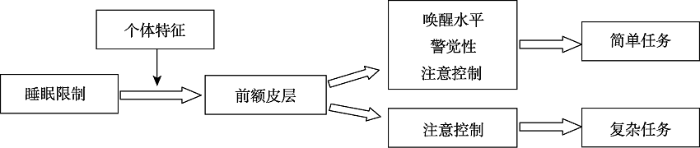
总结而言, 睡眠限制影响认知功能的作用机理存在多种可能假说, 但上述假说并不是相互对立的。例如, 睡眠剥夺后在前额皮层上的损伤可能既是执行功能, 也可能是基本注意功能发生变化的结果, 体现了前额皮层在自上而下和自下而上加工中的重要作用(Boonstra, Stins, Daffertshofer, & Beek, 2007)。因此, 这些假说是从不同的角度解释了同一种现象。另外, 以上四个假说各有优缺点, 解释力和适用范围均存在一定局限。例如, 唤醒假说和警觉性假说难以解释睡眠限制对复杂认知功能的影响; 注意控制假说难以分离持续性操作和睡眠剥夺各自对认知功能的影响。因此, 基于目前这些假说的局限性和片面性, 未来研究可以通过更加先进的技术(如EEG-fMRI同步记录)和更加精准的实验设计检验其可靠性。此外, 这些作用假说看似各自独立, 但可能存在内在关联。例如, 现有研究结果表明, 个体特征(如年龄)是影响前额皮层功能的一个重要因素, 同时fMRI研究也证明, 除了参与高级执行功能加工之外, 前额皮层也是警觉性加工的一个重要脑区(de Dreu, Schouwenaars, Rutten, Ramsey, & Jansma, 2019; Thimm et al., 2006)。因此, 未来研究可以统合睡眠限制后脑活动、生理唤醒和行为反应等多种信号的变化特点, 尝试构建一个如图1所示的整合性理论框架或提出一个适用范围更广的理论假说, 这对于人们深入认识睡眠限制与身心健康之间的密切关系以及睡眠限制相关研究的理论建构具有十分重大的意义。
图1
4 未来研究方向
尽管睡眠限制对认知功能的影响已经得到较为广泛的研究和验证, 但仍然存在一些不足和局限。第一, 已有的睡眠限制研究对不同人群认知功能影响的差异性及其内在机理缺乏深入探讨。实际上, 不同个体间存在着巨大差异, 如睡眠需要、睡眠类型和对睡眠不足的耐受性等等。已有研究发现, 大多数健康个体会随着睡眠限制而出现认知神经缺陷, 但不同个体间的缺陷程度存在显著差异(Tkachenko & Dinges, 2018)。比如, Mu等人(2005)选取了10个易受损和10个不易受损的被试, 将这些被试在正常睡眠和睡眠剥夺下工作记忆任务的脑激活模式进行对比, 结果发现, 在睡眠剥夺后, 两组被试的额顶叶激活均减少, 同时, 相比易受损被试, 不易受损被试在正常睡眠和睡眠剥夺情况下额顶叶激活均明显更多。随后一项研究发现, 在正常睡眠条件下左侧额顶叶脑区激活更强的被试在睡眠剥夺后记忆表现会更好(Chee et al., 2006)。与睡眠内稳态压力和昼夜节律相关的PERIOD3(PER3)基因的差异可能可以解释不同个体受睡眠限制影响的特异性。PER3是一个数量可变的等位基因串联的多态性基因, 大约有10%个体的PER3有5个等位基因, 即为PER35/5, 其余个体所拥有的基因则为PER34/4。已有综述表明, 相比PER34/4基因, PER35/5基因与睡眠内稳态压力和昼夜节律的关系更为紧密, 即PER35/5个体更易受睡眠限制的影响(Dijk & Archer, 2010)。但就目前而言, 睡眠不足易感性的个体间差异的生物学和神经生物学上的机制还不十分明确, 因此, 未来的研究需要系统地探索在睡眠限制影响角度上出现个体间差异的生物学因子。
第二, 由于研究技术局限, 睡眠限制对大脑神经活动与行为表现的影响及三者间关系尚不明晰。关于睡眠限制如何影响脑激活, 以及神经反应如何与行为表现变化相关这些问题, fMRI研究已经在很大程度上丰富了我们的理解, 但是血氧水平依赖(BOLD)对比的方法存在一些本质的技术与方法缺陷, 可能会限制其将来在睡眠限制研究上的应用。血氧水平依赖对比的方法通过含氧血量与缺氧血量之间的变化而测量相对信号变化, 但在测量过程中, 信号中会出现低频噪音(Friston et al., 2000), 影响血氧水平依赖对于长于几分钟的慢速神经活动变化追踪的敏感度(Aguirre, Detre, Zarahn, & Alsop, 2002)。由于信号变化非常微弱, 通常只能使用统计方法判断哪些脑区有信号变化, 因此血氧水平依赖对比方法对睡眠限制引发活性变化的脑区进行定位的精确性欠佳, 故难以全面深入地了解睡眠限制的生理基础。近来, 动脉自旋标记灌注功能磁共振成像技术(ASL Perfusion fMRI)发展迅猛。该神经成像技术利用磁性标记的上游动脉血水作为扩散示踪剂, 待已标记的血水对人脑组织灌注后, 进行全脑快速成像, 然后与非磁化标记的图像进行减影, 从而获得组织灌注参数图。这一技术可以通过非侵入性的成像方法来绝对量化认知任务执行过程和休息状态过程中的脑血流(Rao, Gillihan et al., 2007; Rao, Wang, Tang, Pan, & Detre, 2007)。另外, 动脉自旋标记灌注功能磁共振成像技术也可以在长时间内提供可靠的脑血流测量(Wang et al., 2003), 即信噪比得以提高, 从而可以定位到更加精确的脑区, 增加空间分辨率和敏感度, 克服了血氧水平依赖技术信噪比低、定位精确度差的缺点。这些特性表明, 该技术可能是探究睡眠限制生理基础的一个优势技术。这一技术不仅可以得到睡眠限制后完成认知任务时大脑皮层上绝对量化的生理数据, 还可以更加精确地观察到大脑脑区皮层上的变化。目前该神经成像技术已广泛应用于临床医学研究(Fujiwara et al., 2017; Grade et al., 2015), 具有很大的实用价值和潜力。因此, 未来研究可以使用这项技术更进一步地探究睡眠限制、生理基础与认知功能表现之间的关系。
第三, 已有研究较多关注中重度睡眠限制对个体认知功能的影响, 对轻度睡眠限制的影响缺乏探讨。实际上, 探究轻度睡眠限制对个体身心健康的影响更具现实指导意义。已有元分析发现, 现有大多数睡眠限制研究中操纵的睡眠时长大约为3.83小时(Lowe et al., 2017)。然而, 在现实生活中, 成年人在工作日的平均睡眠时长大约是6.7小时(Hirshkowitz et al., 2015), 所以, 这些中重度睡眠限制的研究结果对在实际生活中经受轻度睡眠限制的人群的参考意义较为有限。Stojanoski等人(2019)的一项研究发现, 当成年被试的夜间睡眠时长缩短2小时(轻度睡眠限制)时, 其认知功能也会受到显著损害, 表现为完成警觉性任务时错误更多、反应更慢, 且信息加工容量减少, 执行动作开始更慢。不过, 这项研究的被试仅经受了1晚的轻度睡眠限制, 中长期的轻度睡眠限制会对个体的身心功能产生何种程度的影响尚不明确。目前关于中长期轻度睡眠限制的研究数量较少, 因此, 未来研究者们可以通过操纵多晚或数周的轻度睡眠限制来考察中长期轻度睡眠限制对个体身心功能的影响。
5 小结
睡眠限制是现代社会人群中普遍存在的现象, 可能会显著危害个体身心健康, 影响其工作与学习效率, 甚至引发严重的生产或交通安全事故, 因此研究睡眠限制对个体认知功能的影响具有重大应用价值。目前, 睡眠限制对认知功能影响的研究结果并不十分一致, 而实验选用的任务类型、睡眠限制的严重程度、个体年龄和性别等因素都会影响睡眠限制对认知功能的作用。研究者们提出了多种可能性的作用假说。未来研究需要通过更加严谨科学的实验设计和更多先进的方法技术, 全面探讨睡眠限制对个体身心健康的影响, 并据此寻求能够有效对抗睡眠限制负性影响的干预措施。
参考文献
Experimental design and the relative sensitivity of BOLD and perfusion fMRI

This paper compares the statistical power of BOLD and arterial spin labeling perfusion fMRI for a variety of experimental designs within and across subjects. Based on theory and simulations, we predict that perfusion data are composed of independent observations in time under the null hypothesis, in contrast to BOLD data, which possess marked autocorrelation. We also present a method (sinc subtraction) of generating perfusion data from its raw source signal that minimizes the presence of oxygen-sensitive signal changes and can be used with any experimental design. Empirically, we demonstrate the absence of autocorrelation in perfusion noise, examine the shape of the hemodynamic response function for BOLD and perfusion, and obtain a measure of signal to noise for each method. This information is then used to generate a model of relative sensitivity of the BOLD and perfusion methods for within-subject experimental designs of varying temporal frequency. It is determined that perfusion fMRI provides superior sensitivity for within-subject experimental designs that concentrate their power at or below approximately 0.009 Hz (corresponding to a
Sleep deprivation: Impact on cognitive performance
Sleepiness enhances distraction during a monotonous task
STUDY OBJECTIVES: Although sleepiness appears to heighten distraction from the task at hand, especially if the latter is dull and monotonous, this aspect of sleep loss has not been assessed in any systematic way. Distractions are a potential cause of performance lapses (as are micro-sleeps). Here, we investigate the effects of sleepiness on a monotonous task, with and without distraction. DESIGN: Repeated Measures 2 x 2 counterbalanced design, comprising Sleepiness (night sleep restricted to 5 hours x normal sleep) and Distraction (distraction x no distraction). SETTING: Participants underwent 30-minute sessions on the Psychomotor Vigilance Test (2:00 PM - 3:10 PM), with or without an attractive distraction to be ignored, under normal and sleep-restricted conditions. PARTICIPANTS: Sixteen healthy young adults (mean age 21.10 years; 21-25 years [8 men; 8 women]) without any sleep or medical problems and without any indication of daytime sleepiness. INTERVENTIONS: Normal sleep versus sleep restricted to 5 hours and distraction versus no distraction. Distraction comprised a television in the visual periphery, showing an attractive video that had to be ignored. MEASUREMENTS AND RESULTS: Psychomotor Vigilance Test performance was monitored, as were the participants' head turns toward the television via videocameras. There was a significant increase in both head turns and lapses during sleep restriction plus distraction. Moreover, sleepiness also increased head turns even during no distraction. Distracting effects of sleepiness were clearly evident during the initial 10 minutes of testing. CONCLUSIONS: Distractibility is an important aspect of sleepiness, which has relevance to safety in the real world, eg, sleepy driving.
Human memory: A proposed system and its control processes
Circadian rhythms, sleep, and the menstrual cycle
Sleep loss and sleepiness: Current issues
Behavioral and physiological consequences of sleep restriction
Neurobehavioral dynamics following chronic sleep restriction: Dose-response effects of one night for recovery
Preliminary fMRI findings in experimentally sleep-restricted adolescents engaged in a working memory task
Here we report preliminary findings from a small-sample functional magnetic resonance imaging (fMRI) study of healthy adolescents who completed a working memory task in the context of a chronic sleep restriction experiment. Findings were consistent with those previously obtained on acutely sleep-deprived adults. Our data suggest that, when asked to maintain attention and burdened by chronic sleep restriction, the adolescent brain responds via compensatory mechanisms that accentuate the typical activation patterns of attention-relevant brain regions. Specifically, it appeared that regions that are normally active during an attention-demanding working memory task in the well-rested brain became even more active to maintain performance after chronic sleep restriction. In contrast, regions in which activity is normally suppressed during such a task in the well-rested brain showed even greater suppression to maintain performance after chronic sleep restriction. Although limited by the small sample, study results provide important evidence of feasibility, as well as guidance for future research into the functional neurological effects of chronic sleep restriction in general, the effects of sleep restriction in children and adolescents, and the neuroscience of attention and its disorders in children.
Short-term total sleep deprivations does not selectively impair higher cortical functioning
Effects of sleep deprivation on neural functioning: An integrative review
Acute restriction of nocturnal sleep in children
Learning and sleep-dependent consolidation of spatial and procedural memories are unaltered in young men under a fixed short sleep schedule
OBJECTIVE: To investigate if a fixed short sleep schedule impairs one of the main functions of sleep, which is to consolidate newly learned memories. METHODS: Sixteen young men participated in two experimental conditions, each of which lasted for 3 consecutive days and nights in our laboratory: a short sleep schedule (4.25-h sleep opportunity per night) versus a normal sleep schedule (8.5h per night). In the evening after two experimental nights, participants learned locations of 15 card pairs (spatial memory task) and a procedural finger tapping sequence task. Post-sleep retrieval of both memory tasks was tested the next morning. RESULTS: The short sleep schedule, compared with the normal sleep schedule, considerably altered sleep characteristics, e.g. the proportion of time in slow-wave sleep increased across the three experimental nights. In contrast, neither learning in the evening of day 2, nor subsequent overnight memory consolidation (i.e. concerning the change in memory performance between pre-sleep learning on day 2 and post-sleep retrieval on day 3) differed between the normal and short sleep schedule conditions. CONCLUSIONS: Our findings suggest that learning in the evening and subsequent sleep-dependent consolidation of procedural and spatial memories are unaltered in young men living under a fixed short sleep schedule. Future studies are warranted to validate our findings in other groups (e.g. adolescents and older subjects) and after more prolonged chronic sleep loss paradigms.
Functional imaging of working memory after 24 hr of total sleep deprivation
The neurobehavioral effects of 24 hr of total sleep deprivation (SD) on working memory in young healthy adults was studied using functional magnetic resonance imaging. Two tasks, one testing maintenance and the other manipulation and maintenance, were used. After SD, response times for both tasks were significantly slower. Performance was better preserved in the more complex task. Both tasks activated a bilateral, left hemisphere-dominant frontal-parietal network of brain regions reflecting the engagement of verbal working memory. In both states, manipulation elicited more extensive and bilateral (L>R) frontal, parietal, and thalamic activation. After SD, there was reduced blood oxygenation level-dependent signal response in the medial parietal region with both tasks. Reduced deactivation of the anterior medial frontal and posterior cingulate regions was observed with both tasks. Finally, there was disproportionately greater activation of the left dorsolateral prefrontal cortex and bilateral thalamus when manipulation was required. This pattern of changes in activation and deactivation bears similarity to that observed when healthy elderly adults perform similar tasks. Our data suggest that reduced activation and reduced deactivation could underlie cognitive impairment after SD and that increased prefrontal and thalamic activation may represent compensatory adaptations. The additional left frontal activation elicited after SD is postulated to be task dependent and contingent on task complexity. Our findings provide neural correlates to explain why task performance in relatively more complex tasks is better preserved relative to simpler ones after SD.
Functional imaging of working memory following normal sleep and after 24 and 35 h of sleep deprivation: Correlations of fronto-parietal activation with performance
Effects of partial sleep deprivation on information processing speed in adolescence
Effect of 38 h of total sleep deprivation on the waking EEG in women: Sex differences
38 h of sleep deprivation in women resulted in decreased alpha, increased theta and increased intrahemispheric correlation during rest and increased theta and reaction time during task. F3-O1 coherent activity was selectively decreased consistent with the role of sleep for recovery of frontal functions. Sleep deprivation effects were milder in women than in men, however, recovery was not complete suggesting that women need more sleep than men to recover.
Physiological arousal and attention during a week of continuous sleep restriction
Waking brain physiology underlying deficits from continuous sleep restriction (CSR) is not well understood. Fourteen good sleepers participated in a 21-day protocol where they slept their usual amount in a baseline week, had their time in bed restricted by 33% in a CSR week, and slept the desired amount in a recovery week. Participants slept at home, completing diaries and wearing activity monitors to verify compliance. Each day participants completed an RT task and mood and sleepiness ratings every 3 h. Laboratory assessment of electrophysiology and performance took place at the end of baseline, three times throughout the CSR week, and at the beginning of recovery. Participants reported less sleep during CSR which was confirmed by activity monitors. Correspondingly, well-being and neurobehavioural performance was impaired. Quantitative EEG analysis revealed significantly reduced arousal between the 1st and 7th days of restriction and linear effects at anterior sites (Fp2, Fz, F8, T8). At posterior sites (P4, P8), reductions occurred only later in the week between the 4th and 7th nights of restriction. Both the immediate linear decline in arousal and precipitous drop later in the week were apparent at central sites (C4, Cz). Thus, frontal regions were affected immediately, while parietal regions showed maintenance of function until restriction was more severe. The P300 ERP component showed evidence of reduced attention by the 7th day of restriction (at Pz, P4). EEG and ERPs deficits were more robust in the right-hemisphere, which may reflect greater vulnerability to sleep loss in the non-dominant hemisphere.
Memory encoding is impaired after multiple nights of partial sleep restriction
Acute sleep restriction has differential effects on components of attention
Inadequate nightly sleep duration can impair daytime functioning, including interfering with attentional and other cognitive processes. Current models posit that attention is a complex function regulated by several separate, but interacting, neural systems responsible for vigilance, orienting, and executive control. However, it is not clear to what extent each of these underlying component processes is affected by sleep loss. The purpose of this study was to evaluate the effects of acute sleep restriction on these attentional components using the Dalhousie Computerized Attention Battery (DalCAB). DalCAB tasks were administered to healthy women (aged 19-25 years) on two consecutive mornings: once after a night with 9 h time in bed (TIB), and once again after either another night with 9 h TIB (control condition, n = 19) or after a night with 3 h TIB (sleep restriction condition, n = 20). Self-ratings of sleepiness and mood were also obtained following each sleep condition. Participants showed increases in self-reported sleepiness and fatigue after the second night only in the sleep restriction group. Sleep restriction primarily affected processing speed on tasks measuring vigilance; however, performance deficits were also observed on some measures of executive function (e.g., go/no-go task, flanker task, working memory). Tasks assessing orienting of attention were largely unaffected. These results indicate that acute sleep restriction has differential effects on distinct components of attention, which should be considered in modeling the impacts of sleep loss on the underlying attentional networks.
Effects of sleep manipulation on cognitive functioning of adolescents: A systematic review
Adolescents are considered to be at risk for deteriorated cognitive functioning due to insufficient sleep. This systematic review examined the effects of experimental sleep manipulation on adolescent cognitive functioning. Sleep manipulations consisted of total or partial sleep restriction, sleep extension, and sleep improvement. Only articles written in English, with participants' mean age between 10 and 19 y, using objective sleep measures and cognitive performance as outcomes were included. Based on these criteria 16 articles were included. The results showed that the sleep manipulations were successful. Partial sleep restriction had small or no effects on adolescent cognitive functioning. Sleep deprivation studies showed decrements in the psychomotor vigilance task as most consistent finding. Sleep extension and sleep improvement contributed to improvement of working memory. Sleep directly after learning improved memory consolidation. Due to the great diversity of tests and lack of coherent results, decisive conclusions could not be drawn about which domains in particular were influenced by sleep manipulation. Small number of participants, not accounting for the role of sleep quality, individual differences in sleep need, compensatory mechanisms in adolescent sleep and cognitive functioning, and the impurity problem of cognitive tests might explain the absence of more distinct results.
Brain activity associated with expected task difficulty
Partial sleep deprivation impacts impulsive action but not impulsive decision-making
Regulation and functional correlates of slow wave sleep
PERIOD3, circadian phenotypes, and sleep homeostasis
Microcomputer analyses of performance on a portable, simple visual RT task during sustained operations
Increasing task difficulty facilitates the cerebral compensatory response to total sleep deprivation
Compensatory recruitment after sleep deprivation and the relationship with performance
Healthy older adults better tolerate sleep deprivation than young adults
Neurocognitive consequences of sleep deprivation
Deficits in daytime performance due to sleep loss are experienced universally and associated with a significant social, financial, and human cost. Microsleeps, sleep attacks, and lapses in cognition increase with sleep loss as a function of state instability. Sleep deprivation studies repeatedly show a variable (negative) impact on mood, cognitive performance, and motor function due to an increasing sleep propensity and destabilization of the wake state. Specific neurocognitive domains including executive attention, working memory, and divergent higher cognitive functions are particularly vulnerable to sleep loss. In humans, functional metabolic and neurophysiological studies demonstrate that neural systems involved in executive function (i.e., prefrontal cortex) are more susceptible to sleep deprivation in some individuals than others. Recent chronic partial sleep deprivation experiments, which more closely replicate sleep loss in society, demonstrate that profound neurocognitive deficits accumulate over time in the face of subjective adaptation to the sensation of sleepiness. Sleep deprivation associated with disease-related sleep fragmentation (i.e., sleep apnea and restless legs syndrome) also results in neurocognitive performance decrements similar to those seen in sleep restriction studies. Performance deficits associated with sleep disorders are often viewed as a simple function of disease severity; however, recent experiments suggest that individual vulnerability to sleep loss may play a more critical role than previously thought.
Biological Basis of Personality
Effects of acute sleep restriction on behavior, sustained attention, and response inhibition in children
To smooth or not to smooth?: Bias and efficiency in fMRI time-series analysis
This paper concerns temporal filtering in fMRI time-series analysis. Whitening serially correlated data is the most efficient approach to parameter estimation. However, if there is a discrepancy between the assumed and the actual correlations, whitening can render the analysis exquisitely sensitive to bias when estimating the standard error of the ensuing parameter estimates. This bias, although not expressed in terms of the estimated responses, has profound effects on any statistic used for inference. The special constraints of fMRI analysis ensure that there will always be a misspecification of the assumed serial correlations. One resolution of this problem is to filter the data to minimize bias, while maintaining a reasonable degree of efficiency. In this paper we present expressions for efficiency (of parameter estimation) and bias (in estimating standard error) in terms of assumed and actual correlation structures in the context of the general linear model. We show that: (i) Whitening strategies can result in profound bias and are therefore probably precluded in parametric fMRI data analyses. (ii) Band-pass filtering, and implicitly smoothing, has an important role in protecting against inferential bias.
Comparison of long-labeled pseudo-continuous arterial spin labeling (ASL) features between young and elderly adults: Special reference to parameter selection
Cognitive workload and sleep restriction interact to influence sleep homeostatic responses
A neuroradiologist's guide to arterial spin labeling MRI in clinical practice
Positive evidence for Eysenck’s arousal hypothesis: A combined EEG and MRI study with multiple measurement occasions
The impact of sleep deprivation on decision making: A review
Prefrontal neuropsychological effects of sleep deprivation in young adults — A model for healthy aging?
Neuropsychological testing and brain imaging show that healthy aging leads to a preferential impairment of the prefrontal cortex (PFC). Interestingly, in young adults sleep deprivation (SD) has similar effects. Psychological tasks not so oriented to the PFC are less sensitive both to SD and aging. The PFC is a cortical region working particularly hard during wakefulness, which may make it more vulnerable to
National Sleep Foundation's sleep time duration recommendations: Methodology and results summary
Sleep deprivation, vigilant attention, and brain function: A review
Vigilant attention is a major component of a wide range of cognitive performance tasks. Vigilant attention is impaired by sleep deprivation and restored after rest breaks and (more enduringly) after sleep. The temporal dynamics of vigilant attention deficits across hours and days are driven by physiologic, sleep regulatory processes-a sleep homeostatic process and a circadian process. There is also evidence of a slower, allostatic process, which modulates the sleep homeostatic setpoint across days and weeks and is responsible for cumulative deficits in vigilant attention across consecutive days of sleep restriction. There are large inter-individual differences in vulnerability to sleep loss, and these inter-individual differences constitute a pronounced human phenotype. However, this phenotype is multi-dimensional; vulnerability in terms of vigilant attention impairment can be dissociated from vulnerability in terms of other cognitive processes such as attentional control. The vigilance decrement, or time-on-task effect-a decline in performance across the duration of a vigilant attention task-is characterized by progressively increasing response variability, which is exacerbated by sleep loss. This variability, while crucial to understanding the impact of sleep deprivation on performance in safety-critical tasks, is not well explained by top-down regulatory mechanisms, such as the homeostatic and circadian processes. A bottom-up, neuronal pathway-dependent mechanism involving use-dependent, local sleep may be the main driver of response variability. This bottom-up mechanism may also explain the dissociation between cognitive processes with regard to trait vulnerability to sleep loss.
Effect of time of day and partial sleep deprivation on the reaction time and the attentional capacities of the handball goalkeeper
Effect of chronic sleep restriction on sleepiness and working memory in adolescents and young adults
OBJECTIVES: To test the feasibility of using a home-based sleep restriction protocol in adolescents and young adults; and to examine the different effects of chronic sleep restriction on a subjective sleepiness scale and working memory task in adolescents and young adults. METHOD: Twenty adolescents (ages 13-16 years) and 20 young adults (ages 18-20 years) underwent a 2-week home-based sleep manipulation protocol consisting of a week of 5 school days with 8 hr spent in bed per night and another week of 5 school days with 6 hr spent in bed per night. The protocol used a counterbalanced crossover experimental design. Subjective sleepiness was scored by the participant each morning, and working memory tests were administered during the weekend corresponding to each experimental week. RESULTS: Adherence to the prescribed protocol was similar in the two groups, and both groups achieved the desired differences in total sleep duration across the two sleep conditions. Subjective sleepiness scores significantly increased in young adults after sleep restriction, but were not accompanied by significant changes in working memory. However, reaction times during simple verbal and arithmetic working memory tasks increased among adolescents after sleep restriction, without affecting accuracy on task, and without eliciting increases in subjective sleepiness scores. CONCLUSION: Mild sleep restriction for 5 days impairs reaction times during working memory tasks in adolescents in the absence of increased perception of sleepiness.
Effects of sleep deprivation on cognition
Sleep deprivation is commonplace in modern society, but its far-reaching effects on cognitive performance are only beginning to be understood from a scientific perspective. While there is broad consensus that insufficient sleep leads to a general slowing of response speed and increased variability in performance, particularly for simple measures of alertness, attention and vigilance, there is much less agreement about the effects of sleep deprivation on many higher level cognitive capacities, including perception, memory and executive functions. Central to this debate has been the question of whether sleep deprivation affects nearly all cognitive capacities in a global manner through degraded alertness and attention, or whether sleep loss specifically impairs some aspects of cognition more than others. Neuroimaging evidence has implicated the prefrontal cortex as a brain region that may be particularly susceptible to the effects of sleep loss, but perplexingly, executive function tasks that putatively measure prefrontal functioning have yielded inconsistent findings within the context of sleep deprivation. Whereas many convergent and rule-based reasoning, decision making and planning tasks are relatively unaffected by sleep loss, more creative, divergent and innovative aspects of cognition do appear to be degraded by lack of sleep. Emerging evidence suggests that some aspects of higher level cognitive capacities remain degraded by sleep deprivation despite restoration of alertness and vigilance with stimulant countermeasures, suggesting that sleep loss may affect specific cognitive systems above and beyond the effects produced by global cognitive declines or impaired attentional processes. Finally, the role of emotion as a critical facet of cognition has received increasing attention in recent years and mounting evidence suggests that sleep deprivation may particularly affect cognitive systems that rely on emotional data. Thus, the extent to which sleep deprivation affects a particular cognitive process may depend on several factors, including the magnitude of global decline in general alertness and attention, the degree to which the specific cognitive function depends on emotion-processing networks, and the extent to which that cognitive process can draw upon associated cortical regions for compensatory support.
Work-related accidents and daylight saving time in Finland
Sustaining attention to simple tasks: A meta-analytic review of the neural mechanisms of vigilant attention
Maintaining attention for more than a few seconds is essential for mastering everyday life. Yet, our ability to stay focused on a particular task is limited, resulting in well-known performance decrements with increasing time on task. Intriguingly, such decrements are even more likely if the task is cognitively simple and repetitive. The attentional function that enables our prolonged engagement in intellectually unchallenging, uninteresting activities has been termed vigilant attention. Here we synthesized what we have learned from functional neuroimaging about the mechanisms of this essential mental faculty. To this end, a quantitative meta-analysis of pertinent neuroimaging studies was performed, including supplementary analyses of moderating factors. Furthermore, we reviewed the available evidence on neural time-on-task effects, additionally considering information obtained from patients with focal brain damage. Integrating the results of both meta-analysis and review, we identified a set of mainly right-lateralized brain regions that may form the core network subserving vigilant attention in humans, including dorsomedial, mid- and ventrolateral prefrontal cortex, anterior insula, parietal areas (intraparietal sulcus, temporoparietal junction), and subcortical structures (cerebellar vermis, thalamus, putamen, midbrain). We discuss the potential functional roles of different nodes of this network as well as implications of our findings for a theoretical account of vigilant attention. It is conjectured that sustaining attention is a multicomponent, nonunitary mental faculty, involving a mixture of (a) sustained/recurrent processes subserving task-set/arousal maintenance and (b) transient processes subserving the target-driven reorienting of attention. Finally, limitations of previous studies are considered and suggestions for future research are provided.
Alcohol and sleep restriction combined reduces vigilant attention, whereas sleep restriction alone enhances distractibility
Multiple nights of partial sleep deprivation do not affect prospective remembering at long delays
Short-term memory deficits correlate with hippocampal-thalamic functional connectivity alterations following acute sleep restriction
Sleep deprivation and vigilant attention
A meta-analysis of the impact of short-term sleep deprivation on cognitive variables
Sleep restriction can attenuate prioritization benefits on declarative memory consolidation
Sleep deprivation increases formation of false memory
Cognitive performance, sleepiness, and mood in partially sleep deprived adolescents: The need for sleep study
STUDY OBJECTIVES: To investigate the effects of sleep restriction (7 nights of 5 h time in bed [TIB]) on cognitive performance, subjective sleepiness, and mood in adolescents. METHODS: A parallel-group design was adopted in the Need for Sleep Study. Fifty-six healthy adolescents (25 males, age = 15-19 y) who studied in top high schools and were not habitual short sleepers were randomly assigned to Sleep Restriction (SR) or Control groups. Participants underwent a 2-w protocol consisting of 3 baseline nights (TIB = 9 h), 7 nights of sleep opportunity manipulation (TIB = 5 h for the SR and 9 h for the control groups), and 3 nights of recovery sleep (TIB = 9 h) at a boarding school. A cognitive test battery was administered three times each day. RESULTS: During the manipulation period, the SR group demonstrated incremental deterioration in sustained attention, working memory and executive function, increase in subjective sleepiness, and decrease in positive mood. Subjective sleepiness and sustained attention did not return to baseline levels even after 2 recovery nights. In contrast, the control group maintained baseline levels of cognitive performance, subjective sleepiness, and mood throughout the study. Incremental improvement in speed of processing, as a result of repeated testing and learning, was observed in the control group but was attenuated in the sleep-restricted participants, who, despite two recovery sleep episodes, continued to perform worse than the control participants. CONCLUSIONS: A week of partial sleep deprivation impairs a wide range of cognitive functions, subjective alertness, and mood even in high-performing high school adolescents. Some measures do not recover fully even after 2 nights of recovery sleep. COMMENTARY: A commentary on this article appears in this issue on page 497.
The neurocognitive consequences of sleep restriction: A meta-analytic review
Effects of tyrosine, phentermine, caffeine D-amphetamine, and placebo on cognitive and motor performance deficits during sleep deprivation
Cognitive and motor performance are critical in many circumstances and are impaired by sleep deprivation. We administered placebo, tyrosine 150 mg/kg, caffeine 300 mg/70 kg, phentermine 37.5 mg and D-amphetamine 20 mg at 15.30 h following overnight sleep deprivation and compare their effects on cognitive and motor performance in healthy young men. Tests of visual scanning, running memory, logical reasoning, mathematical processing, the Stroop task, four-choice serial reaction time, time wall take, pursuit tracking, visual vigilance, Trails (B) task and long-term memory were evaluated at standardized intervals before, during and after sleep deprivation and drugs. Performance decrements with sleep deprivation occurred in visual scanning, running memory, logical reasoning, mathematical processing, the Stroop test, the time wall test, tracking and visual vigilance. Interestingly, with sleep deprivation some tests improved and others did not deteriorate. Improvements with medication following sleep deprivation were seen in running memory, logical reasoning, mathematical processing, tracking and visual vigilance. Although less effective than D-amphetamine, tyrosine improved performance on several tests. We conclude that all drugs tested improved at least some aspects of cognitive and motor performance after sleep deprivation. As a naturally occurring amino acid, and thus amenable to nutritional strategies, tyrosine may deserve further testing.
The cognitive cost of sleep lost
The nature and organization of individual differences in executive functions: Four general conclusions
The unity and diversity of executive functions and their contributions to complex "Frontal Lobe" tasks: A latent variable analysis
Impaired cortical oxygenation is related to mood disturbance resulting from three nights of sleep restriction
Insufficient sleep impairs driving performance and cognitive function
Decreased brain activation during a working memory task at rested baseline is associated with vulnerability to sleep deprivation
STUDY OBJECTIVE: To examine whether differences in patterns of brain activation under baseline conditions relate to the differences in sleep-deprivation vulnerability. DESIGN: Using blood oxygenation level dependent (BOLD) functional magnetic resonance imaging, we scanned 33 healthy young men while they performed the Sternberg working memory task following a normal night of sleep and again following 30 hours of sleep deprivation. From this initial group, based on their Sternberg working memory task performance, we found 10 subjects resilient to sleep deprivation (sleep deprivation-resilient group) and then selected 10 age- and education-matched subjects vulnerable to sleep deprivation (sleep deprivation-vulnerable group). SETTING: Inpatient General Clinical Research Center and outpatient functional magnetic resonance imaging center. PATIENTS OR PARTICIPANTS: Data from 10 young men (mean age 27.8 +/- 1.7 years) in the sleep deprivation-resilient group and 10 young men (mean age 28.2 +/- 1.9 years) in the sleep deprivation-vulnerable group were included in the final analyses. INTERVENTIONS: None. MEASUREMENTS AND RESULTS: We compared functional magnetic resonance imaging BOLD signal at rested baseline and sleep deprivation states in the 2 groups. As hypothesized, following sleep deprivation, both groups showed significant decreases in global brain activation compared to their rested group baseline. At rested baseline and in the sleep-deprivation state, the sleep deprivation-resilient group had significantly more brain activation than did the sleep deprivation-vulnerable group. There were also differences in functional circuits within and between groups in response to sleep deprivation. CONCLUSIONS: These preliminary data suggest that patterns of brain activation during the Sternberg working memory task at the rested baseline and the sleep-deprivation state, differ across individuals as a function of their sleep-deprivation vulnerability.
The frontal predominance in human EEG delta activity after sleep loss decreases with age
Vigilance, alertness, or sustained attention: Physiological basis and measurement
Vigilance is a term with varied definitions but the most common usage is sustained attention or tonic alertness. This usage of vigilance implies both the degree of arousal on the sleep-wake axis and the level of cognitive performance. There are many interacting neural and neurotransmitter systems that affect vigilance. Most studies of vigilance have relied on states where the sleep-wake state is altered, e.g. drowsiness, sleep-deprivation, and CNS-active drugs, but there are factors ranging from psychophysics to motivation that may impact vigilance. While EEG is the most commonly studied physiologic measure of vigilance, various measures of eye movement and of autonomic nervous system activity have also been used. This review paper discusses the underlying neural basis of vigilance and its assessment using physiologic tools. Since, assessment of vigilance requires assessment of cognitive function this aspect is also discussed.
The attention system of the human brain: 20 years after
Human performance under sustained operations and acute sleep deprivation conditions: Toward a model of controlled attention
Distinct neural correlates of time-on-task and transient errors during a visuomotor tracking task after sleep restriction
Genetic variation in serotonin transporter alters resting brain function in healthy individuals
BACKGROUND: Perfusion functional magnetic resonance imaging (fMRI) was used to investigate the effect of genetic variation of the human serotonin transporter (5-HTT) gene (5-HTTLPR, SLC6A4) on resting brain function of healthy individuals. METHODS: Twenty-six healthy subjects, half homozygous for the 5-HTTLPR short allele (s/s group) and half homozygous for the long allele (l/l group), underwent perfusion functional and structural magnetic resonance imaging during a resting state. The two genotype groups had no psychiatric illness and were similar in age, gender, and personality scores. RESULTS: Compared with the l/l group, the s/s group showed significantly increased resting cerebral blood flow (CBF) in the amygdala and decreased CBF in the ventromedial prefrontal cortex. The effect of functional modulation in these regions by 5-HTTLPR genotype cannot be accounted for by variations in brain anatomy, personality, or self-reported mood. CONCLUSIONS: The 5-HTTLPR genotype alters resting brain function in emotion-related regions in healthy individuals, including the amygdala and ventromedial prefrontal cortex. Such alterations suggest a broad role of the 5-HTT gene in brain function that may be associated with the genetic susceptibility for mood disorders such as depression.
Imaging brain activity during natural vision using CASL perfusion fMRI
Functional MRI (fMRI) has begun to be used to explore human brain activity during ecological and natural conditions. Arterial spin labeling (ASL) perfusion fMRI provides an appealing approach for imaging sustained brain activity during natural conditions because of its long-term temporal stability and ability to noninvasively quantify absolute cerebral blood flow (CBF). The present study used ASL perfusion fMRI to measure brain activation patterns associated with natural vision by concurrently recording CBF and blood oxygen level-dependent (BOLD) contrasts while subjects were freely viewing a cartoon movie. Reliable quantitative whole-brain CBF values ( approximately 60 mL/100g/min) as well as regional CBF values (45 approximately 80 mL/100g/min) were measured during movie viewing and resting states. The perfusion contrast revealed CBF increases in multiple visual pathway areas and frontal areas, and CBF decreases in ventromedial frontal cortex and superior temporal cortex during movie viewing compared to resting states. Concurrent BOLD contrast revealed similar but weaker activation and deactivation patterns. Regression analyses of both CBF data and BOLD data showed significant associations between activation in the middle temporal (MT) region and subjects' perception of motion. Region of interest analysis based on a priori literature-defined MT demonstrated significant monotonic stepwise associations between the intensity of motion perception and the CBF and BOLD signal changes. These results demonstrate the feasibility of using ASL perfusion fMRI for imaging both sustained and dynamic effects in neural activation during natural and ecologically valid situations, and support the notion of maintained functional segregation and specialization during natural vision.
Ethanol and sleep loss: A "dose" comparison of impairing effects
Time to talk about work-hour impact on anesthesiologists: The effects of sleep deprivation on Profile of Mood States and cognitive tasks
The effects of sleep restriction and extension on school-age children: What a difference an hour makes
Sex differences in the circadian regulation of sleep and waking cognition in humans
Estimating adolescent sleep need using dose-response modeling
Effects of sleep deprivation, lunch, and personality on performance, mood, and cardiovascular function
Effects of sleep restriction on cognition in women
The aim of the present study was to investigate how three nights of sleep restriction affected cognitive functions in young and aged healthy women. Ten young (20-30 years) and ten aged (55-65 years) women participated to the study. After one baseline night (11 pm-07 am), their sleep was restricted to 4h per night (01-05 am) during three nights. A recovery night of 8h (11 pm-07 am) followed the sleep restriction. The neurobehavioural assessment included evaluation of Attention (Stroop, Trail Making test, Tests of Attentional Performance), Memory (Buschke, Logical Memory, PASAT, Brown Peterson), Addition of numbers and Abstraction (Wisconsin). Sleep restriction decreased significantly the speed of execution, particularly in the young women, without affecting the accuracy of the answers. This effect was the most significant on reaction times in simple tests. The more complex tasks (PASAT, Brown Peterson, Logical Memory, Addition of numbers, Wisconsin) were not affected. However, the inhibition of an automatic activity (Stroop test) and the formation of a memory trace (Buschke memory test) were disturbed in both young and older women.
Reciprocal roles of sleep and diet in cardiovascular health: A review of recent evidence and a potential mechanism
PURPOSE OF REVIEW: This review investigates the potential bi-directional relation between sleep and diet in considering their contribution to cardiovascular health. We further explore the involvement of the gut microbiome in the relationships between poor sleep and dietary intakes and increased cardiovascular disease (CVD) risk. RECENT FINDINGS: There is strong evidence that sleep restriction leads to unhealthy food choices and increased energy intake. The diet may impact sleep, as well. Epidemiological studies show that higher adherence to a Mediterranean dietary pattern predicts healthier sleep. One factor that could underlie these relationships is the gut microbiome. Although data are mixed, there is some evidence that sleep restriction can influence the composition of the gut microbiome in humans. Similarly, Mediterranean diets and other plant-rich diets are related to increased diversity of the microbiota. At present, few studies have investigated the influence of the microbiome on sleep; however, limited evidence from epidemiological and intervention studies suggest that the composition of the microbiome may relate to sleep quality. More research is needed to better understand the role of the microbiome in the multi-directional relationship between sleep, diet, and CVD. There is growing evidence of a bi-directional relationship between sleep and the diet, which could act in concert to influence CVD risk. Diets such as the Mediterranean diet, comprised of high intakes of fruits, vegetables, and other plant-based foods, may promote healthy sleep and beneficial gut microflora. The gut microbiome may then underlie the relation between diet, sleep, and CVD risk.
Sustained vigilance is negatively affected by mild and acute sleep loss reflected by reduced capacity for decision making, motor preparation, and execution
Functions of the frontal lobes: Relation to executive functions
Impact of alertness training on spatial neglect: A behavioural and fMRI study
The effects of a 3-week computerised alertness training on chronic (>3 months) visuospatial hemineglect were investigated prospectively in seven patients by means of neuropsychological tests and functional magnetic resonance imaging (fMRI). Following the alertness training, the group showed improved alertness and a significant improvement in the performance of a neglect test battery over and above any improvement during a 3-week baseline phase. Improvements in the neglect tasks were accompanied by an increase of right hemisphere neural activity in frontal cortex, anterior cingulate cortex, precuneus, cuneus and angular gyrus. These areas have previously been associated with alertness and spatial attention. A similar pattern of increased neural activity was found for the left hemisphere. Four weeks after the end of the training, the patients' neglect test performance had mostly returned to baseline, while the increases in neural activity bilaterally in frontal areas, in the right anterior cingulate cortex, the right angular gyrus and the left temporoparietal cortex remained. The data show that a 3-week computerised alertness training can improve performance both in alertness and neglect tests and that these behavioural improvements are associated with reactivation in areas associated with alerting and visuospatial attention. The limited stability of these effects over time suggests that a 3-week alertness training alone does not result in long lasting improvements in every patient, but refining the treatment protocol may lead to a more stable amelioration of neglect symptoms.
Interindividual variability in neurobehavioral response to sleep loss: A comprehensive review
Chronic sleep reduction is associated with academic achievement and study concentration in higher education students
Inadequate sleep impairs cognitive function and has been associated with worse academic achievement in higher education students; however, studies that control for relevant background factors and include knowledge on sleep hygiene are scarce. This study examined the association of chronic sleep reduction (i.e. symptoms of chronic sleep reduction such as shortness of sleep, sleepiness and irritation), subjective sleep quality and sleep hygiene knowledge with academic achievement (grades and study credits) and study concentration among 1378 higher education students (71% female, mean age 21.73 years, SD = 3.22) in the Netherlands. Demographic, health, lifestyle and study behaviour characteristics were included as covariates in hierarchical regression analyses. After controlling for significant covariates, only chronic sleep reduction remained a significant predictor of lower grades (last exam, average in current academic year). Better sleep quality and sleep hygiene knowledge were associated with better academic achievement, but significance was lost after controlling for covariates, except for a remaining positive association between sleep hygiene beliefs and grades in the current academic year. Moreover, better sleep quality and lower scores on chronic sleep reduction were associated with better study concentration after controlling for significant covariates. To conclude, chronic sleep reduction is associated with academic achievement and study concentration in higher education students. Inadequate sleep hygiene knowledge is moderately associated with worse academic achievement. Future research should investigate whether sleep hygiene interventions improve academic achievement in students of higher education.
Sleep benefits subsequent hippocampal functioning
Sleep before learning benefits memory encoding through unknown mechanisms. We found that even a mild sleep disruption that suppressed slow-wave activity and induced shallow sleep, but did not reduce total sleep time, was sufficient to affect subsequent successful encoding-related hippocampal activation and memory performance in healthy human subjects. Implicit learning was not affected. Our results suggest that the hippocampus is particularly sensitive to shallow, but intact, sleep.
The cumulative cost of additional wakefulness: dose-response effects on neurobehavioral functions and sleep physiology from chronic sleep restriction and total sleep deprivation
Temperament moderates the association between sleep duration and cognitive performance in children
The importance of sufficient sleep for cognitive performance has been increasingly recognized. Individual differences in susceptibility to effects of sleep restriction have hardly been investigated in children. We investigated whether individual differences in temperament moderate the association of sleep duration with sustained attention, inhibition, and working memory in 123 children (42% boys) aged 9 to 11 years. Sleep duration was assessed using parental diaries, and temperament traits of extraversion and negative affectivity were assessed by child self-report (Early Adolescent Temperament Questionnaire-Revised). Computerized assessment of sustained attention (short-form Psychomotor Vigilance Task, PVT), inhibition (PVT Go/No-Go adaptation), and working memory (visual Digit Span) were performed at school. Our findings demonstrate that long-sleeping introverted and negatively affective children show worse sustained attention and working memory than short-sleeping children with these temperaments.
Effects of sleep reduction on spatial attention
Sleep restriction over several days does not affect long-term recall of declarative and procedural memories in adolescents
OBJECTIVES: There is broad evidence that sleep as opposed to waking facilitates the consolidation of both declarative and procedural memory. The current study addressed the question whether different extents of sleep restriction after learning would impair long-term memory consolidation in adolescents. METHODS: Eighty-eight healthy adolescents were randomized to five different sleep protocols with 9, 8, 7, 6 or 5 h of time in bed for four consecutive nights under controlled conditions that excluded daytime sleep. Declarative (word-pair task) and procedural memory (mirror tracing task) encoding was assessed prior to the sleep restriction protocol. Recall was assessed after two recovery nights following the sleep protocol and 4 weeks later. RESULTS: Sleep diaries and actigraphy data demonstrated that the participants closely followed the sleep protocols. There were no differences in demographic parameters or memory encoding at baseline. In contrast to the initial prediction, restriction of nocturnal sleep over four consecutive nights had no significant impact on declarative or procedural memory consolidation. Polysomnographic monitoring after sleep restriction demonstrated a high preservation of the amount of slow wave sleep in the restricted conditions. CONCLUSIONS: The results suggest that adolescents show a high resilience of memory consolidation to substantial sleep curtailment across four nights that might be promoted by increased sleep intensity under conditions of sleep restriction.
Arterial spin labeling perfusion fMRI with very low task frequency
Functional magnetic resonance imaging (fMRI) has become the most widely used modality for visualizing regional brain activation in response to sensorimotor or cognitive tasks. While the majority of fMRI studies have used blood oxygenation level-dependent (BOLD) contrast as a marker for neural activation, baseline drift effects result in poor sensitivity for detecting slow variations in neural activity. By contrast, drift effects are minimized in arterial spin labeling (ASL) perfusion contrast, primarily as a result of successive pairwise subtraction between images acquired with and without labeling. Recent data suggest that ASL contrast shows stable noise characteristics over the entire frequency spectrum, which makes it suitable for studying low-frequency events in brain function. The present study investigates the relative sensitivities of ASL and BOLD contrast in detecting changes in motor cortex activation over a spectrum of frequencies of experimental design, where the alternating period between the resting state and activation is varied from 30 s up to 24 hr. The results demonstrate that 1) ASL contrast can detect differences in motor cortex activation over periods of minutes, hours, and even days; 2) the functional sensitivity of ASL contrast becomes superior to that of BOLD contrast when the alternating period between the resting state and activation is greater than a few minutes; and 3) task activation measured by ASL tends to have less intersubject variability than BOLD contrast. The improved sensitivity of the ASL contrast for low task frequency and longitudinal studies, along with its superior power in group analysis, is expected to extend the range of experimental designs that can be studied using fMRI.
Impaired performance with acute sleep loss
Effects of menstrual cycle phase and oral contraceptives on alertness, cognitive performance, and circadian rhythms during sleep deprivation
The influence of menstrual cycle phase and oral contraceptive use on neurobehavioral function and circadian rhythms were studied in healthy young women (n = 25) using a modified constant routine procedure during 24 h of sleep deprivation. Alertness and performance worsened across sleep deprivation and also varied with circadian phase. Entrained circadian rhythms of melatonin and body temperature were evident in women regardless of menstrual phase or oral contraceptive use. No significant difference in melatonin levels, duration, or phase was observed between women in the luteal and follicular phases, whereas oral contraceptives appeared to increase melatonin levels. Temperature levels were higher in the luteal phase and in oral contraceptive users compared to women in the follicular phase. Alertness on the maintenance of wakefulness test and some tests of cognitive performance were poorest for women in the follicular phase especially near the circadian trough of body temperature. These observations suggest that hormonal changes associated with the menstrual cycle and the use of oral contraceptives contribute to changes in nighttime waking neurobehavioral function and temperature level whereas these factors do not appear to affect circadian phase.
The relation of strength of stimulus to rapidity of habit-formation
A deficit in the ability to form new human memories without sleep
Experimental sleep restriction effect on adult body weight: A meta-analysis
BACKGROUND: Sleep is increasingly recognized as a potential risk for overweight and obesity. Observational studies have shown links between short sleep duration with weight gain. However, the findings from longitudinal studies in adults are conflicting. This review aimed to examine the effectiveness of experimental sleep restriction on adult body weight. METHOD: A systematic search was undertaken in MEDLINE, EMBASE, PsycINFO, and CENTRAL (Cochrane center register of controlled trials) to identify experimental studies examining the effectiveness of sleep restriction on body weight, and search period was from January 2005 to June 2018. Meta-analysis was applied by using the random model. RESULTS: A total of 275 adults from six experimental studies were included. The pooled standard mean difference in body weight and body fat was 0.44 (95% CI - 0.13 to 1.02) (Z = 1.51, p > 0.05) and 0.35 kg (95% CI - 0.19 to 0.88) (Z = 1.27, p > 0.05), respectively. The experimental sleep restriction did not result in significant differences in adult body weight or body fat. Subgroup analysis showed that there were differences in weight gain between genders and races. CONCLUSION: The finding from this review cannot support the hypothesis from observational studies that short sleep leads to weight gain.
Prefrontal cortex and executive functions in healthy adults: A meta-analysis of structural neuroimaging studies
Decreased information replacement of working memory after sleep deprivation: Evidence from an event-related potential study
Working memory (WM) components are altered after total sleep deprivation (TSD), both with respect to information replacement and result judgment. However, the electrophysiological mechanisms of WM alterations following sleep restriction remain largely unknown. To identify such mechanisms, event-related potentials were recorded during the n-back WM task, before and after 36 h sleep deprivation. Thirty-one young volunteers participated in this study and performed a two-back WM task with simultaneous electroencephalography (EEG) recording before and after TSD and after 8 h time in bed for recovery (TIBR). Repeated measures analysis of variance revealed that, compared to resting wakefulness, sleep deprivation induced a decrease in the P200 amplitude and induced longer reaction times. ERP-component scalp topographies results indicated that such decrease primarily occurred in the frontal cortex. The N200 and P300 amplitudes also decreased after TSD. Our results suggest that decreased information replacement of WM occurs after 36 h of TSD and that 8 h TIBR after a long period of TSD leads to partial restoration of WM functions. The present findings represent the EEG profile of WM during mental fatigue.
Young adults are more vulnerable to chronic sleep deficiency and recurrent circadian disruption than older adults
More than a third of US adults report fewer than 6 hours of sleep a night, making chronic sleep restriction a growing public health concern. Sleep curtailment is associated with an increase in industrial accidents, motor vehicle accidents, medical and other occupational errors. Young adults are more vulnerable to acute sleep deprivation than older adults, but less is known about how young vs. older adults respond to the more commonly experienced chronic sleep restriction. To test the hypothesis that young adults are more vulnerable to chronic sleep loss than older adults, we compared data from young and older adults who underwent three weeks of chronic sleep restriction (equivalent to 5.6 hours/24 hours) combined with recurrent circadian disruption in an experiment that enabled us to separate the influences of the sleep-wake homeostatic process, the circadian timing system, and the chronic sleep deficit. We found that while young and older adults reported similar levels of subjective sleepiness, objective measures of sleepiness revealed that young adults were more vulnerable and had more attentional failures than the older adults. These results have important public health implications, particularly related to prevention of sleep-related motor vehicle crashes in young drivers. Further research is needed to understand the neurobiological basis of these age-related differences.
