

1 引言
近年来, 随着认知神经科学的快速发展和对功能磁共振成像(functional Magnetic Resonance Imaging, fMRI)了解的深入, 有关脑信号的频率研究也被广泛关注。fMRI信号来源于大脑血液中脱氧血红蛋白含量的动态变化, 是一种非平稳非线性变化的信号, 具有复杂的时频特征。另外, fMRI信号是大脑复杂的机理性反映, 不同频段节律信号的生理意义尚无定论, 对fMRI信号进行时频分析是当下一种较为热门的研究方向, 但是在已有的研究中, 研究人员多根据主观经验或经典方法将其划分, 缺乏统一的分频标准, 致使该领域内相关分频研究的可重复性较差, 同时由于经典算法对数据的假设较多, 未能充分发掘功能磁共振信号的自身特性, 也阻碍了我们进一步了解它的内在生理意义。例如, 在对fMRI低频成分的脑功能网络研究中, Wu等人(2008)用切比雪夫滤波器将fMRI信号分为12个频段, 并探讨了功能连接的频率特性, 发现具有高功能连接系数的脑功能活动集中在特定的低频段(0.01~0.06 Hz); Salvador, Suckling, Schwarzbauer和Bullmore (2005)以及Salvador等人(2008)基于傅里叶方法对信号分频揭示了较高功能连接系数的脑功能活动分布在低频段(<0.10 Hz); Achard, Salvador, Whitcher, Suckling和Bullmore (2006)利用小波变换方法分为6个频段, 证明了低频成分(0.03~0.06 Hz)的全脑相关性最高; Zuo等人(2010)基于滤波器方法揭示了低频振幅在(0.027~0.073 Hz)频段有更强的幅值; Liang等人(2012)采用滤波器方法验证了低频段(0.027~ 0.073 Hz)形成的脑网络比0.01~0.027频段形成的脑网络更稳定。上述研究均强调了fMRI信号低频成分的功能连接和网络特性, 但是在频率边界的界定上未能形成统一, 为后续的验证性和发现性研究设置了一定障碍。此外, 上述研究中采用的分频方法如傅里叶变换、切比雪夫滤波器等多假设信号线性平稳, fMRI信号并不能满足这一点, 这也限制了对其结果的解释和推广, 没有充分揭示fMRI信号的完整特性。
本文采用了一种数据驱动的多维集合经验模式算法对fMRI信号进行分频, 相比传统分频方法, 该算法以信号自身特性为出发点, 从时间维度和空间维度共同提取信号有效信息, 可以更加准确刻画出信号的物理过程。为展示这一新方法的应用, 我们针对“性别差异”这一生命科学, 特别是心理学, 领域中长期以来感兴趣的科学问题, 应用上述多尺度自适应的分频方法探究了大脑低频成分功能连接的性别差异, 尝试为该算法在脑功能领域的应用提供参考。
2 材料与方法
2.1 数据采集和预处理
本研究招募了66名健康右利手被试, 年龄范围18~35岁, 其中男性37例, 女性29例, 均无高血压、心脏病、脑血管病和神经精神类疾病, 均签署知情同意书。所有脑影像扫描均在中国科学院心理研究所磁共振中心完成, 磁共振扫描仪为GE (3T)MR750扫描仪, 扫描前用海绵垫将其头部固定好, 并告知在静息态磁共振扫描中保持头部不动, 注视屏幕中的十字, 不能入睡。人脑解剖形态的T1加权结构磁共振成像参数设置如下:翻转角为8度; 层厚为1 mm; 层间距为1 mm; 重复时间为6.896 mm; 回波时间为2.992 mm; 视野为256 mm, 采集矩阵为256 × 256。人脑静息态功能的磁共振成像参数设置如下:翻转角为10度; 层厚为3.5 mm; 层间距为0.5 mm; 回波时间为30 mm; 重复时间为2000 mm; 视野为220 mm, 采集矩阵为64 × 64。图像采用中国科学院心理研究所脑功能连接组及发展实验室研制的连接组计算平台(Connectome Computation System, CCS) (https:// github.com/zuoxinian/CCS) (Zuo et al., 2013; Xu et al., 2015)进行预处理, 首先结构像使用volbrain脑影像在线处理平台(http://volbrain.upv.es/)进行图像去噪, 信号值矫正, 颅骨分割等步骤, 再调用FreeSurfer (Fischl, 2012)的Recon-all程序对大脑皮层进行分割和二维表面重建。静息态fMRI图像预处理包括:(1)剔除每次扫描的前5个时间点共10 s的数据。(2)进行野点矫正, 扫描时间矫正、头动矫正, 其中头动使用平均帧平移(meanFD)进行量化, meanFD超过0.5 mm的数据将被排除(Jo, Saad, Simmons, Milbury, & Cox, 2010; Power, Schlaggar, & Petersen, 2015)。(3)使用基于灰白质边界的配准方法将个体的功能像配准至T1加权结构像(FreeSurfer:bbregister) (Greve & Fischl, 2009)。(4)对个体的功能像进行灰质、白质、脑脊液的分割(Fischl et al., 2002)。(5)回归个体白质和脑脊液平均时间序列、使用Frston24头动参数模型进行头动回归(Yan et al., 2013; Zang et al., 2007)。(6)进行时间序列的带通滤波(0.01~0.1 Hz)滤波。
2.2 分频脑网络的构建
本文采用快速多维集合经验模式分解(Fast Multi-Dimensional Ensemble Empirical Mode Decomposition, FMEEMD)方法对fMRI信号进行分频, 不同于传统的Fourier变换和小波变换方法, 完全以信号自身特征为依据进行分解, 分解过程中会产生不同特征尺度的本征模态函数(IMF)并对其进行Hilbert变换可得信号瞬时频率等特性, 是对信号的真实物理过程的反映(Wu, Huang, & Chen, 2009; Wu & Huang, 2009)。在求解fMRI信号的频段时, 对大脑的每个感兴趣区域(Region Of Interest, ROI)的BOLD信号分解得到IMF, 再进行Hilbert变换得到瞬时频率, 同时引入希尔伯特加权频率(Hilbert weighted frequency, HWF) (Xie & Wang, 2006)来反映IMF的平均振荡频率(Song, Zhang, & Liu, 2014)。具体计算方法如下:
(1)对每个IMF进行Hilbert变换如公式(1)所示
$y_{i}(t)=\frac{1}{π}P\int\frac{IMF_{i}(t')}{t-t'}dτ$ (1)
其中P代表柯西准则值。
(2)计算其解析信号:
$Z_{i}(t)=IMF_{i}(t)+iy_{i}(t)=a_{i}(t)e^{-iΦ_{i}(t)}$ (2)
其中$a_{i}(t)$为幅值函数,$Ø_{i}(t)$为相位函数, 分别为:
$a_{i}(t)=\sqrt{IMF_{i}^{2}(t)+y_{i}^{2}(t)}$ (3)
$Φ_{i}(t)=arctan\frac{y_{i}(t)}{IMF_{i}(t)}$ (4)
(3) IMF的瞬时频率计算如公式(5)所示:
$w_{i}(t)=\frac{dΦ_{i}(t)}{dt}$ (5)
(4)每个IMF的m个数据点的HWF计算:
$HWF_{i}=\frac{\sum\limits_{i=1}^{m}w_{i}(t)a_{i}^{2}(t)}{\sum\limits_{i=1}^{m}a_{i}^{2}(t)}$ (6)
本文计算了每个ROI的IMF1到IMF5的HWF值, 得到整个大脑信号的IMF1到IMF5的频率分布, 采用了如图1所示的方法构建不同频段的脑网络, 主要过程包括数据预处理、根据脑图谱提取感兴趣区域的时间序列、时间序列分频处理、计算不同频段范围节点之间的关联矩阵形成脑网络、计算不同频段脑网络全局和局部特征等, 上述算法将随未来CCS的新版本发布。人脑连接组计划(Human Connect Project, HCP) (Glasser et al., 2016)提出的最新多模态皮层分区涵盖了迄今最为全面的多模态脑影像学信息, 基于此我们提取每个ROI内所有体素的平均时间序列作为该ROI的时间序列。HCP皮层分区将全脑皮层数据划分成360个ROI作为脑节点, 左右半脑各180个。由于功能像采集中颞极、眶额等部分的信号丢失, 最终剩余354个脑区。对每个被试的354个脑区的时间序列采用如上的FMEEMD方法进行分频处理, 得到4个频段的ROI的时间序列, 然后在各个频段下计算每对ROI时间序列之间的皮尔逊相关系数, 构建脑功能网络。
图1

2.3 统计检验
将每个频段的功能连接矩阵通过fisher Z变换转换为Z值, 进行双样本(男女) t检验, 使用FDR矫正进行ROI级别的多重比较校正, 校正后的统计性显著性水平为0.05。为便于讨论研究发现, 最后再将功能连接矩阵涉及的HCP分区联系到Yeo的7个脑功能网络(Yeo et al., 2011), 即根据一个HCP的分区所占脑功能网络的比例来决定它的网络归属, 这样就可以结合脑区和网络来共同探讨不同频段的大脑功能连接的性别差异。
3 结果
3.1 不同频段的脑网络构建结果
采用FMEEMD方法对所有被试的数据分频范围显示频率范围0.01~0.10 Hz, 详细频率范围分布如图2所示。其中IMF1的频段范围为0.06~ 0.10 Hz, IMF2范围为0.036~0.06 Hz, IMF3范围为0.025~0.036 Hz, IMF4范围为0.01~0.025 Hz。
图2
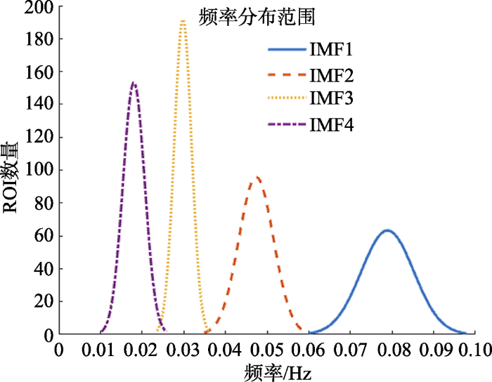
图3展示了4个频段上的组水平平均功能连接矩阵, 按照Yeo的7网络分区将所有脑区重新排序, 矩阵从左至右(从上至下)依次为视觉网络、感觉运动网络、背侧注意网络、腹侧注意网络、边缘网络、额顶控制网络和默认网络内脑区与其他脑区的组水平平均相关系数(z值)。研究结果显示:随着频率段降低, 网络间的功能连接逐步以负连接为主, 而网络内部区域之间也逐步出现较多的负连接, 七大皮层功能网络间模块化逐渐减弱, 在IMF1 (0.06~0.10 Hz)内可发现7个网络内的功能连接明显强于网络间的功能连接, 其中默认网络的网络间功能连接, 特别是与边缘网络之间的功能连接强于其他网络, 在该频段内, 网络间的长程连接数量和强度均多于其他频段; 在IMF2 (0.036~0.06 Hz)内, 全脑功能连接均有增强, 同时边缘网络、额顶控制网络和默认网络之间的网络间功能连接强度上升, 该三网络间的区分度下降; 在IMF3 (0.025~0.036 Hz)内, 网络间的区分度进一步下降, 只有视觉网络、感觉运动网络、背侧注意网络、腹侧注意网络保持了较好的网络间区分度, 同时网络内的功能连接矩阵也逐渐出现分化, 提示在各网络内也存在着具有不同连接特性的子模块; 在IMF4 (0.01~0.025 Hz)内, 只有视觉网络和感觉运动网络保持了较好的网络完整性, 默认网络保持了部分子模块特性, 其他网络则已经很难区分。
图3

3.2 男女差异的统计检验结果
磁共振成像扫描期间, 男女未表现出显著的头动差异(t = -0.58, p = 0.57), 因此未将头动作为影响因素考虑在功能连接比较中。将不同频段下的功能连接矩阵经Fisher Z转换后做独立样本t检验检验性别差异, 发现仅在IMF1 (0.06~0.10 Hz)频段上功能连接在性别间有显著的差异(校正后), 其他频段并无显著性差异。精确显示差异的脑区位置可在HCP脑区ROI分布中定位, 为L_FOP1_ ROI和R_V3CD_ROI, L_6ma_ROI和L_10pp_ROI, L_6ma_ROI与 R_10pp_ROI, L_8BM_ROI与 R_8C_ROI。上述脑区详细位置信息和所属网络如表1所示, 性别差异数据信息如表2所示, 其中p值经FDR校正(p < 0.05, 校正后)。研究结果显示男性较女性组的功能连接较强的脑区在左侧6ma和左右侧10pp区域, 而在左侧FOP1和V3CD、8BM和8C区域间的功能连接在女性中强于男性。进一步从网络视角发现, 男性大于女性的功能连接存在于腹侧注意网络和边缘网络的网络间功能连接, 女性比男性较强的功能连接则在腹侧注意网络和视觉网络、额顶控制网络的网络内功能连接, 在其他频段并未发现显著差异。
表1 脑区分布信息表
| 脑区 | 皮层位置 | 网络 | 脑区 | 皮层位置 | 网络 |
|---|---|---|---|---|---|
| L_FOP1 | 左侧额叶岛盖皮层 | 腹侧注意网络 | R_V3CD | 右侧枕外侧额叶皮层 | 视觉网络 |
| L_6ma | 左侧旁中央小叶、中扣带皮层 | 腹侧注意网络 | R_10pp | 右侧眶额叶皮层 | 边缘网络 |
| L_10pp | 右侧眶额叶皮层 | 边缘网络 | R_8C | 右侧背外侧前额叶皮层 | 额顶控制网络 |
| L_8BM | 左侧前扣带回、内侧前额叶皮层 | 额顶控制网络 |
表2 功能连接性别差异结果
| 连接脑区 | 男性 | 女性 | t | p* | |||
|---|---|---|---|---|---|---|---|
| 均值 | 标准差 | 均值 | 标准差 | ||||
| L_FOP1 | R_V3CD | -0.052 | 0.115 | -0.208 | 0.111 | 5.584 | 0.032 |
| L_6ma | L_10pp | 0.216 | 0.178 | 0.0154 | 0.124 | 5.160 | 0.040 |
| L_6ma | R_10pp | 0.154 | 0.149 | -0.020 | 0.096 | 5.460 | 0.026 |
| L_8BM | R_8C | 0.190 | 0.188 | 0.416 | 0.159 | -5.188 | 0.047 |
注:* p经过FDR矫正
4 讨论
本研究首先使用FMEEMD方法对所有被试的功能磁共振数据进行了频率划分, 根据已有研究表明, fMRI的高频信号可能和头动、血流等因素有关, 同时不同预处理流程对分频结果产生较大影响, 由于本研究的主要目的并不在于使用FMEEMD分频方法区分fMRI原始信号中的噪音成分, 而是探究在不同频率段内fMRI信号可能具有的网络特性和男女差异, 因此使用了通常的数据预处理流程, 同时重点关注已有发现的具有较强生理意义的0.01~0.1 Hz低频段的信号。分频结果发现高频信号内具有较好的脑网络模块化, 同时网络间的功能连接较强, 高级认知皮层的模块化程度最强。而随着频率下降, 网络模块化趋势减弱, 但是局部连通性和网络内子模块化特性逐渐凸显, 最低频频段内, 只保留了初级感知觉皮层如视觉网络和感觉运动网络的模块化, 且网络内的子模块性最强, 如默认网络内的部分区域间功能连接在该频段内强度较高, 提示在该低频频段内的功能连接, 更倾向于反映初级感知觉皮层以及局部大脑区域间的功能连通性。因此, 我们认为, 不同频段内的大脑功能磁共振信号具有不同的功能特性, 这种功能特性可能对应着大脑认知功能的层级性。例如, 在高频成分内的较强的网络间连接可能反映的是不同网络间的信息传递性较强, 对应着高级认知功能协调不同大脑网络协同工作的特征; 随着频率降低, 皮层七大网络间模块化减弱, 可能反映在不同频段下人脑的模块化信息处理的尺度差别, 信息主要是相同认知功能的成分间传递; 而在最低频段内仅保留初级感知觉皮层的模块化, 而网络内不同成分间的连通性都已经较低, 可能预示着在该频段内信息只在负责特定具体功能的区域间具有较好的连通性, 而负责高级认知功能的网络模式已很难观察到。因此随着频段由高至低, 可能对应着大脑功能从高级认知控制(协调负责不同功能的脑区间活动)到复杂认知功能实现(实现特定认知能力)再到单一认知能力(初级感知觉加工)的功能层级性。在此基础上, 我们进一步分析了不同频段下功能连接的性别差异, 发现只在IMF1频段出现了差异, 且多为网络间的长程连接差异, 网络内功能连接差异体现在左右脑间的额顶控制网络内, 一定程度上也佐证了上述分频结果, 即IMF1较好保留了大脑网络特性, 能反映网络间的长程功能连接。
FOP1和V3CD区域间存在负向功能连接, 且具有显著的男女差异。该连接强度在男性中较弱, 女性中较强, 说明男性中该两区域间的功能关联性较低, 而在女性中存在反向的功能同步性。FOP1比周围区域的髓鞘化更强, 一般在进行运动相关任务时激活。该区域从属于腹侧注意网络, 主要负责自下而上的认知加工, 接收初级皮层的感觉信息输入并对其进行反应(Eckert et al., 2009)。V3CD区域属于枕外侧颞叶皮质, 毗邻视觉联络区V4 (Abdollahi et al., 2014; Malikovic et al., 2016), 有更多的髓磷脂和较少的皮层厚度, 负责对复杂的视觉的加工。我们认为, 女性在FOP1和V3CD间的反向功能同步性可能预示着在女性中处理复杂视觉信息的脑区与注意相关的脑区活动间存在一定的竞争关系, 而在男性中这两个区域间的关联性很低, 说明可能仅在女性中存在支持脑功能活动的竞争关系(Sorge & Totsch, 2017; Herlitz & Rehnman, 2008)。
在6ma区域和左右侧10pp区域间的功能连接在男性中显著强于女性, 6ma区域属于辅助运动区(SMA)的一部分, 在人体中SMA的神经元直接投射到脊髓上, 主要控制运动的内部生成规划、运动序列的规划和身体两侧的协调, 起到直接控制运动的作用, 如攀爬和跳跃等具体行为(Serrien, Strens, Oliviero, & Brown, 2002; 张琪涵等, 2014)。该区域也属于腹侧注意网络, 参与自下而上的认知调控。10pp区域位于眶额和额极皮质区域, 在HCP分区中该区域被划分为11个子区域, 10pp是其中一个, 眶额区域被认为是参与许多高级的认知工作, 比如未来行动的规划和组织, 以及类比的能力等, 是基于抽象的规划、情节记忆和短时记忆信息的基础(Bludau et al., 2014), 但是相较于眶额部其他区域, 10pp区在工作记忆、语言故事等任务中表现出了去激活, 仅在运动任务中表现出了一定的激活, 我们推测10pp区域可能在眶额皮层区域负责协调信息传入和认知规划的作用(van Essen, Glasser, Dierker, Harwell, & Coalson , 2012)。我们认为, 6ma和双侧10pp区域间的功能连接在男性中强于女性, 与上述FOP1和V3CD间的功能连接结果相呼应V3CD和FOP1间的功能连接是基本感知觉信息上传至高级认知控制区域的中转, 是信息开始进入腹侧注意系统自下而上加工的开始, 并且FOP1主要受运动相关任务激活, 而6ma区域则恰好是属于辅助运动区, 它和左右侧10pp区域的功能连接很可能是腹侧注意系统加工后的信息进入高级认知控制的通路, 之后再由眶额部分其他区域进行运动规划。该通路体现了从刺激输入, 到引起注意分配, 再到高级皮层形成运动规划的自下而上加工的完整信息加工通路。而该通路的连通性在女性中弱于男性, 暗示男性在接收外界刺激信息, 并由此作出相关反应的功能通路连通性强于女性, 也符合日常经验, 即男性更擅长、可以更迅速对外界输入的视觉信息进行组织并作出应对。
相比于男性, 女性在左侧8BM和右侧8C区域之间的功能连接更强。8BM和8C区域都属于额顶控制网络, 8BM区域主要位于前扣带回和内侧前额叶皮质的顶后侧。该区域在结构上髓鞘化程度较强, 在工作记忆任务中激活, 在数学问题背景中的激活强于听觉理解, 并且具有偏侧化趋势, 如在关联任务强于匹配任务的激活中, 右侧8BM的激活区域更广泛。8C区域主要位于背外侧前额叶皮质, 背外侧前额叶皮质是人类大脑皮层中负责高级认知功能的皮层, 主要负责执行和认知功能, 包括工作记忆和认知灵活性(Barbey, Koenigs, & Grafman, 2013)等, 是实施认知控制的最重要脑区之一。8C区域在结构上髓鞘化程度较低, 在工作记忆任务上局部激活较强, 我们推测, 左侧8BM和右侧8C之间的功能连接, 特别是半球间连接, 体现了大脑在高级认知成分, 特别是在工作记忆和数字加工上的脑区间协调及整合性上, 女性要强于男性。
综上所述, 我们认为FMEEMD方法可以有效地提取大脑功能磁共振时间信号的频率特性, 同时, 基于FMEEMD方法所得到的男女功能连接差异表明, 男性在由下至上的加工中具有较强的功能连通性, 而女性则在工作记忆相关的高级脑区间表现出了较强的半球间整合性。本研究具有一定的局限性, 例如样本量较少, 影像学结果无法通过多重比较校正, 可能会导致未在其他频段发现性别差异; 同时本研究结果基于静息态功能磁共振数据, 缺少行为数据和相关实验结果的佐证, 未能充分讨论脑影像结果在行为中的表现; 此外在方法学上, 我们使用了较为保守传统的数据预处理方式, 可能会导致一定的假阴性结果, 未来可以使用FMEEMD方法对原始高质量(如multiband)序列采集的功能磁共振数据进行分析, 探讨该方法在数据预处理上的应用前景。
5 结束语
本文采用FMEEMD分解方法有效避免了经验驱动方式中需要提前定义频段的缺陷。对大脑的静息态fMRI低频成分(< 0.1 Hz)进行分频, 得到了4个频率段范围, 从高到低依次为0.06~0.10 Hz, 0.036~0.06 Hz, 0.025~0.036 Hz和0.01~0.025 Hz, 并构建了4个频段的大脑功能网络, 讨论了网络内和网络间的功能连接属性, 随频率提高, 功能连接矩阵中的网络化程度上升, 网络内功能连接下降, 网络间功能连接增强。在此基础上, 我们进一步比较了不同频段内功能连接的性别差异, 结果显示在0.06~0.10 Hz频段上, 男性比女性较强的功能连接分别集中在腹侧注意网络和边缘网络间, 体现了自下而上的加工方式; 女性比男性较强在腹侧注意网络和视觉网络、以及额顶控制网络内的功能连接, 体现了高级皮层在认知功能上的协调和整合性。本研究展示了FMEEMD分频算法在功能磁共振数据上的应用, 为男女差异提供了基于影像的人脑功能连接组学证据。
参考文献
主运动区与辅助运动区在运动执行与运动想象任务中的作用:一个近红外光谱技术的研究
见功能性近红外光谱技术是近期发展起来的脑功能成像的方法。它主要是通过监测大脑活动时相关生物组织对近红外光的吸收情况来反映氧合血红蛋白、脱氧血红蛋白的浓度变化,继而获得大脑皮层的激活情况。功能性近红外光谱技术具有高时间分别率、无创伤性、成本低、易便携、可用于低龄个体等特点,目前在认知神经科学领域中被广泛应用。部分功能性磁共振成像研究发现主运动区与辅助运动区在运动任务中存在功能性分离的现象,然而在另一些研究中没有观察到这一现象。另外,随着运动想象疗法在运动功能康复治疗领域的迅速发展,越来越多的人关注运动想象疗法的作用效果与内部机制。为了检测主运动区与辅助运动区在运动任务中是否存在功能性分离现象;比较运动执行任务与运动想象任务大脑皮质激活情况探究运动想象的加工过程;探索运动想象任务中是否也可够发现主运动区与辅助运动区的功能分离。本研究用近红外光谱技术监测20名健康大学生在不同运动任务中大脑皮质的激活情况。采用2任务类型(运动执行,运动想象)×2持续时间(2s,5s)的被试内设计,考察健康大学生在不同任务条件下激活脑区的血液动力学变化特征,以及激活程度的差异。结果发现:在运动执行任务中,主运动区的激活情况与任务的持续时间存在正相关关系,而辅助运动区与任务的持续时间没有相关。这表明在运动执行任务中主运动区与辅助运动区在功能上分离,辅助运动区更多的参与最初的运动准备计划阶段,而主运动区更多的参与运动执行阶段。另外在持续时间为5s的实验条件下,运动执行任务与运动想象任务的大脑皮质激活情况基本相同。这说明运动想象疗法是一种有效地康复治疗手段。
Correspondences between retinotopic areas and myelin maps in human visual cortex
DOI:10.1016/j.neuroimage.2014.06.042
URL
PMID:4121090
[本文引用: 1]

We generated probabilistic area maps and maximum probability maps (MPMs) for a set of 18 retinotopic areas previously mapped in individual subjects (Georgieva et al., 2009 and Kolster et al., 2010) using four different inter-subject registration methods. The best results were obtained using a recently developed multimodal surface matching method. The best set of MPMs had relatively smooth borders between visual areas and group average area sizes that matched the typical size in individual subjects. Comparisons between retinotopic areas and maps of estimated cortical myelin content revealed the following correspondences: (i) areas V1, V2, and V3 are heavily myelinated; (ii) the MT cluster is heavily myelinated, with a peak near the MT/pMSTv border; (iii) a dorsal myelin density peak corresponds to area V3D; (iv) the phPIT cluster is lightly myelinated; and (v) myelin density differs across the four areas of the V3A complex. Comparison of the retinotopic MPM with cytoarchitectonic areas, including those previously mapped to the fs_LR cortical surface atlas, revealed a correspondence between areas V1–3 and hOc1–3, respectively, but little correspondence beyond V3. These results indicate that architectonic and retinotopic areal boundaries are in agreement in some regions, and that retinotopy provides a finer-grained parcellation in other regions. The atlas datasets from this analysis are freely available as a resource for other studies that will benefit from retinotopic and myelin density map landmarks in human visual cortex. 61 Maximum probability maps for 18 retinotopic areas were generated. 61 Multimodal surface matching was used to compare with myelin and cytoarchitectonic maps. 61 Early areas V1–3 areas are heavily myelinated, as are V3D and most of areas MT/pMSTv. 61 The phPIT cluster is lightly myelinated compared to other retinotopic areas. 61 Early areas V1–3 correspond to areas hOc1–3, with little correspondence beyond V3. Maximum probability maps for 18 retinotopic areas were generated. Multimodal surface matching was used to compare with myelin and cytoarchitectonic maps. Early areas V1–3 areas are heavily myelinated, as are V3D and most of areas MT/pMSTv. The phPIT cluster is lightly myelinated compared to other retinotopic areas. Early areas V1–3 correspond to areas hOc1–3, with little correspondence beyond V3.
A resilient, low-frequency, small-world human brain functional network with highly connected association cortical hubs
DOI:10.1523/JNEUROSCI.3874-05.2006 URL [本文引用: 1]
Dorsolateral prefrontal contributions to human working memory
DOI:10.1016/j.cortex.2012.05.022 URL [本文引用: 1]
Cytoarchitecture, probability maps and functions of the human frontal pole
DOI:10.1016/j.neuroimage.2013.05.052
URL
PMID:23702412
[本文引用: 1]
61The human frontopolar cortex consists of two cytoarchitectonically distinct areas.613D probabilistic maps of frontopolar area 1 and 2 (Fp1,Fp2) were created.61Quantitative inference showed involvement in cognition (Fp1) and emotion (Fp2).61Significant difference in task-based functional connectivity between Fp1 and Fp2.
At the heart of the ventral attention system: The right anterior insula
DOI:10.1002/hbm.20688
URL
PMID:2712290
[本文引用: 1]
The anterior insula has been hypothesized to provide a link between attention-related problem solving and salience systems during the coordination and evaluation of task performance. Here, we test the hypothesis that the anterior insula/medial frontal operculum (aI/fO) provides linkage across systems supporting task demands and attention systems by examining the patterns of functional connectivity during word recognition and spatial attention functional imaging tasks. A shared set of frontal regions (right aI/fO, right dorsolateral prefrontal cortex, bilateral anterior cingulate) were engaged, regardless of perceptual domain (auditory or visual) or mode of response (word production or button press). We present novel evidence that: (1) the right aI/fO is functionally connected with other frontal regions implicated in executive function and not just brain regions responsive to stimulus salience; and (2) that the aI/fO, but not the ACC, exhibits significantly correlated activity with other brain regions specifically engaged by tasks with varying perceptual and behavioral demands. These results support the hypothesis that the right aI/fO aids in the coordination and evaluation of task performance across behavioral tasks with varying perceptual and response demands. Hum Brain Mapp 2009. 2008 Wiley-Liss, Inc.
FreeSurfer
DOI:10.1016/j.neuroimage.2012.01.021 URL [本文引用: 1]
Whole brain segmentation: Automated labeling of neuroanatomical structures in the human brain
DOI:10.1016/S0896-6273(02)00569-X URL [本文引用: 1]
A multi- modal parcellation of human cerebral cortex
DOI:10.1038/nature18933
URL
PMID:4990127
[本文引用: 1]
Understanding the amazingly complex human cerebral cortex requires a map (or parcellation) of its major subdivisions, known as cortical areas. Making an accurate areal map has been a century-old objective in neuroscience. Using multi-modal magnetic resonance images from the Human Connectome Project (HCP) and an objective semi-automated neuroanatomical approach, we delineated 180 areas per hemisphere bounded by sharp changes in cortical architecture, function, connectivity, and/or topography in a precisely aligned group average of 210 healthy young adults. We characterized 97 new areas and 83 areas previously reported using post-mortem microscopy or other specialized study-specific approaches. To enable automated delineation and identification of these areas in new HCP subjects and in future studies, we trained a machine-learning classifier to recognize the multi-modal ingerprint of each cortical area. This classifier detected the presence of 96.6% of the cortical areas in new subjects, replicated the group parcellation, and could correctly locate areas in individuals with atypical parcellations. The freely available parcellation and classifier will enable substantially improved neuroanatomical precision for studies of the structural and functional organization of human cerebral cortex and its variation across individuals and in development, aging, and disease.
Accurate and robust brain image alignment using boundary-based registration
DOI:10.1016/j.neuroimage.2009.06.060 URL [本文引用: 1]
Sex differences in episodic memory
DOI:10.1111/j.1467-8721.2008.00547.x URL [本文引用: 1]
Mapping sources of correlation in resting state fMRI, with artifact detection and removal
DOI:10.1016/j.neuroimage.2010.04.246
URL
PMID:20420926
[本文引用: 1]
Many components of resting-state (RS) FMRI show non-random structure that has little to do with neural connectivity but can covary over multiple brain structures. Some of these signals originate in physiology and others are hardware-related. One artifact discussed herein may be caused by defects in the receive coil array or the RF amplifiers powering it. During a scan, this artifact results in small image intensity shifts in parts of the brain imaged by the affected array components. These shifts introduce artifactual correlations in RS time series on the spatial scale of the coil's sensitivity profile, and can markedly bias RS connectivity results. We show that such a transient artifact can be substantially removed from RS time series by using locally formed regressors from white matter tissue. This is particularly important in arrays with larger numbers of coils, which may generate smaller artifact zones. In such a case, brain-wide average noise estimates would fail to capture the artifact. We also examine the anatomical structure of artifactual variance in RS FMRI time series, by identifying sources that contribute to these signals and where in the brain are they manifested. We consider current methods for reducing confounding sources (or noises) and their effects on connectivity maps, and offer an improved approach (ANATICOR) that can also reduce hardware artifacts. The methods described herein are currently available with AFNI, in addition to tools for rapid, interactive generation of seed-based correlation maps at single-subject and group levels.
Effects of different correlation metrics and preprocessing factors on small-world brain functional networks: A resting-state functional MRI study
DOI:10.1371/journal.pone.0032766 URL [本文引用: 1]
Cytoarchitecture of the human lateral occipital cortex: Mapping of two extrastriate areas hOc4la and hOc4lp
DOI:10.1007/s00429-015-1009-8 URL [本文引用: 1]
Recent progress and outstanding issues in motion correction in resting state fMRI
DOI:10.1016/j.neuroimage.2014.10.044
URL
PMID:25462692
[本文引用: 1]
61Reviews post-2011 research on motion artifact in resting state fMRI61Explains analyses to detect and quantify motion artifact61Presents evidence for removal of artifact by various processing strategies
A simple view of the brain through a frequency-specific functional connectivity measure
DOI:10.1016/j.neuroimage.2007.08.018
URL
PMID:17919927
[本文引用: 1]
Here we develop a measure of functional connectivity describing the degree of covariability between a brain region and the rest of the brain. This measure is based on previous formulas for the mutual information (MI) between clusters of regions in the frequency domain. Under the current scenario, the MI can be given as a simple monotonous function of the multiple coherence and it leads to an easy visual representation of connectivity patterns. Computationally efficient formulas, adequate for short time series, are presented and applied to functional magnetic resonance imaging (fMRI) data measured in subjects (02=0234) performing a working memory task or being at rest. While resting state coherence in high (0.17–0.25 Hz) and middle (0.08–0.17 Hz) frequency intervals is bilaterally salient in several limbic and temporal areas including the insula, the amygdala, and the primary auditory cortex, low frequencies (
Undirected graphs of frequency-dependent functional connectivity in whole brain networks
DOI:10.1098/rstb.2005.1645
URL
PMID:1854928
[本文引用: 1]
We explored properties of whole brain networks based on multivariate spectral analysis of human functional magnetic resonance imaging (fMRI) time-series measured in 90 cortical and subcortical subregions in each of five healthy volunteers studied in the (no-task) resting state. We note that undirected graphs representing conditional independence between multivariate time-series can be more readily approached in the frequency domain than the time domain. Estimators of partial coherency and normalized partial mutual information 0, an integrated measure of partial coherence over an arbitrary frequency band, are applied. Using these tools, we replicate the prior observations that bilaterally homologous brain regions tend to be strongly connected and functional connectivity is generally greater at low frequencies [0.0004, 0.1518 Hz]. We also show that long-distance intrahemispheric connections between regions of prefrontal and parietal cortex were more salient at low frequencies than at frequencies greater than 0.3 Hz, whereas many local or short-distance connections, such as those comprising segregated dorsal and ventral paths in posterior cortex, were also represented in the graph of high-frequency connectivity. We conclude that the partial coherency spectrum between a pair of human brain regional fMRI time-series depends on the anatomical distance between regions: long-distance (greater than 7 cm) edges represent conditional dependence between bilaterally symmetric neocortical regions, and between regions of prefrontal and parietal association cortex in the same hemisphere, are predominantly subtended by low-frequency components.
Repetitive transcranial magnetic stimulation of the supplementary motor area (SMA) degrades bimanual movement control in humans
DOI:10.1016/S0304-3940(02)00499-8 URL [本文引用: 1]
Frequency specificity of regional homogeneity in the resting-state human brain
DOI:10.1371/journal.pone.0086818
URL
PMID:24466256
[本文引用: 1]
Resting state-fMRI studies have found that the inter-areal correlations in cortical networks concentrate within ultra-low frequencies (0.01–0.04 Hz) while long-distance connections within subcortical networks distribute over a wider frequency range (0.01–0.14 Hz). However, the frequency characteristics of regional homogeneity (ReHo) in different areas are still unclear. To examine the ReHo properties in different frequency bands, a data-driven method, Empirical Mode Decomposition (EMD), was adopted to decompose the time series of each voxel into several components with distinct frequency bands. ReHo values in each of the components were then calculated. Our results showed that ReHo in cortical areas were higher and more frequency-dependent than those in the subcortical regions. BOLD oscillations of 0.02–0.04 Hz mainly contributed to the cortical ReHo, whereas the ReHo in limbic areas involved a wider frequency range and were dominated by higher-frequency BOLD oscillations (>0.08 Hz). The frequency characteristics of ReHo are distinct between different parts of the striatum, with the frequency band of 0.04–0.1 Hz contributing the most to ReHo in caudate nucleus, and oscillations lower than 0.02 Hz contributing more to ReHo in putamen. The distinct frequency-specific ReHo properties of different brain areas may arise from the assorted cytoarchitecture or synaptic types in these areas. Our work may advance the understanding of the neural-physiological basis of local BOLD activities and the functional specificity of different brain regions.
Sex differences in pain
DOI:10.1002/jnr.v95.6 URL [本文引用: 1]
Parcellations and hemispheric asymmetries of human cerebral cortex analyzed on surface-based atlases
DOI:10.1093/cercor/bhr291 URL [本文引用: 1]
Frequency specificity of functional connectivity in brain networks
DOI:10.1016/j.neuroimage.2008.05.035
URL
PMID:2612530
[本文引用: 1]
Synchronized low-frequency spontaneous fluctuations of the functional MRI (fMRI) signal have been shown to be associated with electroencephalography (EEG) power fluctuations in multiple brain networks within predefined frequency bands. However, it remains unclear whether frequency-specific characteristics exist in the resting-state fMRI signal. In this study, fMRI signals in five functional brain networks (sensorimotor, ‘default mode’, visual, amygdala, and hippocampus) were decomposed into various frequency bands within a low-frequency range (0–0.2402Hz). Results show that the correlations in cortical networks concentrate within ultra-low frequencies (0.01–0.0602Hz) while connections within limbic networks distribute over a wider frequency range (0.01–0.1402Hz), suggesting distinct frequency-specific features in the resting-state fMRI signal within these functional networks. Moreover, the connectivity decay rates along the frequency bands are positively correlated with the physical distances between connected brain regions and seed points. This distance-frequency relationship might be attributed to a larger attenuation of synchrony of brain regions separated with longer distance and/or connected with more synaptic steps.
Ensemble empirical mode decomposition: A noise-assisted data analysis method
DOI:10.1142/S1793536909000047 URL
The multi- dimensional ensemble empirical mode decomposition method
DOI:10.1142/S1793536909000187
URL
[本文引用: 2]
A multi-dimensional ensemble empirical mode decomposition (MEEMD) for multi-dimensional data (such as images or solid with variable density) is proposed here. The decomposition is based on the applications of ensemble empirical mode decomposition (EEMD) to slices of data in each and every dimension involved. The final reconstruction of the corresponding intrinsic mode function (IMF) is based on a comparable minimal scale combination principle. For two-dimensional spatial data or images, f(x,y), we consider the data (or image) as a collection of one-dimensional series in both x-direction and y-direction. Each of the one-dimensional slices is decomposed through EEMD with the slice of the similar scale reconstructed in resulting two-dimensional pseudo-IMF-like components. This new two-dimensional data is further decomposed, but the data is considered as a collection of one-dimensional series in y-direction along locations in x-direction. In this way, we obtain a collection of two-dimensional components. These directly resulted components are further combined into a reduced set of final components based on a minimal-scale combination strategy. The approach for two-dimensional spatial data can be extended to multi-dimensional data. EEMD is applied in the first dimension, then in the second direction, and then in the third direction, etc., using the almost identical procedure as for the two-dimensional spatial data. A similar comparable minimal-scale combination strategy can be applied to combine all the directly resulted components into a small set of multi-dimensional final components. For multi-dimensional temporal-spatial data, EEMD is applied to time series of each spatial location to obtain IMF-like components of different time scales. All the ith IMF-like components of all the time series of all spatial locations are arranged to obtain ith temporal-spatial multi-dimensional IMF-like component. The same approach to the one used in temporal-spatial data decomposition is used to obtain the resulting two-dimensional IMF-like components. This approach could be extended to any higher dimensional temporal-spatial data.
Mean frequency derived via Hilbert-Huang transform with application to fatigue EMG signal analysis
DOI:10.1016/j.cmpb.2006.02.009 URL [本文引用: 1]
A connectome computation system for discovery science of brain
DOI:10.1007/s11434-014-0698-3
URL
[本文引用: 1]
Much like genomics, brain connectomics has rapidly become a core component of most national brain projects around the world. Beyond the ambitious aims of these projects, a fundamental challenge is the need for an efficient, robust, reliable and easy-to-use pipeline to mine such large neuroscience datasets. Here, we introduce a computational pipeline—namely the Connectome Computation System (CCS)—for discovery science of human brain connectomes at the macroscale with multimodal magnetic resonance imaging technologies. The CCS is designed with a three-level hierarchical structure that includes data cleaning and preprocessing, individual connectome mapping and connectome mining, and knowledge discovery. Several functional modules are embedded into this hierarchy to implement quality control procedures, reliability analysis and connectome visualization. We demonstrate the utility of the CCS based upon a publicly available dataset, the NKI–Rockland Sample, to delineate the normative trajectories of well-known large-scale neural networks across the natural life span (6–8502years of age). The CCS has been made freely available to the public via GitHub ( https://github.com/zuoxinian/CCS ) and our laboratory’s Web site ( http://lfcd.psych.ac.cn/ccs.html ) to facilitate progress in discovery science in the field of human brain connectomics.
A comprehensive assessment of regional variation in the impact of head micromovements on functional connectomics
DOI:10.1016/j.neuroimage.2013.03.004 URL [本文引用: 1]
The organization of the human cerebral cortex estimated by intrinsic functional connectivity
DOI:10.1152/jn.00338.2011 URL [本文引用: 1]
Altered baseline brain activity in children with ADHD revealed by resting-state functional MRI
DOI:10.1016/j.braindev.2006.07.002
URL
PMID:16919409
[本文引用: 1]
In children with attention deficit hyperactivity disorder (ADHD), functional neuroimaging studies have revealed abnormalities in various brain regions, including prefrontal-striatal circuit, cerebellum, and brainstem. In the current study, we used a new marker of functional magnetic resonance imaging (fMRI), amplitude of low-frequency (0.01 0.08 Hz) fluctuation (ALFF) to investigate the baseline brain function of this disorder. Thirteen boys with ADHD (13.0 1.4 years) were examined by resting-state fMRI and compared with age-matched controls. As a result, we found that patients with ADHD had decreased ALFF in the right inferior frontal cortex, left sensorimotor cortex, and bilateral cerebellum and the vermis as well as increased ALFF in the right anterior cingulated cortex, left sensorimotor cortex, and bilateral brainstem. This resting-state fMRI study suggests that the changed spontaneous neuronal activity of these regions may be implicated in the underlying pathophysiology in children with ADHD.
The oscillating brain: Complex and reliable
DOI:10.1016/j.neuroimage.2009.09.037
URL
PMID:19782143
[本文引用: 1]
The human brain is a complex dynamic system capable of generating a multitude of oscillatory waves in support of brain function. Using fMRI, we examined the amplitude of spontaneous low-frequency oscillations (LFO) observed in the human resting brain and the test–retest reliability of relevant amplitude measures. We confirmed prior reports that gray matter exhibits higher LFO amplitude than white matter. Within gray matter, the largest amplitudes appeared along mid-brain structures associated with the “default-mode” network. Additionally, we found that high-amplitude LFO activity in specific brain regions was reliable across time. Furthermore, parcellation-based results revealed significant and highly reliable ranking orders of LFO amplitudes among anatomical parcellation units. Detailed examination of individual low frequency bands showed distinct spatial profiles. Intriguingly, LFO amplitudes in the slow-4 (0.027–0.07302Hz) band, as defined by Buzsáki et al., were most robust in the basal ganglia, as has been found in spontaneous electrophysiological recordings in the awake rat. These results suggest that amplitude measures of LFO can contribute to further between-group characterization of existing and future “resting-state” fMRI datasets.
Toward reliable characterization of functional homogeneity in the human brain: Preprocessing, scan duration, imaging resolution and computational space
DOI:10.1016/j.neuroimage.2012.10.017 URL [本文引用: 1]
