

1 引言
人类视网膜上存在视锥细胞和视杆细胞两种感光细胞。当光线进入人眼后, 视锥细胞和视杆细胞接受光信号, 并通过视神经传递给大脑视觉皮层, 形成视觉体验。随后研究者发现视网膜上还存在一种新型的感光细胞(Berson, Dunn, & Takao, 2002), 这种细胞能够合成感光蛋白——黑视蛋白, 并且具备自主感光的能力, 又被称为自主感光神经节细胞(intrinsically photosensitive retinal ganglion cells, ipRGCs)。ipRGCs的发现逐渐让人们认识到, 眼睛不仅具有传统感光细胞的视觉效应, 还具有了非视觉效应。ipRGCs分布在整个视网膜上, 占视网膜神经细胞的1% ~3%, 它们将接受到的光信号传递到大脑的生物钟——下丘脑视交叉上核(suprachiasmatic nucleus, SCN), 而SCN又与控制人体某些激素分泌的松果体相连, 由此实现对生理节律的调节以及激素分泌的控制(曾强, 何士刚, 2011), 如图1所示。随着光照对SCN昼夜节律调节作用的深入研究, 研究者开始将目光转向光照对警觉性的影响(Vandewalle et al., 2013)。警觉性指人在完成任务时保持注意力或警惕程度的水平。SCN参与启动生命的代谢、生化及物理过程。在清醒之前, SCN激活皮质醇大量分泌, 同时启动其他影响觉醒的重要环节。通常, 当机体晨起接受日光照射后, 核心体温、警觉程度、认知水平均提高, 同时激发大脑产生更多的5-羟色胺, 提高情绪及机体活动力。到了夜间, SCN抑制皮质醇的分泌, 促使松果体分泌褪黑素, 降低机体的警觉度及反应能力, 降低核心体温(董栩然, 王旻舒, 刘梦媛, 王薇, 2014)。
图1

图1
光照激活大脑非视觉区域的路径图(资料来源:Cajochen, 2007)
光照的非视觉生物效应包括重置昼夜节律的相位、抑制褪黑素的分泌和提高即时警觉性等。这三者之间又相互影响, 且均取决于光照强度、时间点、时长、光谱成分及光照之前的照明体验这些特征(Cajochen, 2007; Adamsson, Laike, & Morita, 2017)。目前, 关于光照警觉性研究大都只是考察了光照强度、光照时长、光照色温、光照波长、光照时间点等众多影响因素中的一个或几个, 并且结果也存在一定的分歧。基于此, 我们将从警觉性的测量工具、光照的警觉性的影响因素、光照在临床和非临床上的应用等方面对前人的文献进行梳理总结, 以期为后人的研究提供参考依据。
1 警觉性的测量工具
1.1 主观测量工具
对健康个体来说, 警觉性可以通过简单的询问或者主观评定得到。目前应用比较广泛的主观测试量表有卡罗林斯卡困倦度量表(Karolinska sleepiness scale, KSS) (Horne & Burley, 2010)和斯坦福嗜睡量表(Stanford sleepiness scale, SSS) (Martinez et al., 2011)。KSS包括1个题目, 9点记分:1=极度清醒, 2=非常清醒, 3=清醒, 4=比较清醒, 5=既不清醒也不困乏, 6=有点困乏, 7=有点困乏但无须费力保持清醒, 8=非常困乏, 需努力保持清醒, 9=极度困乏, 需极力保持清醒。SSS从1到7 七点记分。1指完全清醒、精力旺盛, 7指基本无意识、接近睡眠。
1.2 客观测量工具
主观评定的缺点是, 个体在较亮的光环境中可能会直观地认为更加警觉, 并且对于光来说没有真正的安慰剂对照组。因此, 在光照实验中就需要更多客观的测量工具, 最常用的就是精神运动警觉性测验(psychomotor vigilance test, PVT) (Dinges & Powell, 1985)。PVT是一个简单的反应时任务。在测试中, 视觉线索以伪随机方式反复呈现, 刺激时间间隔范围是2~10秒。当线索呈现时, 被试需要尽可能快速地按键, 然后激活下一次测试。它的优点是对反应时的增加和注意脱漏非常敏感, 而且学习效应很小; 缺点是它测量的是持续性注意而非警觉性或疲劳本身。
1.3 生理指标
在警觉性研究的早期阶段, 眨眼频率、皮肤阻抗、体温以及血压等生理信号都曾被用来评估警觉性。目前研究使用的神经生理学指标主要包括脑电前额叶低活性的波(1~7 Hz)、眼电的慢速眼动和眨眼速率(Cajochen, Khalsa, Wyatt, Czeisler, & Dijk, 1999)。这种高时间分辨率的测量可以在秒的范围内检测是微睡眠还是任务失效。但是, 这种测量在实际操作时会干扰被试本来的性质, 例如在现场研究中会干扰晚上值班工人的脑电活动。除此之外, 收集唾液来测定褪黑素、皮质醇、淀粉酶也是常用的一种生理指标, 且通常需要收集1~2 ml唾液, 然后把唾液冷冻起来送到实验室测定分析(Figueiro & Rea, 2010)。
2 光照的警觉性效应的影响因素
2.1 昼夜节律对光照警觉性的影响
昼夜节律(circadian rhythm)是指生物体各种生理机能为适应外界环境的昼夜变化而建立起的规律周期。在长期生物进化过程中, 生物体内发育分化出一个特殊的“器官”—生物钟来协调不同组织与器官的昼夜节律。哺乳类动物调控昼夜节律的中枢位于下丘脑的SCN。内源性的调节中枢由光照进行调控。昼夜节律的光协同过程使得生物体的昼夜节律与自然节律同步, 并不断调整生理周期及行为表现。警觉性和生理节律中的核心体温有紧密的时间关系:晚上核心体温最高, 清晨核心体温最低。刚睡醒时对警觉性的不利影响在接近最低核心体温时最强烈。稳定且高水平的警觉性只有在内源性昼夜计时系统和睡眠/觉醒周期处于特定关系下才可能实现。这种关系就是内源性昼夜计时系统与觉醒有关的警觉性下降相对立, 也被概括为“对立过程模型” (Dijk & Czeisler, 1994)。生理过程在觉醒阶段表现出一种促进觉醒的动力, 以平衡觉醒阶段累积的睡眠驱力(Cajochen, Blatter, & Wallach, 2004)。睡眠惯性是指刚睡醒之后开始的一段低唤醒的状态, 它对认知过程有不利的影响, 这种影响最长能持续4个小时, 具体则取决于之前的睡眠时长(Jewett et al., 1999)。综上可以看出, 光照在生理节律睡眠驱力最大(在核心体温最低点的清晨)、较高的睡眠稳态的压力(觉醒后16小时以上)、睡眠惯性(刚睡醒时)时对警觉性的影响最大。
2.2 不同光照时间点对光照警觉性的影响
光照的视觉效应不受光照时间点的影响, 但是光照的非视觉效应却受到光照时间点的影响。所以, 光照的警觉性也受到光照时间点的影响。例如, 有研究发现光照的警觉性作用在晚上或者睡眠剥夺之后特别强烈, 在白天也依然存在, 只是似乎比较平和(Smolders & De Kort, 2014)。夜间研究表明, 与昏暗灯光的控制条件相比, 不同光照强度和光照时长条件下均能显示出光照的警觉性作用。尽管光照的直接作用看起来似乎是在晚上通过视网膜投影到SCN从而产生的褪黑素的抑制。但是有研究发现, 即使在白天, 几乎检测不到褪黑素的时候, 光照也能提高警觉性(Vandewalle et al., 2006; Sahin & Figueiro, 2013)。而且, 正电子发射型计算机断层显像(Positron Emission Computed Tomography, PET)结果显示, 警觉性的反应不仅局限于视交叉神经上核附近的下丘脑区域, 而且拓展到包括注意网络在内的大规模的皮质区域, 尤其是包含右侧顶内沟的大规模的枕顶叶注意网络(Perrin et al., 2004)。一项fMRI的研究也发现类似结果, 即在白天短时间的亮光条件下也可以短暂阻止由于持续性处在黑暗中而产生的困意。光照引起的主观警觉性的提高与后侧丘脑, 包括丘脑核的增强反应相一致, 这与视觉模式识别、视觉注意和警觉性调节有关。光照暴露还可以导致大面积皮层网络的反应增强, 包括顶骨、颞骨、枕骨以及包含注意任务对象的岛叶(Vandewalle et al., 2006)。总之, 目前很少研究比较了白天和晚上光照对警觉性的影响, 而且这些研究大都用的是强光复合白光(≥1000 lux), 这个照度大到足以产生最大的警觉性效应, 因此可能表现不出时间的主效应。然而, 目前还没有用室内典型照明照度(90~180 lux)比较白天和晚上的警觉性, 故不能片面认为光照的警觉性作用独立于时间点。
2.3 不同照度的光照对警觉性的影响
Lewy, Wehr, Goodwin, Newsome和Markey (1980)首次证明了光照的生理效应, 研究指出健康人类至少需要1000 lux的白光才能抑制褪黑素的产生。之后很多研究也都证明了这一点, 并且经常用大于1000 lux的光照作为减少倒班工人的睡意、提高倒班工人工作绩效的方法(Cajochen, Zeitzer, Czeisler, & Dijk, 2000; Figueiro, Rea, Boyce, White, & Kolberg, 2001)。光照提高警觉性和工作绩效的路径之一是通过对褪黑素激素分泌的抑制。在通常条件下, 一般室内照明的照度(100~200 lux)不会抑制褪黑素的产生。但是, Brainard等人(1988)在严格的实验室控制下发现, 眼睛接收的辐照度在1.03~5.50 lux时也会抑制褪黑素的分泌。为了探讨光照的警觉性作用中是否存在类似剂量与反应的关系这一问题, Cajochen等人(2000)让被试晚上在实验室中暴露在小于3 lux的光照几小时之后, 接受6.5小时从3 lux到9100 lux的光照。结果发现被试的慢速眼球运动减少、theta-alpha频率的脑电活动减少、主观睡意减少, 表现出了光照的即时警觉性反应。光照警觉性的指数随照度的变化呈现出了一种对数剂量反应曲线, 在9100 lux时警觉性最高, 在100 lux时出现中值。这个剂量反应函数的特征描绘了光照警觉性的效应。同时, Smith, Schoen和Czeisler (2004)发现如果个体之前处于一个较高强度的光环境中, 则会导致由于光照而减少褪黑素抑制的量减少10%~15%。但由于光照抑制褪黑素的分泌只是一条提高警觉性的通道, 因此减少褪黑素抑制是否意味着更少的警觉性反应还需要进一步探讨。
2.4 不同波长的光照对警觉性的影响
传统的感光细胞对555 nm的中间波长的光比较敏感, 而新型的感光细胞对460 nm的短波长的光比较敏感。例如研究发现相比波长更短的紫光(430 nm)或者波长更长的绿光(550 nm, 555 nm)和红光(600 nm, 630 nm), 暴露在460nm的短波长蓝光下个体的警觉性更高(Rahman et al, 2014; Okamoto & Nakagawa, 2015)。并且, 较低照度下的短波单色光便可以影响个体的警觉性(Figueiro, Bullough, Bierman, Fay, & Rea, 2007; Chellappa et al., 2014; Papamichael, Skene, & Revell, 2012)。Revell, Arendt, Fogg和Skene (2006)对5种不同波长(420 nm, 440 nm, 470 nm, 600 nm)的光进行了比较, 也发现低照度的光对个体主观警觉性的影响在短波长的光照中效应最大。其中ipRGCs对480 nm波长的光最为敏感, 因此在480 nm光条件下会抑制褪黑素的分泌。而波长更短的光(420 nm)很有可能通过视锥细胞影响非视觉成像反应(Hattar et al., 2006; Panda et al., 2005)。Chellappa等人(2011)发现增加复合光中的短波长成分可以提高警觉性。因此, 暴露在短波长的光照下或许是提高夜班工作人员警觉性的可行方法。但是, Güler等人(2008)在啮齿动物中发现, 光照在不存在视黑蛋白的条件下通过视锥细胞和视杆细胞的作用同样可以影响非视觉成像的功能。后来在人类实验中也发现了光照并不单单是通过内在光敏感性神经节细胞抑制褪黑素这一通道来提高警觉性的。例如Sahin和Figueiro (2013)在中午饭后1~1.5小时探讨被试在红光(630 nm, 40 lux)、蓝光(470 nm, 40 lux)和暗光(< 0.01 lux)环境中的脑电波的变化, 结果发现, 蓝光条件下的警觉性的提高并没有达到显著性, 而红光条件下的警觉性显著提高。还有研究者发现, 当困意很高时, 即使暴露在屏蔽掉短波光的光照下30分钟, 依然可以提高人们在夜晚中的警觉性(Sasseville, Martin, Houle, & Hébert, 2015)。
2.5 不同色温的光照对警觉性的影响
自大量研究证实了蓝光对警觉性具有很大影响后, 研究者开始探讨短波成分较多的复合白光对个体警觉性的影响, 即探讨不同色温对光照警觉性的影响。目前大多数研究都发现, 色温越高, 警觉性越高(Mills, Tomkins, & Schlangen, 2007; Viola, James, Schlangen, & Dijk, 2008; Iskra-Golec, Wazna, & Smith, 2012)。例如, 有研究发现, 相比正常光照条件, 在短波成分较多的复合白光的照明环境下, 睡意和褪黑素节律均显著下降(Motamedzadeh, Golmohammadi, Kazemi, & Heidarimoghadam, 2017)。在某些环境中, 使用短波成分较多的白光或许是提高工作绩效和警觉性的一个人体工程学策略。Chellappa等(2011)比较了两种不同色温的节能灯(40 lux, 6500 K和40 lux, 2500 K)和一种白炽灯(40 lux, 3000 K)在晚上对警觉性的影响, 结果发现6500 K的光照在减少睡意、提高认知任务尤其是提高持续性注意任务上的效果更优。但也有部分研究得出了不一致的结论。例如Smolders和De Kort (2017)探讨了不同时间点(上午vs.下午)在不同色温照明环境(2700 K, 500 lux vs. 6000 K, 500 lux)下暴露1小时对个体的活力、任务绩效和唤醒度的影响。结果发现, 除了早上在6000 K色温的光照下被试有较高的主观活力之外, 在唤醒度等方面的影响均没有显著差异。因此, 晚上的结果几乎一致, 高色温引发高警觉性; 白天的结果尚且不太一致。除此之外, 大多数关于色温的研究中所用到的色温水平不一致, 而色温是否和照度一样存在反应剂量曲线, 还有待进一步的研究。
3 光照的应用领域
3.1 光照在生理节律中的应用
光照可以解决昼夜节律相关的睡眠障碍和昼夜中断相关的时差问题、轮班工作问题和航空飞行问题(Lucas et al., 2014)。Smith和Eastman (2012)研究发现晚上光照可以延迟生理节律时相, 能够在轮班工作中提高工作绩效。但夜间的亮光或蓝光会抑制褪黑素的分泌, 导致昼夜中断, 这两者都与增加健康风险有关, 使用蓝光或亮光似乎不是一个好的选择。而波长较长的红光在不抑制褪黑素分泌的条件下依然能够提高警觉性, 保持绩效水平(Figueiro, Bierman, Plitnick, & Rea, 2009; Plitnick, Figuerio, Wood, & Rea, 2010), 因此红光或许可以作为晚上照光的一种替代光源。例如有研究发现, 夜间暴露在红光环境下可以让护士在不抑制褪黑素分泌和改变生理节律的同时保持工作的警觉性(Figueiro, Sahin, Wood, & Plitnick, 2016)。还有研究发现, 含短波成分较少的复合白光可以在不损害认知功能和警觉性的同时, 对倒班工人的心脏机能有积极的作用(Canazei, Pohl, Bliem, & Weiss, 2017)。同时需要注意的是, 光照对生理节律系统的敏感性受到短期内光照强度的影响, 白天光照强度越大, 光照对生理节律的影响的敏感性越低(Hanford & Figueiro, 2013)。
3.2 光照疗法在精神障碍中的应用
光疗由于较短的潜伏期和较小的副作用, 逐渐成为治疗睡眠问题、阿尔茨海默病和相关痴呆(Alzheimer’s disease and related dementias, ADRD)、季节性情感障碍(seasonal affective disorder, SAD)等神经和精神障碍的有效方法(Shirani & St. Louis, 2009; Hanford & Figueiro, 2013)。除了正常人群会由于轮班或跨时区旅行等原因产生睡眠问题外, 某些特殊疾病如ADRD等也会引发一系列睡眠问题(Figueiro, Plitnick, & Rea, 2016; Figueiro, 2017)。一项关于光疗对睡眠问题的元分析中, 纳入了53个研究共计1154名被试, 结果发现, 总体上光疗对治疗诸如昼夜节律睡眠障碍、失眠、阿尔茨海默症和相关痴呆有关的睡眠障碍都很有效。其中, 光疗对昼夜节律睡眠障碍和失眠症状的治疗效果效应量最大; 在失眠症的治疗中, 使用光疗的光照度越大, 效应量越大; 在治疗阿尔茨海默症和相关痴呆有关的睡眠障碍中, 女被试越多效应量越大(van Maanen, Meijer, van der Heijden, & Oort, 2016)。同时, 很多研究还发现恰当的光照暴露可以提高ADRD患者的夜间睡眠效率, 减少日间游荡, 缓解晚上焦躁不安, 是治疗ADRD的一种有效的非药物治疗方法。通常, 治疗ADRD时所用光照都是2500~8000 lux的亮白光, 一般是清晨光照1 h以上, 至少持续2周(Figueiro et al., 2014)。有研究者发现利用一个自发光的桌子来进行光照干预也可以提高ADRD病人的睡眠、情绪和行为(Figueiro, Plitnick et al., 2016)。Hanford等人甚至提出未来可以制定一个治疗ADRD病人的24小时光照计划(Hanford & Figueiro, 2013)。除此之外, 光疗也是有效治疗SAD的非药物手段。Terman等(1989)对来自14个不同研究中心的332例SAD患者进行系统分析后发现, 接受光疗之后, 轻度抑郁症患者的抑郁症状缓解率高达67%, 重度抑郁症患者的缓解率高达40%。Golden等人(2005)的元分析结果也表明, 光照能有效缓解SAD患者的抑郁症状, 其效果大小相当于大多数抗抑郁药物治疗试验中的效果大小。在SAD的治疗中, 一般每日用10000 lux的光照30 min, 或用2500 lux的光照2 h; 相较于夜间, 清晨醒后进行光疗的效果更好(Lewy et al., 1998; Figueiro, 2017)。随后, 众多学者发现光疗对于非季节性抑郁、产前抑郁症、双向抑郁等情感障碍的治疗也有一定的促进作用(Even, SchrÖder, Friedman, & Rouillon, 2008; Wirz-Justice et al., 2011; Tseng et al.,2016; Al-Karawi & Jubair, 2016)。如在一项非季节性抑郁的强光疗法的元分析研究中, 纳入了9个临床实验共419名病人, 结果发现强光治疗对非季节性抑郁非常有效。其中强光治疗2~5周时抗抑郁的效果量最大; 作为单一疗法时效果显著, 作为抗抑郁药物的附属疗法时则效果不显著(Al-Karawi & Jubair,2016)。还有研究对比了普通室内光照和自然强光下21名轻度抑郁的老年住院患者的情绪、行为和心脏的压力反应, 结果发现即使是自然强光也能够减轻消极情绪和生理症状的压力(Canazei, Pohl, Bauernhofer et al., 2017)。总之, 光疗作为一种非药物疗法, 在众多神经和精神障碍的治疗中都可能卓有成效, 但目前除了在治疗SAD时有较明确的光照设置外, 在治疗其他精神障碍时的最佳光照时长、长度、时间点和疗程时长等尚不明确, 未来研究中需要进一步的深入探讨。
3.3 光照在办公照明中的应用
有益健康的照明环境可以减弱眼部疲劳、改善人的情绪并提高工作效率。光照的警觉性作用和对提高绩效的影响在建筑中的室内光设计中越来越普遍。一般来说, 办公环境的照度越高, 员工警觉性越高, 工作效率也越高。例如, Smolders, De Kort和Cluitmans (2012)比较了32名被试分别在上午和下午暴露在200 lux和1000 lux的光照下1小时, 结果发现即使在正常白天条件下(即没有睡眠剥夺), 光照强度越大, 个体的警觉性和活力也越高, 生理唤醒和工作绩效也越高。但现代的办公室照明仍然有很多都不能满足光照非视觉效应的标准。Aries (2005)研究发现办公室照明中的垂直照度大于1000 lux的比例不到20%, 在控制了性别、年龄、红眼校正、季节敏感性和睡眠类型之后, 相比高照度环境, 在低照度环境中工作疲劳显著增加, 睡眠质量显著下降。传统的建筑照明主要关注视觉绩效、视觉舒适度和审美。现在需要在建筑照明中考虑到光的非视觉作用。
4 总结与展望
光是调节生理、激素和行为表现的较强环境刺激。光疗是治疗某些情感障碍、睡眠问题和生理节律紊乱的有效方式。可以说, 对光进行的研究具有重要的意义。ipRGCs的发现推动研究者进一步发现了非图像形成的新型感光系统及与其联结的神经回路。但光照警觉性的研究才刚刚起步, 并且大多是变量分散的研究, 缺少系统全面的研究。未来应该着重从以下几个方向展开研究。
4.1 探讨光照警觉性的神经机制
随着认知神经科学的发展, 研究者们也开始利用这种技术来探究环境光照对个体警觉性影响的神经机制。早期, Perrin等人(2004)利用正电子断层扫描技术(PET)来探测夜间不同复合光照度环境下被试执行听觉注意任务的脑区激活情况, 发现被试在高照度复合光下, 与警觉、注意相关的顶-枕区注意网络(特别是顶内沟)被显著激活。随后Vandewalle等人(2006)使用功能性磁共振成像技术(fMRI)比较了白天不同复合光照度下个体执行注意任务时的脑区活动, 结果发现高照度条件下显著激活背侧丘脑、右侧上顶叶和顶内沟等与注意相关的大脑皮层区, 而低照度条件下相关脑区激活并不明显。除了高照度复合光, 在单色光环境下执行注意任务时的大脑活动也有显著变化。例如, Okamoto和Nakagawa (2015)使用ERPs技术发现, 相比无光照的照明环境, 被试在短波长的光照环境下执行注意相关的任务时P300振幅会显著增大, 而中波长和长波长光照环境下的P300振幅则无显著差异。虽然前人已经发现光的警觉性相应脑区的激活, 但是关于光照通过何种路径而对警觉性产生作用尚未定论。光对生理和行为的影响除了受到传统的视锥细胞和视杆细胞的影响之外, 还受到一种新型的感光细胞——自主感光神经节细胞(ipRGCs)的影响(Lucas et al., 2014)。ipRGCs有一种特殊的非视觉视网膜下丘脑束, 用来提供和视交叉上核的直接神经连接, 并且通过视交叉上核直接或间接的投射到和觉醒相关的大脑区域。此外, 视交叉上核还与调节褪黑素的松果体相连, 同时与外侧膝状体核、前顶盖、上丘等与视觉感光系统共享的区域相连(Lockley & Gooley, 2006)。所以通常认为, 光照的警觉性作用是通过光敏感性神经节细胞抑制褪黑素的分泌而作用, 但有一些研究者得出了不同的结果, 没有褪黑素的抑制也能产生光照的警觉性作用(Sahin & Figueiro, 2013; Sasseville et al., 2015; Figueiro, Sahin et al., 2016), 甚至有结果得出传统感光细胞和ipRGCs影响光照警觉性的条件不一致。例如, Gooley等(2010)发现视锥细胞在光照开始和较低照度水平下时对光照的非视觉作用起重要作用, 而新型自主感光神经节细胞则在长时间光照和高照度时对光照的非视觉性作用有重要影响。因此, 光照如何作用于大脑的唤醒系统, 以何种路径激活大脑的唤醒系统目前尚不清楚。为了进一步解释光照的警觉性作用机制, Rautkylä, Puolakka和Halonen (2012)基于神经元连接的角度提出光照影响警觉性的双通道模型(图2), 他认为光照除了通过生物节律系统(即SCN调控褪黑素分泌)影响警觉性, 还能直接作用于边缘系统(如杏仁核)产生对光信号的情绪反应, 在神经元连接的作用下到达唤醒系统, 从而提高个体的警觉性。双通道模型从宏观层面上为我们解释日间光照影响警觉性的神经机制提供了重要的理论依据, 即光照可能通过影响情绪状态, 从而影响警觉性。然而双通道模型尚处于理论建构水平, 还未得到相关的实验验证, 因此未来研究还需进一步验证该模型的适用性。综上, 我们可以看出目前已知的光照警觉性的神经机制只是冰山一角, 未来需要对不同光照(照度高/低, 单色光/复合光, 作用时间长/短)达到视网膜后由何种感光细胞通过何种路径影响警觉性的神经机制进行深入探讨。
图2
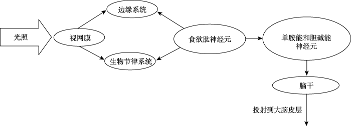
图2
光照警觉性的双通道模型(资料来源:Rautkylä et al, 2012)
4.2 优化光照警觉性的参数特征
高照度的灯光照射或蓝光照明均可在短时间内起到调节内在生理节律、促进个体清醒、保持正常的活动状态等作用(朱莹莹, 汝涛涛, 周国富, 2015)。但在一项研究中, 60个成年被试经过慢性睡眠限制后, 暴露在不同波长的光照下(458~ 480 nm的蓝光、551~555 nm的绿光, 0 lux的暗光控制)。研究人员对光照暴露前、暴露中、暴露后的警觉性进行了测量, 结果发现均没有显著差异(Segal, Sletten, Flynn-Evans, Lockley, & Rajaratnam, 2016)。所以, 睡眠压力、生理阶段和光照反应曲线之间可能存在一定的交互作用。虽然研究者们一致认为夜晚照射蓝光或富含短波长的白光会促进警觉性, 但对白天警觉性的敏感波长的研究仍存在分歧。例如Sahin和Figueiro (2013)证实白天的红光比蓝光对人的警觉性的作用更大。另外还有研究考察了动态光对人警觉性和表现的影响, 结果发现相比静态光, 动态光更让人警觉(罗明, 郑诗琪, 叶鸣, 2016)。因此未来的研究可以从动态角度去探讨随着24 h的变化光照警觉性的光谱敏感性的变化, 进一步优化在白天能够引起警觉性的光照系统的参数特征。
4.3 探讨光照警觉性的混淆变量
现在的研究大都强调光照在任务中对认知神经功能的影响。比如有研究认为在亮白光或蓝光成分较多的白光环境中可以提高警觉性和神经活动过程(Cajochen et al., 2005; Lockley et al., 2006)。但是这种光照警觉性的影响并不总是促进行为表现。实际上, 有研究报告了光照可能会对认知任务产生不利的作用或者没有显著差异的影响(Gabel et al., 2015; Kaida, Takeda, & Tsuzuki, 2013)。同时也有研究发现, 光照效应在抑制控制任务上的表现有很大的个体差异性, 并且取决于被试实验前的警觉性状态(Correa, Barba, & Padilla, 2016)。实验前处于较高警觉性状态的被试在经过蓝光成分较多的白光照射之后, 在高级认知任务上的成绩更好。这提示我们在探讨光照效应的时候要注意实验之前的警觉性。所以, 对光照警觉性的混淆变量进行研究, 排除或控制一些混淆变量, 对我们更好的理解光照的非视觉效应至关重要。
参考文献
光照对昼夜节律与警觉度的影响
DOI:10.1360/n972014-00104
URL
[本文引用: 1]

近年研究发现,光照可以显著影响人类健康.光敏感视网膜神经节细胞的发现将研究者的关注点从传统的视觉信息传导通路转移至非自觉视觉信息传导通路——即将光照信息传递到非形觉相关的大脑功能区,如下丘脑视交叉上核,进而调解昼夜节律、神经内分泌等生理功能.这条通路生理功能的发挥与光照强度、性质、时间等有着密切联系.年龄相关性因素对实际入眼光照产生各方面的影响.随着不断深入研究光照对下丘脑视交叉上核昼夜节律调节作用,学者开始关注光照对警觉度的影响.我们根据国内外最新研究进展及课题组的研究成果:(1)对比了传统的自觉视觉信息传导通路与非自觉视觉信息传导通路,总结了下丘脑视交叉上核在哺乳类动物生理及行为节律调控中的重要作用;(2)汇总了光照强度、作用时间、波段等因素对非自觉视觉信息传导通路功能的影响——调解昼夜节律、认知功能、警觉度;(3)分析了老龄化群体入眼光照减少的原因、产生的后果、以及白内障摘除手术的益处;(4)总结了脑电图技术作为一种评价警觉度的指标如何客观地反映光照对非自觉视觉信息传导通路的调解作用.
动态光对人警觉度与表现的影响
DOI:10.3969/j.issn.1004-440X.2016.06.001
URL
[本文引用: 1]
本实验研究了照明对警觉性与舒适度的影响.实验使用全面的评价体系,包括主观量表(疲劳问卷、主观睡意、灯光评价)、生理监测(心电、眼电、脑电、CFF)、工作表现数据统计和生理化学指标,来探究色温随时间变化的动态光是否优于照度相同、平均色温相同的静态光.本研究共分成了两个实验.10位被试者参与了此次研究.实验1包含四种光照环境:6000K静态光、9000 K静态光、变化周期为4小时且平均色温为9000 K的长周期动态光和变化周期为2小时且平均色温为9 000 K的短周期动态光.结果显示被试者在两种动态光内的脑电显著优于静态光下.实验2依据实验1的结果而设计,包含六种光环境:色温变化范围4000 K~10000 K,变化频率分别为2h,1h,0.5h的动态光,以及色温变化范围6 000 K~12 000 K,变化频率分别为2h,1h,0.5h的动态光.结果表明高色温,高频率的动态光环境更容易让人警觉.结果亦了解各种的评价系统对健康光源的研究的有效性.
视网膜中的自主感光神经节细胞
DOI:10.3724/SP.J.1260.2011.00387
URL
[本文引用: 1]
视网膜中少数神经节细胞能够合成感光蛋白——黑视素(melanopsin),因此具备了自主感光的能力,被称为自主感光神经节细胞(intrinsically photosensitive retinal ganglion cells,ipRGCs)。ipRGCs可根据树突形态和分层位置的差异分为五个不同的亚型,其轴突主要投射到视交叉上核、橄榄顶盖前核等脑区,参与调控昼夜节律、瞳孔对光反射等非成像视觉功能。此外,部分ipRGCs的轴突投射到外侧膝状体和上丘,可能在调节成像视觉中发挥功能。本文将概述ipRGCs的发现过程和最新研究进展。
照明的非视觉作用及其脑神经机制
DOI:10.3724/SP.J.1042.2015.01348
URL
[本文引用: 1]
照明在人类生活、工作和学习中发挥着举足轻重的作用,除了提供基本的视觉作用(对周围事物的大小、颜色、形状等方面的视觉感知)外,还会对人的生理、心理功能产生显著影响,如调节褪黑素分泌、影响生物节律,促进认知加工、调节由季节变化引起的情绪情感障碍(SAD)等。这种对生理、心理活动产生直接或间接的影响即为照明的非视觉作用。近年来,照明的非视觉作用及其背后的神经机制得到了研究者的广泛关注和大量实证研究,并取得了丰硕成果。未来研究需从模型建构、动态照明等角度入手进一步探索照明的非视觉作用及其脑神经机制。
Annual variation in daily light exposure and circadian change of melatonin and cortisol concentrations at a northern latitude with large seasonal differences in photoperiod length
DOI:10.1186/s40101-016-0103-9
URL
PMID:4952149
[本文引用: 1]
Seasonal variations in physiology and behavior have frequently been reported. Light is the major zeitgeber for synchronizing internal circadian rhythms with the external solar day. Non-image forming effects of light radiation, for example, phase resetting of the circadian rhythms, melatonin suppression, and acute alerting effects, depend on several characteristics of the light exposure including intensity, timing and duration, spectral composition and previous light exposure, or light history. The aim of the present study was to report on the natural pattern of diurnal and seasonal light exposure and to examine seasonal variations in the circadian change of melatonin and cortisol concentrations for a group of Swedish office workers. Fifteen subjects participated in a field study that was carried out in the south of Sweden. Ambulatory equipment was used for monthly measurements of the daily exposure to light radiation across the year. The measurements included illuminance and irradiance. The subjects collected saliva samples every 402h during 102day of the monthly measuring period. The results showed that there were large seasonal differences in daily amount of light exposure across the year. Seasonal differences were observed during the time periods 04:00–08:00, 08:00–12:00, 12:00–16:00, 16:00–20:00, and 20:00–24:00. Moreover, there were seasonal differences regarding the exposure pattern. The subjects were to a larger extent exposed to light in the afternoon/evening in the summer. During the winter, spring, and autumn, the subjects received much of the daily light exposure in the morning and early afternoon. Regarding melatonin, a seasonal variation was observed with a larger peak level during the winter and higher levels in the morning at 07:00. This study adds to the results from other naturalistic studies by reporting on the diurnal and seasonal light exposure patterns for a group living at a northern latitude of 56° N, with large annual variations in photoperiod length. It seems to be seasonal variation in the lighting conditions, both concerning intensities as well as regarding the pattern of the light exposure to which people living at high latitudes are exposed which may result in seasonal variation in the circadian profile of melatonin.
Bright light therapy for nonseasonal depression: Meta-analysis of clinical trials
DOI:10.1016/j.jad.2016.03.016
URL
PMID:27011361
[本文引用: 2]
61Bright light therapy seems efficacious in improving nonseasonal depression.61It is particularly efficacious as monotherapy and for 2-5 weeks07 duration of exposure.61The exposure intensity; treatment duration and study design need to be standardized.61The efficacy of light therapy in perinatal depression needs to be further explored.
Human lighting demands: Healthy lighting in an office environment
DOI:10.1002/erv.2315
URL
[本文引用: 1]
dissertations the and tu/e, lighting. daylighting, civil engineering: dissertations. architecture: dissertations, interior lighting, lighting - office buildings, workplace improvement, dissertations the and tu/e, lighting. daylighting, civil engineering: dissertations. architecture: dissertations, interior lighting, lighting - office buildings, workplace improvement
Phototransduction by retinal ganglion cells that set the circadian clock
DOI:10.1126/science.1067262
URL
PMID:11834835
[本文引用: 1]
Light synchronizes mammalian circadian rhythms with environmental time by modulating retinal input to the circadian pacemaker-the suprachiasmatic nucleus (SCN) of the hypothalamus. Such photic entrainment requires neither rods nor cones, the only known retinal photoreceptors. Here, we show that retinal ganglion cells innervating the SCN are intrinsically photosensitive. Unlike other ganglion cells, they depolarized in response to light even when all synaptic input from rods and cones was blocked. The sensitivity, spectral tuning, and slow kinetics of this light response matched those of the photic entrainment mechanism, suggesting that these ganglion cells may be the primary photoreceptors for this system.
Dose-response relationship between light irradiance and the suppression of plasma melatonin in human volunteers
DOI:10.1016/0006-8993(88)90820-7
URL
PMID:3409004
[本文引用: 1]
Abstract This study tested the capacity of different irradiances of monochromatic light to reduce plasma melatonin in normal humans. Six healthy male volunteers, 24-34 years old, were exposed to 0.01, 0.3, 1.6, 5, or 13 microW/cm2 of 509 nm monochromatic light for 1 h during the night on separate occasions. Light irradiance depressed plasma melatonin in a dose-response pattern. The data indicate that the mean threshold irradiance for suppressing melatonin is between 1.6 and 5 microW/cm2. Individual variations in threshold responses to monochromatic light were observed among the volunteers.
Alerting effects of light
DOI:10.1016/j.smrv.2007.07.009 URL [本文引用: 2]
Circadian and sleep-wake dependent impact on neurobehavioral function
DOI:10.1002/pon.778
URL
[本文引用: 1]
ABSTRACT Va riations in waking neurobehavioral or cognitive functioning are closely lin- ked to endogenous 24-h rhythm (circadian pacemaker) and time awake. We summarize studies in which the contribution of the circadian pacemaker and time awake on neurobehavioral function was investigated. Stable and high levels of attention and vigilance can only be maintained when the circadian timing system opposes the wake-dependent deterioration of alertness and per- formance. Planning performance in a maze tracing task was also affected by time awake, whereas circadian modulation was less pronounced. Additional to circadian phase position and the level of sleep pressure, rapid eye movement sleep may play a role in acquiring specific procedural skills in a sequence lear- ning task. We conclude that circadian phase and time awake have a substantial impact on short and stimulating planning tasks, which are related to the pre- frontal cortex, and on sequence learning that requires activation of striatal brain regions. One of the distinguishing characteristics of sleep throughout much of the animal kingdom is that the periods of sleep and wake occur at specific times of the day and/or night. In mammals, the central circadian (i.e., 24 hours) pacemaker that regulates the timing of sleep and wake as all 24-hours rhythms, is located in a small region of the hypothalamus (the suprachias- matic nuclei (SCN)), whereas the control of sleep and wake per se appears to involve many diverse regions of the brain. Waking neurobehavioral func- tion is directly affected by the timing of sleep and wakefulness. In the pre- sent review, we emphasize two facets of "time", the effect of "internal bio- logical time" and "time spent within a vigilance state" (i.e., wakefulness, sleep). "Internal time" is driven by the endogenous circadian pacemaker (cir- cadian clock-like process) which requires daily synchronization with "exter-
EEG and ocular correlates of circadian melatonin phase and human performance decrements during sleep loss
DOI:10.1016/S0034-5687(99)00052-3
URL
PMID:10484479
[本文引用: 1]
The aim of this study was to quantify the associations between slow eye movements (SEMs), eye blink rate, waking electroencephalogram (EEG) power density, neurobehavioral performance, and the circadian rhythm of plasma melatonin in a cohort of 10 healthy men during up to 32 h of sustained wakefulness. The time course of neurobehavioral performance was characterized by fairly stable levels throughout the first 16 h of wakefulness followed by deterioration during the phase of melatonin secretion. This deterioration was closely associated with an increase in SEMs. Frontal low-frequency EEG activity (1 7 Hz) exhibited a prominent increase with time awake and little circadian modulation. EEG alpha activity exhibited circadian modulation. The dynamics of SEMs and EEG activity were phase locked to changes in neurobehavioral performance and lagged the plasma melatonin rhythm. The data indicate that frontal areas of the brain are more susceptible to sleep loss than occipital areas. Frontal EEG activity and ocular parameters may be used to monitor and predict changes in neurobehavioral performance associated with sleep loss and circadian misalignment.
High sensitivity of human melatonin, alertness, thermoregulation, and heart rate to short wavelength light
DOI:10.1210/jc.2004-0957
URL
PMID:15585546
[本文引用: 1]
Abstract Light can elicit acute physiological and alerting responses in humans, the magnitude of which depends on the timing, intensity, and duration of light exposure. Here, we report that the alerting response of light as well as its effects on thermoregulation and heart rate are also wavelength dependent. Exposure to 2 h of monochromatic light at 460 nm in the late evening induced a significantly greater melatonin suppression than occurred with 550-nm monochromatic light, concomitant with a significantly greater alerting response and increased core body temperature and heart rate ( approximately 2.8 x 10(13) photons/cm(2)/sec for each light treatment). Light diminished the distal-proximal skin temperature gradient, a measure of the degree of vasoconstriction, independent of wavelength. Nonclassical ocular photoreceptors with peak sensitivity around 460 nm have been found to regulate circadian rhythm function as measured by melatonin suppression and phase shifting. Our findings-that the sensitivity of the human alerting response to light and its thermoregulatory sequelae are blue-shifted relative to the three-cone visual photopic system-indicate an additional role for these novel photoreceptors in modifying human alertness, thermophysiology, and heart rate.
Dose-response relationship for light intensity and ocular and electroencephalographic correlates of human alertness
DOI:10.1016/S0166-4328(00)00236-9
URL
PMID:10996410
[本文引用: 2]
Light can elicit both circadian and acute physiological responses in humans. In a dose response protocol men and women were exposed to illuminances ranging from 3 to 9100 lux for 6.5 h during the early biological night after they had been exposed to <3 lux for several hours. Light exerted an acute alerting response as assessed by a reduction in the incidence of slow-eye movements, a reduction of EEG activity in the theta–alpha frequencies (power density in the 5–9 Hz range) as well as a reduction in self-reported sleepiness. This alerting response was positively correlated with the degree of melatonin suppression by light. In accordance with the dose response function for circadian resetting and melatonin suppression, the responses of all three indices of alertness to variations in illuminance were consistent with a logistic dose response curve. Half of the maximum alerting response to bright light of 9100 lux was obtained with room light of 65100 lux. This sensitivity to light indicates that variations in illuminance within the range of typical, ambient, room light (90–180 lux) can have a significant impact on subjective alertness and its electrophysiologic concomitants in humans during the early biological night.
Acute effects of different light spectra on simulated night- shift work without circadian alignment
DOI:10.1080/07420528.2016.1222414
URL
PMID:27579732
[本文引用: 1]
Short-wavelength and short-wavelength-enhanced light have a strong impact on night-time working performance, subjective feelings of alertness and circadian physiology. In the present study, we investigated acute effects of white light sources with varied reduced portions of short wavelengths on cognitive and visual performance, mood and cardiac output.Thirty-one healthy subjects were investigated in a balanced cross-over design under three light spectra in a simulated night-shift paradigm without circadian adaptation.Exposure to the light spectrum with the largest attenuation of short wavelengths reduced heart rate and increased vagal cardiac parameters during the night compared to the other two light spectra without deleterious effects on sustained attention, working memory and subjective alertness. In addition, colour discrimination capability was significantly decreased under this light source.To our knowledge, the present study for the first time demonstrates that polychromatic white light with reduced short wavelengths, fulfilling current lighting standards for indoor illumination, may have a positive impact on cardiac physiology of night-shift workers without detrimental consequences for cognitive performance and alertness.
Psychophysiological effects of a single, short, and moderately bright room light exposure on mildly depressed geriatric inpatients: A pilot study
DOI:10.1159/000455231
URL
PMID:28103597
[本文引用: 1]
Abstract Background: Light interventions typically exert their mood-related effects during morning bright light exposures over several weeks. Evidence about immediate ambient room light effects on depressed individuals is still sparse. Objective: The present study aimed at examining the acute effects of a single moderately bright room light exposure on mood, and behavioural and cardiac stress reactions of mildly depressed geriatric inpatients during a short cognitive stimulation and while resting. Methods: Twenty-one inpatients were tested in a balanced cross-over design on 2 consecutive days under either conventional room light (standard light) or artificial sunlight conditions for 30 min. Room illumination was implemented with an artificial skylight, which perfectly imitated solar indoor illumination (e.g., cloudless sky and bright artificial sun). Light-induced changes of mood, heart rate, and heart rate variability were recorded while performing a perseveration test (acted as cognitive stimulation) twice. Additionally, light-related behaviour was observed during a resting period between the cognitive tests and various subjective ratings were obtained. Results: Compared to standard light, exposure to artificial sunlight had a subjective calming effect over time (p = 0.029) as well as decreased heart rate and increased vagal tone (root mean squared of successive inter-beat intervals), both under cognitive workload and in resting conditions. Effect sizes of reported cardiac reactions were large. Cognitive variables were not influenced by light. Additionally, under the higher corneal illuminance of the artificial sunlight, patients perceived stronger glare (p = 0.030) and kept their eyes closed for longer times (p = 0.033) during the resting period. However, patients did not avoid bright light exposure while resting but voluntarily stayed within the area directly lit by the artificial sun nearly all the time (97%). Conclusion: To our knowledge, this study for the first time demonstrated immediate psychophysiological effects of a single, short room light exposure in mildly depressed geriatric inpatients during a short cognitive stimulation and while resting. The findings complement reported evidence on immediate alerting and mood-related effects of bright light exposures.
Non-visual effects of light on melatonin, alertness and cognitive performance: Can blue-enriched light keep us alert?
DOI:10.1371/journal.pone.0016429
URL
PMID:21298068
[本文引用: 2]
Background: Light exposure can cascade numerous effects on the human circadian process via the non-imaging forming system, whose spectral relevance is highest in the short-wavelength range. Here we investigated if commercially available compact fluorescent lamps with different colour temperatures can impact on alertness and cognitive performance. Methods: Sixteen healthy young men were studied in a balanced cross-over design with light exposure of 3 different light settings (compact fluorescent lamps with light of 40 lux at 6500K and at 2500K and incandescent lamps of 40 lux at 3000K) during 2 h in the evening. Results: Exposure to light at 6500K induced greater melatonin suppression, together with enhanced subjective alertness, well-being and visual comfort. With respect to cognitive performance, light at 6500K led to significantly faster reaction times in tasks associated with sustained attention (Psychomotor Vigilance and GO/NOGO Task), but not in tasks associated with executive function (Paced Visual Serial Addition Task). This cognitive improvement was strongly related with attenuated salivary melatonin levels, particularly for the light condition at 6500K. Conclusions: Our findings suggest that the sensitivity of the human alerting and cognitive response to polychromatic light at levels as low as 40 lux, is blue-shifted relative to the three-cone visual photopic system. Thus, the selection of commercially available compact fluorescent lights with different colour temperatures significantly impacts on circadian physiology and cognitive performance at home and in the workplace.
Light modulation of humansleep depends on a polymorphism in the clock gene Period3
DOI:10.1016/j.bbr.2014.05.050
URL
PMID:24893318
[本文引用: 1]
Non-image-forming (NIF) responses to light powerfully modulate human physiology. However, it remains scarcely understood how NIF responses to light modulate human sleep and its EEG hallmarks, and if there are differences across individuals. Here we investigated NIF responses to light on sleep in individuals genotyped for the PERIOD3 (PER3) variable-number tandem-repeat (VNTR) polymorphism. Eighteen healthy young men (20–28 years; mean±SEM: 25.9±1.2) homozygous for the PER3 polymorphism were matched by age, body-mass index, and ethnicity. The study protocol comprised a balanced cross-over design during the winter, during which participants were exposed to either light of 40lx at 6500K (blue-enriched) or light at 2500K (non-blue enriched), during 2h in the evening. Compared to light at 2500K, light at 6500K induced a significant increase in all-night NREM sleep slow-wave activity (SWA: 1.0–4.5Hz) in the occipital cortex for PER35/5 individuals, but not for PER34/4 volunteers. Dynamics of SWA across sleep cycles revealed increased occipital NREM sleep SWA for virtuallyall sleep episode only for PER35/5 individuals. Furthermore, they experienced light at 6500K as significantly brighter. Intriguingly, this subjective perception of brightness significantly predicted their increased occipital SWA throughout the sleep episode. Our data indicate that humans homozygous for the PER35/5 allele are more sensitive to NIF light effects, as indexed by specific changes in sleep EEG activity. Ultimately, individual differences in NIF light responses on sleep may depend on a clock gene polymorphism involved in sleep–wake regulation.
Light effects on behavioural performance depend on the individual state of vigilance
DOI:10.1371/journal.pone.0164945
URL
[本文引用: 1]
Research has shown that exposure to bright white light or blue-enriched light enhances alertness, but this effect is not consistently observed in tasks demanding high-level cognition (e.g., Sustained Attention to Response Task ART, which measures inhibitory control). Individual differences in sensitivity to light effects might be mediated by variations in the basal level of arousal. We tested this hypothesis by measuring the participants behavioural state of vigilance before light exposure, through the Psychomotor Vigilance Task. Then we compared the effects of a blue-enriched vs. dim light at nighttime on the performance of the auditory SART, by controlling for individual differences in basal arousal. The results replicated the alerting effects of blue-enriched light, as indexed by lower values of both proximal temperature and distal-proximal gradient. The main finding was that lighting effects on SART performance were highly variable across individuals and depended on their prior state of vigilance. Specifically, participants with higher levels of basal vigilance before light exposure benefited most from blue-enriched lighting, responding faster in the SART. These results highlight the importance of considering basal vigilance to define the boundary conditions of light effects on cognitive performance. Our study adds to current research delineating the complex and reciprocal interactions between lighting effects, arousal, cognitive task demands and behavioural performance.
Paradoxical timing of the circadian rhythm of sleep propensity serves to consolidate sleep and wakefulness in humans
DOI:10.1016/0304-3940(94)90841-9
URL
PMID:8190360
[本文引用: 1]
Abstract The contribution of the circadian pacemaker and the sleep homeostat to sleep tendency and consolidation was quantified by forced desynchrony of the sleep-wake cycle from the circadian pacemaker in eight men who lived in time-isolation for 33-36 days. Analysis of 175 polygraphically recorded sleep episodes revealed that the circadian pacemaker and the sleep homeostat contribute about equally to sleep consolidation, and that the phase relationship between these oscillatory processes during entrainment to the 24-h day is uniquely timed to facilitate the ability to maintain a consolidated bout of sleep at night and a consolidated bout of wakefulness throughout the day.
Microcomputer analyses of performance on a portable, simple visual RT task during sustained operations
DOI:10.3758/BF03200977
URL
[本文引用: 1]
The Institute of Pennsylvania Hospital and University of Pennsylvania, Philadelphia, Pennsylvania There is a need for brief, portable performance measures that are free of practice effects but that reliably show the impact of sleep loss on performance during sustained work. Reaction time (RT) tasks hold considerable promise in meeting this need, if the extensive number of responses they typically yield can be processed in ways that quickly provide the essential analyses. While testing the utility of a portable visual RT task during a sustained, quasi-continuous work schedule of 54 h, we developed a microcomputer software system that inputs, edits, transforms, analyzes, and reduces the data from the RT portable audiotapes, for each 10-min trial on the task. With relatively minor modifications, the software system can be used on a minimally configured microcomputer system that supports BASIC. The software is flexible and capable of retrieving distorted data, and it generates a variety of dependent variables reflecting intratrial optimum response capacity, lapsing, and response slowing.
Efficacy of light therapy in nonseasonal depression: A systematic review
DOI:10.1016/S0925-3467(01)00135-5
URL
PMID:17950467
[本文引用: 1]
BACKGROUND: The efficacy of bright light therapy is well established for winter depression but its status in depression without seasonal pattern is unclear. METHODS: We systematically evaluated available data on the efficacy of light therapy in nonseasonal depression. RESULTS: We identified 62 reports among which 15 met our predefined selection criteria. The available data show evidence for the efficacy of light therapy as an adjuvant treatment to antidepressants. Trials that evaluated light therapy alone (without antidepressants) in nonseasonal depression yielded inconsistent results. LIMITATIONS: Most of the studies extracted poorly controlled the issue of blindness and were limited by small sample sizes. Publication bias may have distorted our estimation of the effect of light therapy. CONCLUSIONS: Overall, bright light therapy is an excellent candidate for inclusion into the therapeutic inventory available for the treatment of nonseasonal depression today, as adjuvant therapy to antidepressant medication. Future clinical trials of light therapy should distinguish homogenous subgroups of depressed patients in order to evaluate whether light therapy may eventually be considered as stand-alone treatment for specific subgroups of patients with nonseasonal depression.
Light, sleep and circadian rhythms in older adults with Alzheimer's disease and related dementias
DOI:10.2217/nmt-2016-0060
URL
PMID:28534696
[本文引用: 2]
Alzheimer's disease and related dementias (ADRD) can cause sleep and behavioral problems that are problematic for ADRD patients and their family caregivers. Light therapy has shown promise as a nonpharmacological treatment, and preliminary studies demonstrate that timed light exposure can consolidate and improve nighttime sleep efficiency, increase daytime wakefulness and reduce evening agitation without the adverse effects of pharmacological solutions. Compliance with light treatment and the accurate measurement of light exposures during treatment, however, have presented barriers for the adoption of light therapy for ADRD. Recent research showing that the circadian system is maximally sensitive to short-wavelength light opens the way for the potential application of lower, more-targeted light intensities to maximize compliance and individualize light dose/timing in therapeutic settings.
Preliminary evidence that both blue and red light can induce alertness at night
DOI:10.1186/1471-2202-10-105
URL
PMID:2744917
[本文引用: 1]
Background A variety of studies have demonstrated that retinal light exposure can increase alertness at night. It is now well accepted that the circadian system is maximally sensitive to...
On light as an alerting stimulus at night
Abstract Light exposure at night increases alertness; however, it is not clear if light affects nocturnal alertness in the same way that it affects measures of circadian regulation. The purpose of this study was to determine if a previously established functional relationship between light and nocturnal melatonin suppression was the same as that relating light exposure and nocturnal alertness. Four levels of narrow-band blue light at the cornea were presented during nighttime sessions. The ratio of electroencephalographic alpha power density with eyes closed to eyes open (alpha attenuation coefficient, AAC) and the Norris mood scale were used. The AAC and ratings of alertness increased monotonically with irradiance and were highly correlated. Both measures of alertness were highly correlated with model predictions of nocturnal melatonin suppression for the same circadian light stimulus, consistent with the inference that the suprachiasmatic nuclei play an important role in nocturnal alertness as well as circadian regulation.
Tailored lighting intervention improves measures of sleep, depression, and agitation in persons with Alzheimer's disease and related dementia living in long-term care facilities
DOI:10.2147/CIA.S68557
URL
[本文引用: 1]
Mariana G Figueiro,1 Barbara A Plitnick,1 Anna Lok,1 Geoffrey E Jones,1 Patricia Higgins,2,3 Thomas R Hornick,3,4 Mark S Rea1 1Lighting Research Center, Rensselaer Polytechnic Institute, Troy, NY, USA; 2School of Nursing, 3School of Medicine, Case Western Reserve University, 4Geriatric Research Education and Clinical Center, Louis Stokes Cleveland Veterans Affairs Medical Center, Cleveland, OH, USABackground: Light therapy has shown great promise as a nonpharmacological method to improve symptoms associated with Alzheimerrsquo;s disease and related dementias (ADRD), with preliminary studies demonstrating that appropriately timed light exposure can improve nighttime sleep efficiency, reduce nocturnal wandering, and alleviate evening agitation. Since the human circadian system is maximally sensitive to short-wavelength (blue) light, lower, more targeted lighting interventions for therapeutic purposes, can be used. Methods: The present study investigated the effectiveness of a tailored lighting intervention for individuals with ADRD living in nursing homes. Low-level ldquo;bluish-whiterdquo; lighting designed to deliver high circadian stimulation during the daytime was installed in 14 nursing home resident rooms for a period of 4 weeks. Lightndash;dark and restndash;activity patterns were collected using a Daysimeter. Sleep time and sleep efficiency measures were obtained using the restndash;activity data. Measures of sleep quality, depression, and agitation were collected using standardized questionnaires, at baseline, at the end of the 4-week lighting intervention, and 4nbsp;weeks after the lighting intervention was removed. Results: The lighting intervention significantly (Plt;0.05) decreased global sleep scores from the Pittsburgh Sleep Quality Index, and increased total sleep time and sleep efficiency. The lighting intervention also increased phasor magnitude, a measure of the 24-hour resonance between lightndash;dark and restndash;activity patterns, suggesting an increase in circadian entrainment. The lighting intervention significantly (Plt;0.05) reduced depression scores from the Cornell Scale for Depression in Dementia and agitation scores from the Cohenndash;Mansfield Agitation Inventory. Conclusion: A lighting intervention, tailored to increase daytime circadian stimulation, can be used to increase sleep quality and improve behavior in patients with ADRD. The present field study, while promising for application, should be replicated using a larger sample size and perhaps using longer treatment duration. Keywords: sleep disorders, light therapy, circadian rhythms, ADRD
Research note: A self-luminous light table for persons with Alzheimer's disease
The effects of red and blue lights on circadian variations in cortisol, Alpha amylase, and melatonin
DOI:10.1155/2010/829351
URL
PMID:20652045
[本文引用: 1]
The primary purpose of the present study was to expand our understanding of the impact of light exposures on the endocrine and autonomic systems as measured by acute cortisol, alpha amylase, and melatonin responses. We utilized exposures from narrowband long-wavelength (red) and from narrow-band short-wavelength (blue) lights to more precisely understand the role of the suprachiasmatic nuclei (SCN) in these responses. In a within-subjects experimental design, twelve subjects periodically received one-hour corneal exposures of 40 lux from the blue or from the red lights while continuously awake for 27 hours. Results showed-that, as expected, only the blue light reduced nocturnal melatonin. In contrast, both blue and red lights affected cortisol levels and, although less clear, alpha amylase levels as well. The present data bring into question whether the nonvisual pathway mediating nocturnal melatonin suppression is the same as that mediating other responses to light exhibited by the endocrine and the autonomic nervous systems.
The effects of bright light on day and night shift nurses’ performance and well-being in the NICU
react-text: 471 Lighting technologies are rapidly evolving, creating many opportunities for good lighting within the NICU. With the widespread adoption of advanced solid-state lighting technologies, lighting no longer needs to be static. Rather, lighting systems can be more easily adjusted to the different and changing visual and non-visual needs of the professional staff, infants and family members... /react-text react-text: 472 /react-text [Show full abstract]
Light at night and measures of alertness and performance: Implications for shift workers
DOI:10.1177/1099800415572873 URL [本文引用: 2]
Dawn simulation light impacts on different cognitive domains under sleep restriction
DOI:10.1016/j.bbr.2014.12.043
URL
PMID:25549858
[本文引用: 1]
Chronic sleep restriction (SR) has deleterious effects on cognitive performance that can be counteracted by light exposure. However, it is still unknown if naturalistic light settings (dawn simulating light) can enhance daytime cognitive performance in a sustainable matter. Seventeen participants were enrolled in a 24-h balanced cross-over study, subsequent to SR (6-h of sleep). Two different light settings were administered each morning: a) dawn simulating light (DsL; polychromatic light gradually increasing from 0 to 250lx during 30min before wake-up time, with light around 250lx for 20min after wake-up time) and b) control dim light (DL; <8lx). Cognitive tests were performed every 2h during scheduled wakefulness and questionnaires were completed hourly to assess subjective mood. The analyses yielded a main effect of ight condition for the motor tracking task, sustained attention to response task and a working memory task (visual 1 and 3-back task), as well as for the Simple Reaction Time Task, such that participants showed better task performance throughout the day after morning DsL exposure compared to DL. Furthermore, low performers benefited more from the light effects compared to high performers. Conversely, no significant influences from the DsL were found for the Psychomotor Vigilance Task and a contrary effect was observed for the digit symbol substitution test. No light effects were observed for subjective perception of sleepiness, mental effort, concentration and motivation. Our data indicate that short exposure to artificial morning light may significantly enhance cognitive performance in a domain-specific manner under conditions of mild SR.
The efficacy of light therapy in the treatment of mood disorders: A review and meta-analysis of the evidence
DOI:10.1176/appi.ajp.162.4.656
URL
PMID:15800134
[本文引用: 1]
ABSTRACT The purpose of this study was to assess the evidence base for the efficacy of light therapy in treating mood disorders. The authors systematically searched PubMed (January 1975 to July 2003) to identify randomized, controlled trials of light therapy for mood disorders that fulfilled predefined criteria. These articles were abstracted, and data were synthesized by disease and intervention category. Only 13% of the studies met the inclusion criteria. Meta-analyses revealed that a significant reduction in depression symptom severity was associated with bright light treatment (eight studies, having an effect size of 0.84 and 95% confidence interval [CI] of 0.60 to 1.08) and dawn simulation in seasonal affective disorder (five studies; effect size=0.73, 95% CI=0.37 to 1.08) and with bright light treatment in nonseasonal depression (three studies; effect size=0.53, 95% CI=0.18 to 0.89). Bright light as an adjunct to antidepressant pharmacotherapy for nonseasonal depression was not effective (five studies; effect size=-0.01, 95% CI=-0.36 to 0.34). Many reports of the efficacy of light therapy are not based on rigorous study designs. This analysis of randomized, controlled trials suggests that bright light treatment and dawn simulation for seasonal affective disorder and bright light for nonseasonal depression are efficacious, with effect sizes equivalent to those in most antidepressant pharmacotherapy trials. Adopting standard approaches to light therapy's specific issues (e.g., defining parameters of active versus placebo conditions) and incorporating rigorous designs (e.g., adequate group sizes, randomized assignment) are necessary to evaluate light therapy for mood disorders.
Spectral responses of the human circadian system depend on the irradiance and duration of exposure to light
DOI:10.1126/scitranslmed.3000741
URL
PMID:4414925
[本文引用: 1]
In humans, modulation of circadian rhythms by light is thought to be mediated primarily by melanopsin-containing retinal ganglion cells, not rods or cones. Melanopsin cells are intrinsically blue light-sensitive but also receive input from visual photoreceptors. We therefore tested in humans whether cone photoreceptors contribute to the regulation of circadian and neuroendocrine light responses. Dose-response curves for melatonin suppression and circadian phase resetting were constructed in subjects exposed to blue (460 nm) or green (555 nm) light near the onset of nocturnal melatonin secretion. At the beginning of the intervention, 555-nm light was equally effective as 460-nm light at suppressing melatonin, suggesting a significant contribution from the three-cone visual system ( max = 555 nm). During the light exposure, however, the spectral sensitivity to 555-nm light decayed exponentially relative to 460-nm light. For phase-resetting responses, the effects of exposure to low-irradiance 555-nm light were too large relative to 460-nm light to be explained solely by the activation of melanopsin. Our findings suggest that cone photoreceptors contribute substantially to nonvisual responses at the beginning of a light exposure and at low irradiances, whereas melanopsin appears to be the primary circadian photopigment in response to long-duration light exposure and at high irradiances. These results suggest that light therapy for sleep disorders and other indications might be optimized by stimulating both photoreceptor systems.
Melanopsin cells are the principal conduits for rod-cone input to non-image- forming vision
DOI:10.1038/nature06829
URL
PMID:18432195
[本文引用: 1]
Abstract Rod and cone photoreceptors detect light and relay this information through a multisynaptic pathway to the brain by means of retinal ganglion cells (RGCs). These retinal outputs support not only pattern vision but also non-image-forming (NIF) functions, which include circadian photoentrainment and pupillary light reflex (PLR). In mammals, NIF functions are mediated by rods, cones and the melanopsin-containing intrinsically photosensitive retinal ganglion cells (ipRGCs). Rod-cone photoreceptors and ipRGCs are complementary in signalling light intensity for NIF functions. The ipRGCs, in addition to being directly photosensitive, also receive synaptic input from rod-cone networks. To determine how the ipRGCs relay rod-cone light information for both image-forming and non-image-forming functions, we genetically ablated ipRGCs in mice. Here we show that animals lacking ipRGCs retain pattern vision but have deficits in both PLR and circadian photoentrainment that are more extensive than those observed in melanopsin knockouts. The defects in PLR and photoentrainment resemble those observed in animals that lack phototransduction in all three photoreceptor classes. These results indicate that light signals for irradiance detection are dissociated from pattern vision at the retinal ganglion cell level, and animals that cannot detect light for NIF functions are still capable of image formation.
Light therapy and Alzheimer’s disease and related dementia: Past, present, and future
DOI:10.3233/JAD-2012-121645
URL
PMID:23099814
[本文引用: 4]
Sleep disturbances are common in persons with Alzheimer's disease or related dementia (ADRD), resulting in a negative impact on the daytime function of the affected person and on the wellbeing of caregivers. The sleep/wake pattern is directly driven by the timing signals generated by a circadian pacemaker, which may or may not be perfectly functioning in those with ADRD. A 24-hour light/dark pattern incident on the retina is the most efficacious stimulus for entraining the circadian system to the solar day. In fact, a carefully orchestrated light/dark pattern has been shown in several controlled studies of older populations, with and without ADRD, to be a powerful non-pharmacological tool to improve sleep efficiency and consolidation. Discussed here are research results from studies looking at the effectiveness of light therapy in improving sleep, depression, and agitation in older adults with ADRD. A 24-hour lighting scheme to increase circadian entrainment, improve visibility, and reduce the risk of falls in those with ADRD is proposed, and future research needs are discussed.
Central projections of melanopsin-expressing retinal ganglion cells in the mouse
DOI:10.1002/(ISSN)1096-9861 URL [本文引用: 1]
We know when we are sleepy: Subjective versus objective measurements of moderate sleepiness in healthy adults
DOI:10.1016/j.biopsycho.2009.12.011
URL
PMID:20064579
[本文引用: 1]
The ability to monitor one's sleepiness has obvious implications for safety critical procedures. Laboratory findings indicate that we may be poor at doing this compared with objective measurements (e.g. reaction times (RT)). However, the respective testing situations usually differ, to favour objective measures. These typically entail longer test durations with less distractions; both factors facilitate sleepiness. Using the Karolinska Sleepiness Scale (KSS) we compared subjective responses with RTs, in 2min epochs, over 10min periods in identical quiet settings, early afternoon, in 21 healthy volunteers with 5h prior night's sleep restriction. Whereas the initial KSS score was unrelated to 10min RT, the KSS subsequently showed a similar, significant increase, comparable with RT. Changes in both scores were very significantly correlated. KSS scores indicated that 5min was an effective ‘settling down’ period. Participants were good at estimating their sleepiness if presented with a procedure equivalent to that of the objective measure.
Effects of blue-enriched light on the daily course of mood, sleepiness and light perception: A field experiment
DOI:10.1177/1477153512447528
URL
[本文引用: 1]
Abstract The aim of this study was to investigate the effect of blue-enriched white light on the course of mood, sleepiness and light perception over the working day in a real work setting. The participants were 30 female office workers who were divided into two groups and exposed to two light conditions (blue-enriched white light and white light) in a counterbalanced order. Mood, sleepiness and light perception were measured three times a day on 2 days of each week. More pronounced effects of blue-enriched white light on energetic arousal were found in the morning and at midday when compared to the other times of the working day. At the beginning and the end of the work day, the workers seemed to be more sensitive to the brightness of blue-enriched white light while throughout the whole work day they seemed to be similarly sensitive to colour temperature.
Time course of sleep inertia dissipation in human performance and alertness
DOI:10.1111/j.1365-2869.1999.00128.x
URL
PMID:10188130
[本文引用: 1]
Summary Alertness and performance on a wide variety of tasks are impaired immediately upon waking from sleep due to sleep inertia, which has been found to dissipate in an asymptotic manner following waketime. It has been suggested that behavioural or environmental factors, as well as sleep stage at awakening, may affect the severity of sleep inertia. In order to determine the time course of sleep inertia dissipation under normal entrained conditions, subjective alertness and cognitive throughput were measured during the first 4 h after habitual waketime from a full 8-h sleep episode on 3 consecutive days. We investigated whether this time course was affected by either sleep stage at awakening or behavioural/environmental factors. Sleep inertia dissipated in an asymptotic manner and took 2–4 h to near the asymptote. Saturating exponential functions fitted the sleep inertia data well, with time constants of 0.67 h for subjective alertness and 1.17 h for cognitive performance. Most awakenings occurred out of stage rapid eye movement (REM), 2 or 1 sleep, and no effect of sleep stage at awakening on either the severity of sleep inertia or the time course of its dissipation could be detected. Subjective alertness and cognitive throughput were significantly impaired upon awakening regardless of whether subjects got out of bed, ate breakfast, showered and were exposed to ordinary indoor room light (≈150 lux) or whether subjects participated in a constant routine (CR) protocol in which they remained in bed, ate small hourly snacks and were exposed to very dim light (10–15 lux). These findings allow for the refinement of models of alertness and performance, and have important implications for the scheduling of work immediately upon awakening in many occupational settings.
The effects of short afternoon nap and bright light on task switching performance and error-related negativity
DOI:10.1111/sbr.12013
URL
[本文引用: 1]
The aim of the present study was to demonstrate the effects of a short nap (20 min) and bright light (>2000 lux) in the afternoon on task switching ability (i.e., switch cost and mixing cost) and error-related negativity (Ne/ERN). The effects of a short nap and bright light on task switching performance have been less reported despite the importance and frequency of task switching as a common activity in many occupational situations. Sixteen volunteers performed a switching task twice a day (1200 1330 h and 1430 21600 h). Participants completed a total of four experimental conditions (control, short nap, bright light, and short nap with bright light). A short nap was taken during the break between the first and second tasks. During the second task, bright light treatment was applied in two of the four conditions. A short afternoon nap improved task-switching performance (i.e., switch cost) and enlarged Ne/ERN amplitude. No effects were found for light conditions on task-switching performance and Ne/ERN amplitude. The results suggest that: (i) a short afternoon nap improves executive functions and self-monitoring that involve the prefrontal cortex; and (ii) the effect of bright light on task-switching performance is smaller compared to a short nap.
Morning vs evening light treatment of patients with winter depression
DOI:10.1001/archpsyc.55.10.890
URL
PMID:9783559
[本文引用: 1]
According to the phase-shift hypothesis for winter depression, morning light (which causes a circadian phase advance) should be more antidepressant than evening light (which causes a delay). Although no studies have shown evening light to be more antidepressant than morning light, investigations have shown either no difference or morning light to be superior. The present study assesses these light-exposure schedules in both crossover and parallel-group comparisons.Fifty-one patients and 49 matched controls were studied for 6 weeks. After a prebaseline assessment and a light/dark and sleep/wake adaptation baseline week, subjects were exposed to bright light at either 6 to 8 AM or 7 to 9 PM for 2 weeks. After a week of withdrawal from light treatment, they were crossed over to the other light schedule. Dim-light melatonin onsets were obtained 7 times during the study to assess circadian phase position.Morning light phase-advanced the dim-light melatonin onset and was more antidepressant than evening light, which phase-delayed it. These findings were statistically significant for both crossover and parallel-group comparisons. Dim-light melatonin onsets were generally delayed in the patients compared with the controls.These results should help establish the importance of circadian (morning or evening) time of light exposure in the treatment of winter depression. We recommend that bright-light exposure be scheduled immediately on awakening in the treatment of most patients with seasonal affective disorder.
Light suppresses melatonin secretion in humans
DOI:10.1126/science.7434030
URL
PMID:7434030
[本文引用: 1]
Bright artificial light suppressed nocturnal secretion of melatonin in six normal human subjects. Room light of less intensity, which is sufficient to suppress melatonin secretion in other mammals, failed to do so in humans. In contrast to the results of previous experiments in which ordinary room light was used, these findings establish that the human response to light is qualitatively similar to that of other mammals.
Short- wavelength sensitivity for the direct effects of light on alertness, vigilance, and the waking electroencephalogram in humans
DOI:10.1007/s00213-005-0296-9
URL
PMID:16494083
[本文引用: 1]
To assess the wavelength-dependent sensitivity of the acute effects of ocular light exposure on alertness, performance, waking electroencephalogram (EEG), and cortisol.
Circadian photoreception: Spotlight on the brain
DOI:10.1016/j.cub.2006.08.039
URL
PMID:16979545
[本文引用: 1]
Abstract In addition to its visual function, the mammalian eye detects light for a range of behavioral and physiological responses that are separate and apart from sight. Recent studies have begun to shed light on the areas of the brain that respond to such 'non-visual' photoreception in the human eye.
Measuring and using light in the melanopsin age
DOI:10.1016/j.tins.2013.10.004 URL [本文引用: 2]
Repeating administration of Epworth Sleepiness Scale is clinically useful. S
DOI:10.1007/s11325-010-0434-4
URL
PMID:21063794
[本文引用: 1]
Purpose We aimed to verify whether it is clinically useful to repeat the Epworth Sleepiness Scale (ESS) in individuals with suspected sleep-disordered breathing (SDB). Methods In this cross-sectional, prospective study, results of the repeated administration of the ESS were analyzed. In 929 consecutive patients, ESS was obtained as usual in the laboratory routine, immediately before the sleep study (ESS1) and was repeated in the morning, after the polysomnography (ESS2). ROC curve, classical psychometry, and item response theory (IRT) Rasch analysis were used to assess measurement properties of ESS. Results The ESS1 score was (mean ± SD), 1165±655.1, and the ESS2, 1365±654.7 ( p 6510 to predict apnea–hypopnea index ≥5 increased from 56% (ESS1) to 72% (ESS2). IRT psychometric properties (unidimensionality, invariance, local independence) were maintained in ESS2. Conclusions Repeating the administration of the Epworth Sleepiness Scale in a clinical setting increases its score and diagnostic accuracy and correlation with SDB variables, without changing the psychometric properties of the scale. This experiment indicates the clinical usefulness of repeating the ESS. The scale can be repeated at a negligible cost, before dismissing individual patients on the basis of a low ESS score, discontinuing a potentially lifesaving diagnostic and therapeutic process.
The effect of high correlated colour temperature office lighting on employee well-being and work performance
DOI:10.1186/1740-3391-5-2
URL
PMID:17217543
[本文引用: 1]
Abstract BACKGROUND: The effects of lighting on the human circadian system are well-established. The recent discovery of 'non-visual' retinal receptors has confirmed an anatomical basis for the non-image forming, biological effects of light and has stimulated interest in the use of light to enhance wellbeing in the corporate setting. METHODS: A prospective controlled intervention study was conducted within a shift-working call centre to investigate the effect of newly developed fluorescent light sources with a high correlated colour temperature (17000 K) upon the wellbeing, functioning and work performance of employees. Five items of the SF-36 questionnaire and a modification of the Columbia Jet Lag scale, were used to evaluate employees on two different floors of the call centre between February and May 2005. Questionnaire completion occurred at baseline and after a three month intervention period, during which time one floor was exposed to new high correlated colour temperature lighting and the other remained exposed to usual office lighting. Two sided t-tests with Bonferroni correction for type I errors were used to compare the characteristics of the two groups at baseline and to evaluate changes in the intervention and control groups over the period of the study. RESULTS: Individuals in the intervention arm of the study showed a significant improvement in self-reported ability to concentrate at study end as compared to those within the control arm (p < 0.05). The mean individual score on a 5 point Likert scale improved by 36.8% in the intervention group, compared with only 1.7% in the control group. The majority of this improvement occurred within the first 7 weeks of the 14 week study. Substantial within group improvements were observed in the intervention group in the areas of fatigue (26.9%), alertness (28.2%), daytime sleepiness (31%) and work performance (19.4%), as assessed by the modified Columbia Scale, and in the areas of vitality (28.4%) and mental health (13.9%), as assessed by the SF-36 over the study period. CONCLUSION: High correlated colour temperature fluorescent lights could provide a useful intervention to improve wellbeing and productivity in the corporate setting, although further work is necessary in quantifying the magnitude of likely benefits.
The effect of blue-enriched white light on cognitive performances and sleepiness of night-shift workers: A field study
DOI:10.1016/j.physbeh.2017.05.008
URL
PMID:28495465
[本文引用: 1]
Abstract INTRODUCTION: Night-shift works are basically accompanied by reduced cognitive performance, sleepiness, and higher possibility for human error and related incidents. It is therefore crucial to improve individuals' performance and alertness in sensitive places like industries' control room with the ultimate goal of increasing efficiency and reducing the number of possible incidents. Previous research has indicated that blue light is a critical cue for entraining circadian rhythm. As a result, the present study was an attempt to investigate whether blue-enriched white light illumination was a practical strategy to decrease sleepiness and improve cognitive performance during night shifts. MARTIAL AND METHODS: The study, which adopted a before-after interventional design, was conducted among 30 control room staff members of petrochemical industry. After baseline assessments under existing lighting conditions, every participant was exposed to two new lighting conditions (namely, 17,000K and 6500K blue-enriched white light), each lasting for a week. Assessments were conducted again at the end of these treatments. In order to measure the subjective sleepiness, Karolinska Sleepiness Scale (KSS) was utilized. Subjects also performed the Conners' Continuous Performance Test II (CPT-II) and 1-back test in order to gauge their cognitive performance, and melatonin assessment was carried out using salivary and Eliza technique. The data was analyzed using two-way repeated measure ANOVA. RESULTS: The results indicated that, compared to normal lighting conditions, participants' sleepiness and melatonin rhythm significantly declined when they were exposed to blue-enriched white light. Furthermore, the experimental condition had a significant effect on the reduction of working memory errors. It also decreased omission errors and the reaction time during the sustained attention task. CONCLUSIONS: Thus, using blue-enriched white light may be a proper ergonomic strategy for improving performance and alertness, especially during night, in sensitive environments like control rooms. Copyright 2017 Elsevier Inc. All rights reserved.
Effects of daytime light exposure on cognitive brain activity as measured by the ERP P300
DOI:10.1016/j.physbeh.2014.10.013
URL
PMID:25447474
[本文引用: 2]
Abstract Exposure to light modulates not only human alertness but also cognitive functions. The present study examined the temporal dynamics of the effects of light exposure on cortical activity related to cognitive processes. Event-related potentials (ERPs) were measured while participants performed an auditory oddball task during exposure to short-, medium- or long-wavelength light or darkness. Experiments were conducted in the daytime. After a 10-min period of darkness, one of the three lights was presented for 28 min. In the control condition, darkness was maintained for the entire session. The ERP component observed approximately 300 ms after the onset of the target stimulus (P300) was analyzed. The amplitude of P300 was larger after 5-20 min of exposure to short-wavelength light than at equivalent time points in the darkness. No differences were observed in the amplitude of P300 between the medium- or long-wavelength light condition and darkness at any time point. These results suggest that the amount of attentional resource allocated to the oddball task was increased by daytime exposure to short-wavelength light, and that following approximately 5 min of exposure the impact of light on cortical activity related to cognitive processes was able to be detected. Copyright 2014 Elsevier Inc. All rights reserved.
Illumination of the melanopsin signaling pathway
DOI:10.1126/science.1105121 URL [本文引用: 1]
Human nonvisual responses to simultaneous presentation of blue and red monochromatic light
DOI:10.1177/0748730411431447
URL
PMID:22306975
[本文引用: 1]
Blue light sensitivity of melatonin suppression and subjective mood and alertness responses in humans is recognized as being melanopsin based. Observations that long-wavelength (red) light can potentiate responses to subsequent short-wavelength (blue) light have been attributed to the bistable nature of melanopsin whereby it forms stable associations with both 11-cis and all-trans isoforms of retinaldehyde and uses light to transition between these states. The current study examined the effect of concurrent administration of blue and red monochromatic light, as would occur in real-world white light, on acute melatonin suppression and subjective mood and alertness responses in humans. Young healthy men (18-35 years; n = 21) were studied in highly controlled laboratory sessions that included an individually timed 30-min light stimulus of blue (位(max) 479 nm) or red ( (max) 627 nm) monochromatic light at varying intensities (10(13)-10(14) photons/cm(2)/sec) presented, either alone or in combination, in a within-subject randomized design. Plasma melatonin levels and subjective mood and alertness were assessed at regular intervals relative to the light stimulus. Subjective alertness levels were elevated after light onset irrespective of light wavelength or irradiance. For melatonin suppression, a significant irradiance response was observed with blue light. Co-administration of red light, at any of the irradiances tested, did not significantly alter the response to blue light alone. Under the current experimental conditions, the primary determinant of the melatonin suppression response was the irradiance of blue 479 nm light, and this was unaffected by simultaneous red light administration.
Nonvisual responses to light exposure in the human brain during the circadian night
DOI:10.1016/j.cub.2004.09.082
URL
PMID:15498492
[本文引用: 2]
The brain processes light information to visually represent the environment but also to detect changes in ambient light level. The latter information induces non-image-forming responses and exerts powerful effects on physiology such as synchronization of the circadian clock and suppression of melatonin [1, 2, 3]. In rodents, irradiance information is transduced from a discrete subset of photosensitive retinal ganglion cells via the retinohypothalamic tract to various hypothalamic and brainstem regulatory structures including the hypothalamic suprachiasmatic nuclei, the master circadian pacemaker [4, 5, 6]. In humans, light also acutely modulates alertness [7, 8], but the cerebral correlates of this effect are unknown. We assessed regional cerebral blood flow in 13 subjects attending to auditory and visual stimuli in near darkness following light exposures (>8000 lux) of different durations (0.5, 17, 16.5, and 0 min) during the biological night. The bright broadband polychromatic light suppressed melatonin and enhanced alertness. Functional imaging revealed that a large-scale occipito-parietal attention network, including the right intraparietal sulcus, was more active in proportion to the duration of light exposures preceding the scans. Activity in the hypothalamus decreased in proportion to previous illumination. These findings have important implications for understanding the effects of light on human behavior.
The effects of red and blue light on alertness and mood at night
DOI:10.1177/1477153509360887
URL
[本文引用: 1]
This study was designed to explore the roles that long- and short-wavelength lights have on momentary mood and alertness at night. Twenty-two subjects participated in a mixed-design experiment, where we measured the impact of two levels of long- and short-wavelength lights on brain activity and on self-assessments of alertness, sleepiness and mood. Measurements were obtained 60 minutes prior to, during and after light exposure. Results showed that the red and the blue lights increased electroencephalographic beta power (12 30 Hz), reduced sleepiness, and increased positive affect relative to the previous dim-light period indicating that alertness and mood can be affected by light without necessarily stimulating the melatonin pathway. The impact of light was modest, however, compared to the increase in fatigue over the course of the night.
Diurnal spectral sensitivity of the acute alerting effects of light
DOI:10.5665/sleep.3396
URL
PMID:24501435
[本文引用: 1]
Previous studies have demonstrated short-wavelength sensitivity for the acute alerting response to nocturnal light exposure. We assessed daytime spectral sensitivity in alertness, performance, and waking electroencephalogram (EEG). Between-subjects (n = 8 per group). Inpatient intensive physiologic monitoring unit. Sixteen healthy young adults (mean age standard deviation = 23.8 2.7 y). Equal photon density exposure (2.8 10(13) photons/cm(2)/s) to monochromatic 460 nm (blue) or 555 nm (green) light for 6.5 h centered in the middle of the 16-h episode of wakefulness during the biological day. Results were compared retrospectively to 16 individuals who were administered the same light exposure during the night. Daytime and nighttime 460-nm light exposure significantly improved auditory reaction time (P < 0.01 and P < 0.05, respectively) and reduced attentional lapses (P < 0.05), and improved EEG correlates of alertness compared to 555-nm exposure. Whereas subjective sleepiness ratings did not differ between the two spectral conditions during the daytime (P > 0.05), 460-nm light exposure at night significantly reduced subjective sleepiness compared to 555-nm light exposure at night (P < 0.05). Moreover, nighttime 460-nm exposure improved alertness to near-daytime levels. The alerting effects of short-wavelength 460-nm light are mediated by counteracting both the circadian drive for sleepiness and homeostatic sleep pressure at night, but only via reducing the effects of homeostatic sleep pressure during the day.
Alerting effects of daytime light exposure: A proposed link between light exposure and brain mechanisms
DOI:10.1177/1477153511409294
URL
[本文引用: 1]
ABSTRACT The effects of light on alertness have been shown several times and the proposed cause has been suppressed melatonin levels. The relation of melatonin and alertness applies at night but not by day when there is hardly any melatonin. Still, light can be used to improve daytime alertness, but how? This paper describes the brain mechanisms involved in light-induced daytime alertness and proposes a novel model of two parallel mechanisms. In addition to the well-established circadian pathway, it is suggested that light can use the amygdala in the limbic system to send signals to the cerebral cortex. The participation of the amygdala in light-induced alertness means that light is provoking and modulating emotions that induce alerting responses. The model is assembled from known relations but has not yet been verified as a functional system. The paper proposes methods to test the model.
Alerting effects of light are sensitive to very short wavelengths
DOI:10.1016/j.neulet.2006.01.032
URL
PMID:16490309
[本文引用: 1]
Abstract In humans a range of non-image-forming (NIF) light responses (melatonin suppression, phase shifting and alertness) are short wavelength sensitive (440-480 nm). The aim of the current study was to assess the acute effect of three different short wavelength light pulses (420, 440 and 470 nm) and 600 nm light on subjective alertness. Healthy male subjects (n = 12, aged 27 +/- 4 years, mean +/- S.D.) were studied in 39, 4-day laboratory study sessions. The subjects were maintained in dim light (470 nm>600 nm) suggest that subjective alertness may be maximally sensitive to very short wavelength light.
Alerting effects of short-wavelength (blue) and long-wavelength (red) lights in the afternoon
DOI:10.1016/j.physbeh.2013.03.014
URL
PMID:23535242
[本文引用: 4]
61Light can be used to increase alertness in the afternoon, close to the post-lunch dip hours.61Red light is a stronger alerting stimulus in the afternoon than blue light.61Long-wavelength cones mediate the alerting effects of red light during the daytime.
Investigating the contribution of short wavelengths in the alerting effect of bright light
DOI:10.1016/j.physbeh.2015.06.028
URL
[本文引用: 2]
61The alerting effect of light was observed at night on subjective, but not objective measures.61The subjective alerting effect of light does not depend on the presence of short-wavelengths.61Wearing blue-blockers do not hinder subjective vigilance improvement by bright light.
Daytime exposure to short- and medium-wavelength light did not improve alertness and neurobehavioral performance
DOI:10.1177/0748730416659953
URL
PMID:27474192
[本文引用: 1]
While previous studies have demonstrated short-wavelength sensitivity to the acute alerting effects of light during the biological night, fewer studies have assessed the alerting effect of light during the daytime. This study assessed the wavelength-dependent sensitivity of the acute alerting effects of daytime light exposure following chronic sleep restriction in 60 young adults (29 men, 31 women; 22.5 ± 3.1 mean ± SD years). Participants were restricted to 5 h time in bed the night before laboratory admission and 3 h time in bed in the laboratory, aligned by wake time. Participants were randomized for exposure to 3 h total of either narrowband blue (λmax 458-480 nm, n = 23) or green light (λmax 551-555 nm, n = 25) of equal photon densities (2.8-8.4 × 10(13) photons/cm(2)/sec), beginning 3.25 h after waking, and compared with a darkness control (0 lux, n = 12). Subjective sleepiness (Karolinska Sleepiness Scale), sustained attention (auditory Psychomotor Vigilance Task), mood (Profile of Mood States Bi-Polar form), working memory (2-back task), selective attention (Stroop task), and polysomnographic and ocular sleepiness measures (Optalert) were assessed prior to, during, and after light exposure. We found no significant effect of light wavelength on these measures, with the exception of a single mood subscale. Further research is needed to optimize the characteristics of lighting systems to induce alerting effects during the daytime, taking into account potential interactions between homeostatic sleep pressure, circadian phase, and light responsiveness.08 2016 The Author(s).
Illuminating rationale and uses for light therapy
Light therapy is increasingly applied in a variety of sleep medicine and psychiatric conditions including circadian rhythm sleep disorders, seasonal affective disorder, and dementia. This article reviews the neural underpinnings of circadian neurobiology crucial for understanding the influence of light therapy on brain function, common mood and sleep disorders in which light therapy may be effectively used, and applications of light therapy in clinical practice.
Shift work: Health, performance and safety problems, traditional countermeasures, and innovative management strategies to reduce circadian misalignment
DOI:10.2147/NSS.S10372
URL
PMID:3630978
[本文引用: 1]
There are three mechanisms that may contribute to the health, performance, and safety problems associated with night-shift work: (1) circadian misalignment between the internal circadian clock and activities such as work, sleep, and eating, (2) chronic, partial sleep deprivation, and (3) melatonin suppression by light at night. The typical countermeasures, such as caffeine, naps, and melatonin (for its sleep-promoting effect), along with education about sleep and circadian rhythms, are the components of most fatigue risk-management plans. We contend that these, while better than nothing, are not enough because they do not address the underlying cause of the problems, which is circadian misalignment. We explain how to reset (phase-shift) the circadian clock to partially align with the night-work, day-sleep schedule, and thus reduce circadian misalignment while preserving sleep and functioning on days off. This involves controlling light and dark using outdoor light exposure, sunglasses, sleep in the dark, and a little bright light during night work. We present a diagram of a sleep-and-light schedule to reduce circadian misalignment in permanent night work, or a rotation between evenings and nights, and give practical advice on how to implement this type of plan.
Adaptation of human pineal melatonin suppression by recent photic history
DOI:10.1210/jc.2003-032100
URL
PMID:15240654
[本文引用: 1]
Abstract The human circadian pacemaker controls the timing of the release of the pineal hormone melatonin, which promotes sleep, decreases body temperature, and diminishes cognitive performance. Abnormal melatonin secretion has been observed in psychiatric and circadian disorders. Although melatonin secretion is directly suppressed by exposure to light in a nonlinear intensity-dependent fashion, little research has focused on the effect of prior photic history on this response. We examined eight subjects in controlled laboratory conditions using a within-subjects design. Baseline melatonin secretion was monitored under constant routine conditions and compared with two additional constant routines with a fixed light stimulus for 6.5 h of 200 lux (50 microW/cm(2)) after approximately 3 d of photic exposure during the subjective day of either about 200 lux (50 microW/cm(2)) or about 0.5 lux (0.15 microW/cm(2)). We found a significant increase in melatonin suppression during the stimulus after a prior photic history of approximately 0.5 lux compared with approximately 200 lux, revealing that humans exhibit adaptation of circadian photoreception. Such adaptation indicates that translation of a photic stimulus into drive on the human circadian pacemaker involves more complex temporal dynamics than previously recognized. Further elucidation of these properties could prove useful in potentiating light therapies for circadian and affective disorders.
A higher illuminance induces alertness even during office hours: Findings on subjective measures, task performance and heart rate measures
DOI:10.1016/j.physbeh.2012.04.028
URL
PMID:22564492
[本文引用: 1]
Abstract Nocturnal white light exposure has shown marked results on subjective and objective indicators of alertness, vitality and mood, yet effects of white light during daytime and under usual office work conditions have not been investigated extensively. The current study employed a mixed-group design (N=32), testing effects of two illuminance levels (200lx or 1000lx at eye level, 4000K) during one hour of morning versus afternoon exposure. In four repeated blocks, subjective reports, objective performance and physiological arousal were measured. Results showed effects of illuminance on subjective alertness and vitality, sustained attention in tasks, and heart rate and heart rate variability. Participants felt less sleepy and more energetic in the high versus the low lighting condition, had shorter reaction times on the psychomotor vigilance task and increased physiological arousal. Effects of illuminance on the subjective measures, as well as those on heart rate were not dependent on time of day or duration of exposure. Performance effects were most pronounced in the morning sessions and towards the end of the one-hour exposure period. The effect on heart rate variability was also most pronounced at the end of the one-hour exposure. The results demonstrate that even under normal, i.e., neither sleep nor light deprived conditions, more intense light can improve feelings of alertness and vitality, as well as objective performance and physiological arousal. Copyright 2012 Elsevier Inc. All rights reserved.
Bright light and mental fatigue: Effects on alertness, vitality, performance and physiological arousal
DOI:10.1016/j.jenvp.2013.12.010
URL
[本文引用: 1]
61We investigated effects of diurnal exposure to bright light after mental fatigue.61Participants felt less sleepy, more vital and happier when exposed to bright light.61Effects on subjective sleepiness and self-control were moderated by mental fatigue.61Physiological and performance-based measures showed mixed results.61Daytime bright light effects need not to be identical to effects reported at night.
Investigating daytime effects of correlated colour temperature on experiences, performance, and arousal
DOI:10.1016/j.jenvp.2017.02.001
URL
[本文引用: 1]
Research in the late evening and at night has shown that acute activating effects of light are particularly sensitive to short-wavelength light. Yet, findings on such effects during daytime are still inconclusive. This study (N=39) investigated effects of correlated colour temperature (CCT; 2700K vs. 6000 K, 500 lx on the desk) on individuals' experiences, performance, and physiology during one hour of exposure in the morning versus afternoon. Except for a higher subjective vitality in the 6000K condition in the morning, results showed no significant activating effects, and even subtle performance-undermining effects in the relatively high CCT condition. Moreover, participants rated both their mood and the light settings as less positive in the 6000K vs. 2700 K condition. It is therefore questionable whether lighting solutions with commonly experienced intensity levels should provide a higher CCT during daytime office hours.
Light therapy for seasonal affective disorder
DOI:10.1016/0893-133X(89)90002-X URL [本文引用: 1]
Light therapy in the treatment of patients with bipolar depression: A meta-analytic study
DOI:10.1016/j.euroneuro.2016.03.001
URL
PMID:26993616
[本文引用: 1]
Light therapy (LT) has been widely used in the treatment of seasonal affective disorder. Recently some evidence indicated that LT may play a role in bipolar depression, either as monotherapy or in combination with total sleep deprivation (TSD). However, the studies examining the treatment effect of LT in bipolar depression resulted in inconsistent findings. To clarify the role of LT in the disorder, we conducted a meta-analysis to compare the efficacy of LT in the treatment of bipolar depression. The results of individual studies were synthesized by a random effects model. Nine studies including 489 patients with bipolar depression were included in this current meta-analysis. We found that disease severity was significantly decreased after LT, in both with and without TSD, and with concomitant medication ( p <0.001). Augmentation treatment with LT significantly decreased disease severity compared to treatment without LT ( p =0.024). Our results highlight the significant efficacy of LT, either as monotherapy or in combination with TSD, in the treatment of bipolar depression. However, the detailed mechanism of LT still remains elusive. Further well-designed controlled trials are required to investigate the optimal intensity and frequency of LT in the treatment of bipolar depression.
Daytime light exposure dynamically enhances brain responses
DOI:10.1016/j.cub.2006.06.031
URL
PMID:16920622
[本文引用: 3]
Abstract In humans, light enhances both alertness and performance during nighttime and daytime [1-4] and influences regional brain function [5]. These effects do not correspond to classical visual responses but involve a non-image forming (NIF) system, which elicits greater endocrine, physiological, neurophysiological, and behavioral responses to shorter light wavelengths than to wavelengths geared toward the visual system [6-11]. During daytime, the neural changes induced by light exposure, and their time courses, are largely unknown. With functional magnetic resonance imaging (fMRI), we characterized the neural correlates of the alerting effect of daytime light by assessing the responses to an auditory oddball task [12-15], before and after a short exposure to a bright white light. Light-induced improvement in subjective alertness was linearly related to responses in the posterior thalamus. In addition, light enhanced responses in a set of cortical areas supporting attentional oddball effects, and it prevented decreases of activity otherwise observed during continuous darkness. Responses to light were remarkably dynamic. They declined within minutes after the end of the light stimulus, following various region-specific time courses. These findings suggest that light can modulate activity of subcortical structures involved in alertness, thereby dynamically promoting cortical activity in networks involved in ongoing nonvisual cognitive processes.
Blue light stimulates cognitive brain activity in visually blind individuals
DOI:10.1162/jocn_a_00450
URL
PMID:4497579
[本文引用: 1]
Light regulates multiple non-image-forming (or nonvisual) circadian, neuroendocrine, and neurobehavioral functions, via outputs from intrinsically photosensitive retinal ganglion cells (ipRGCs). Exposure to light directly enhances alertness and performance, so light is an important regulator of wakefulness and cognition. The roles of rods, cones, and ipRGCs in the impact of light on cognitive brain functions remain unclear, however. A small percentage of blind individuals retain non-image-forming photoreception and offer a unique opportunity to investigate light impacts in the absence of conscious vision, presumably through ipRGCs. Here, we show that three such patients were able to choose nonrandomly about the presence of light despite their complete lack of sight. Furthermore, 2 sec of blue light modified EEG activity when administered simultaneously to auditory stimulations. fMRI further showed that, during an auditory working memory task, less than a minute of blue light triggered the recruitment of supplemental prefrontal and thalamic brain regions involved in alertness and cognition regulation as well as key areas of the default mode network. These results, which have to be considered as a proof of concept, show that non-image-forming photoreception triggers some awareness for light and can have a more rapid impact on human cognition than previously understood, if brain processing is actively engaged. Furthermore, light stimulates higher cognitive brain activity, independently of vision, and engages supplemental brain areas to perform an ongoing cognitive process. To our knowledge, our results constitute the first indication that ipRGC signaling may rapidly affect fundamental cerebral organization, so that it could potentially participate to the regulation of numerous aspects of human brain function.
The effects of light therapy on sleep problems: A systematic review and meta-analysis
DOI:10.1016/j.smrv.2015.08.009
URL
PMID:26606319
[本文引用: 1]
Abstract Although bright light therapy seems a promising treatment for sleep problems, research shows inconclusive results. This meta-analysis is the first to systematically review the effect of light therapy on sleep problems in general and on specific types of sleep problems in particular (circadian rhythm sleep disorders, insomnia, sleep problems related to Alzheimer's disease and dementia). Fifty-three studies with a total of 1154 participants were included. Overall effects and effects on separate circadian and sleep outcomes were examined. We calculated Hedges' g effect sizes and we investigated the effects of twelve moderators (design-related, treatment-related, participant-related). Light therapy was found effective in the treatment of sleep problems in general (g0002=00020.39), and for circadian rhythm sleep disorders (g0002=00020.41), insomnia (g0002=00020.47), and sleep problems related to Alzheimer's disease/dementia (g0002=00020.30) specifically. For circadian rhythm sleep disorders, effects were smaller for randomised controlled trials. For insomnia, we found larger effects for studies using a higher light intensity, and for sleep problems related to Alzheimer's disease/dementia larger effects were found for studies with more female participants. There was indication of publication bias. To conclude, light therapy is effective for sleep problems in general, particularly for circadian outcomes and insomnia symptoms. However, most effect sizes are small to medium. Copyright 0008 2015 Elsevier Ltd. All rights reserved.
Blue-enriched white light in the workplace improves self-reported alertness, performance and sleep quality
DOI:10.5271/sjweh.1268
URL
PMID:18815716
[本文引用: 1]
Objectives Specifications and standards for lighting installations in occupational settings are based on the spectral sensitivity of the classical visual system and do not take into account the recently discovered melanopsin-based, blue-light-sensitive photoreceptive system. The authors investigated the effects of exposure to blueenriched white light during daytime workhours in an office setting. Methods The experiment was conducted on 104 white-collar workers on two office floors. After baseline assessments under existing lighting conditions, every participant was exposed to two new lighting conditions, each lasting 4 weeks. One consisted of blue-enriched white light (17 000 K) and the other of white light (4000 K). The order was balanced between the floors. Questionnaire and rating scales were used to assess alertness, mood, sleep quality, performance, mental effort, headache and eye strain, and mood throughout the 8-week intervention. Results Altogether 94 participants [mean age 36.4 (SD 10.2) years] were included in the analysis. Compared with white light (4000 K), blue-enriched white light (17 000 K) improved the subjective measures of alertness (P<0.0001), positive mood (P=0.0001), performance (P<0.0001), evening fatigue (P=0.0001), irritability (P=0.004), concentration (P<0.0001), and eye discomfort (P=0.002). Daytime sleepiness was reduced (P=0.0001), and the quality of subjective nocturnal sleep (P=0.016) was improved under blue-enriched white light. When the participants' expectation about the effect of the light treatments was entered into the analysis as a covariate, significant effects persisted for performance, alertness, evening fatigue, irritability, difficulty focusing, concentrating, and blurred vision. Conclusions Exposure to blue-enriched white light during daytime workhours improves subjective alertness, performance, and evening fatigue.
A randomized, double-blind, placebo-controlled study of light therapy for antepartum depression
DOI:10.4088/JCP.10m06188blu
URL
PMID:21535997
[本文引用: 1]
Affective disorder during pregnancy is a common condition requiring careful judgment to treat the depression while minimizing risk to the fetus. Following up on promising pilot trials, we studied the efficacy of light therapy.Twenty-seven pregnant women with nonseasonal major depressive disorder according to DSM-IV (outpatients, university polyclinic) were randomly assigned to 7,000 lux fluorescent bright white or 70 lux dim red (placebo) light administered at home in the morning upon awakening for 1 h/d in a 5-week double-blind trial carried out between October 2004 and October 2008. Clinical state was monitored weekly with the 29-item Structured Interview Guide for the Hamilton Depression Rating Scale (HDRS) with Atypical Depression Supplement (SIGH-ADS). Changes of rating scale scores over time were analyzed with the general linear model. Differences from baseline of SIGH-ADS and 17-item HDRS scores at every time point were the dependent variables, time was the within-subjects factor, and treatment was the between-subjects factor. The model also included baseline score of depression and gestational age at intervention start.The superiority of bright light over dim light placebo was shown for both SIGH-ADS (R = 0.251; F(3,23) = 3.91; P < .05) and HDRS (R = 0.338; F(3,23) = 5.42; P < .01) when analyzing the week-by-week change from baseline, and HDRS scores showed a significant interaction of treatment with time (F(4,92) = 2.91; P < .05). Categorical analysis revealed that the response rate (HDRS 50% improvement) at week 5 was significantly greater for bright light (81.3%, n = 16) than for placebo light (45.5%, n = 11) (P < .05). Remission (final score 8) was attained by 68.6% versus 36.4%, respectively (P < .05). Expectation ratings did not differ significantly between groups.Bright white light treatment for 5 weeks improved depression during pregnancy significantly more than placebo dim red light. The study provides evidence that light therapy, a simple, cost-effective antidepressant modality with minimal side effects for the mother and no known risk for the unborn child, may be a useful nonpharmacologic approach in this difficult situation.clinicaltrials.gov Identifier: NCT01043289.
