

1 引言
大脑的可塑性受多种因素影响, 其中长期学习训练是其结构改变的重要因素(Draganski et al., 2004)。舞蹈或音乐训练是指依据科学原理, 计划性地对受训者进行机体结构机能重建和技术加工, 使其通过长期练习掌握常人不易获得的特殊技能的过程(覃嫔, 2018; 马清, 2000)。两类训练的特殊性与长期性为研究脑可塑性提供了良好模型。舞蹈训练与音乐训练具有许多异同点:两者都是复杂感觉运动过程, 涉及大脑对多种感知觉信息的加工整合; 都是情感表达性艺术训练, 需要脑对内部躯体变化与外部刺激信息进行交互加工, 实现艺术情感的表现。不同在于舞蹈训练是以舞者自身为工具的韵律性动觉训练, 它强调本体感觉与身体控制, 强调视-听-动的高度整合。舞蹈学习往往通过大量的动作观察与动作模仿而实现(平心, 2004)。音乐训练特别是器乐训练, 是以掌握特殊演奏技能为目标的训练, 它强调听觉与手指精细运动的高度协调与快速反馈, 要求听-动的高度整合。音乐学习通过听觉信息修正动作, 从而达到对演奏技能的修正(Mutschler et al., 2007; 蒋存梅, 2016)。
目前, 相关研究主要从脑功能与脑结构的改变来刻画两种训练的可塑性影响。关于舞蹈的研究主要聚焦于动作观察网络(action observation network, AON)以及感觉运动网络。研究AON主要是为探索舞蹈专业性对动作知觉以及对目标导向性行为的影响。这些研究发现个体通过舞蹈训练所获得的真实动作经验, 与动作观察所获得的间接经验都会影响其动作观察网络, 具体来讲两种动作经验都会增进该网络的激活(Calvo-Merino, Glaser, Grèzes, Passingham, & Haggard, 2005; Cross, Hamilton, & Grafton, 2006; Jola et al., 2013)。与此同时, 研究者们通过扫描舞者执行下肢舞蹈动作时的脑功能活动情况, 进而研究舞蹈动作中独特的感觉运动过程。其结果显示舞蹈动作执行时与本体感觉、动作控制、节奏保持等功能相关的皮层、皮层下结构及小脑等广泛区域被激活(Brown, Martinez, & Parsons, 2006; Ono et al., 2014)。关于脑结构方面的研究也表明长期舞蹈训练将导致感觉运动区域的灰质与白质结构发生广泛改变。Hänggi, Koeneke, Bezzola和Jäncke (2010)首次开展芭蕾舞蹈家的大脑结构研究。他们发现舞蹈家的左侧前运动皮层、辅助运动皮层、壳核与额上回的灰质体积显著降低; 双侧皮质脊髓束、双侧内囊与胼胝体的白质体积显著降低; 连接前运动皮层的白质纤维微结构出现各向异性降低。舞蹈家大脑的广泛结构改变引起了其他研究者的关注。Giacosa, Karpati, Foster, Penhune和Hyde (2016)通过进一步对比舞蹈家与音乐家的白质纤维微结构, 发现舞蹈家感觉运动网络内的白质纤维结构(如皮质脊髓束、上纵束和胼胝体)变化明显。较之音乐家与对照组, 舞蹈家的白质纤结构更为发散和长程, 连接了更为广泛的感觉运动皮层。综上所述, 舞蹈训练对脑结构的特异性影响主要表现在涉及感觉运动的广泛脑区发生改变, 具体表现为功能皮层灰质体积的减小, 且连接这些皮层的白质纤维微结构发生改变(各项异性降低)。关于音乐的研究主要集中于听-动整合相关网络以及音乐认知的神经过程。研究发现音乐家的感觉运动皮层、听觉皮层及小脑的功能增强, 说明音乐训练可能提升了大脑对听-动信息的处理与整合(Baumann et al., 2007; Bangert et al., 2006)。另一类研究则揭示音乐训练中的句法加工、工作记忆和音乐情绪加工的神经过程, 结果发现音乐训练增强了额下回、顶下小叶、脑岛等相关脑区的功能(Groussard et al., 2010; Koelsch & Siebel, 2005)。关于音乐家脑结构的研究结果基本与其脑功能的特异性改变一致。相关研究发现灰质体积的增加与音乐专业性呈显著正相关(Bermudez, Lerch, Evans, & Zatorre, 2009), 且分布在与音乐技能训练高度相关的脑区, 如颞横回(Schneider et al., 2002)、左侧颞平面(Schlaug, Jancke, Huang, & Steinmetz, 1995)、颞上回(Bermudez et al., 2009)、中央沟内手指感觉区域(Li et al., 2010)、小脑(Hutchinson, Lee, Gaab, & Schlaug, 2003)等。也有研究发现音乐家的手指运动区(位于中央前回)的灰质结构更清晰和典型(Bangert & Schlaug, 2006)。相应的, 音乐家大脑中连接以上灰质区域的白质结构也发生特异性改变, 出现各项异性的增高, 如连接前运动皮层与感觉运动皮层的胼胝体前部和后部(Öztürk, Tasçioglu, Aktekin, Kurtoglu, & Erden, 2002)、连接皮层-脊髓的皮质脊髓束(Rüber, Lindenberg, & Schlaug, 2015)等。总的来说, 音乐训练对脑结构的特异性影响主要表现在与听觉、手指运动与语义分析高度相关的皮层发生改变, 具体表现为皮层灰质体积的增大, 且连接这些区域的白质纤维微结构发生改变(各项异性增高)。
综上所述, 舞蹈与音乐的脑可塑性研究呈现以下几点不足:对音乐的关注远远多于舞蹈, 舞蹈的相关研究还有待于进一步丰富与深入。结构研究远远少于功能研究, 且有限的结构研究结果并不稳定。单独研究舞蹈或音乐的可塑性改变较普遍, 而比较两种训练导致的差异非常有限, 仅有两篇文章报道了舞蹈家与音乐家的白质与灰质结构的异同(Giacosa et al., 2016; Karpati, Giacosa, Foster, Penhune, & Hyde, 2017)。对两种训练导致的灰质体积变化结果存在争议, 如Giacosa等人(2016)发现舞蹈家与音乐家灰质体积在颞叶上部有共同的增加, 但最近发表的一项研究则报告舞蹈训练对灰质结构的改变并无显著影响(Burzynska, Finc, Taylor, Knecht, & Kramer, 2017)。除了上述不足, 我们的前期研究结果还无法完整描述舞蹈训练的特异性影响。我们虽然发现舞蹈训练对大脑皮层-基底节回路的连接具有增强作用(Li et al., 2015), 说明舞蹈训练提高了受训者的感觉运动整合功能, 但这种改变也发生在全身性运动的体育训练中(Huang et al., 2017)。同时由于舞蹈与音乐关系最为密切, 它往往在音乐节奏中进行, 因此要刻画舞蹈的特征就涉及对音乐效应的考虑。所以我们将研究重点放在两种训练的脑结构对比上, 希望一定程度上分离舞蹈中的音乐影响, 进一步探索舞蹈训练相关的脑结构特异性改变。基于前述的研究概况与深入探索舞蹈特征的需要, 我们选取接受现代舞与弦乐训练的被试, 对两种被试的脑灰质体积进行比较研究。选择现代舞是因为它更接近舞蹈本质。相对于单纯追求形式与技巧的芭蕾, 现代舞更强调人体动作本身。这正如表现主义现代舞创始人玛丽.魏格曼在关于舞蹈本体论中所述, 舞蹈的核心因素是人体动作, 即人体动作构成的“力”的因素, 因此没有音乐要素的舞蹈也能独立存在(吕艺生, 2013)。选择弦乐是因为它更典型地体现了音乐训练中的听-动整合。相对于吹奏类与键盘类乐器的训练, 弦乐训练对演奏者音准要求非常高。弦乐演奏音高的变化是演奏者根据听觉反馈, 精准确定手指在琴弦上的位置而实现(马清, 2000)。因此弦乐训练涉及快速、密集和精确的听-动整合。
本文的创新与意义在于:训练组的类型齐整统一。我们选取相同舞蹈风格与乐器类型的被试, 在脑结构可塑性分析研究中降低了舞蹈训练风格与乐器不一致因素的干扰, 进而更好地描绘两种训练对脑结构的特异性影响。研究的问题具有探索性。关于两种训练对灰质结构影响的研究非常有限且存在争议, 我们的研究能为这一问题提供新的证据。研究具有延续性。基于课题组前期研究发现, 我们通过比较进一步刻画舞蹈训练对脑结构的独特影响。
本研究采用基于体素的形态学分析方法(voxel- based morphometry, VBM)进行组间灰质体积对比。我们预测训练组的脑灰质体积可能存在如下改变:现代舞与弦乐训练组的变化范围主要位于感觉运动网络; 现代舞训练组可能出现灰质体积的减小, 而弦乐训练组则可能是增加; 两组被试还可能出现颞叶上部灰质体积的共同增高。
2 材料与方法
2.1 研究对象及纳入标准
本研究招募了3组被试, 包括18名现代舞被试(现代舞训练组)、20名弦乐被试(弦乐训练组)及25名对照被试(对照组) (见表1)。训练组被试均为大学艺术特长生或艺术专业学生, 其中现代舞训练组主修现代舞, 弦乐训练组主修西洋弦乐(小提琴、中提琴或大提琴)。在纳入本实验前, 两组专业被试分别接受两名舞蹈或两名音乐专业教师的综合评估, 其专业性被评价为达到舞蹈专长或音乐专长条件。舞蹈教师对现代舞训练组被试进行如下评估:舞蹈模仿。向被试示范3次长度为4个八拍的全新舞蹈动作(被试之前从未训练过), 要求被试根据自己的记忆重复舞蹈片段, 评估其动作完整性与准确性。舞蹈基本功。要求被试完成下横竖叉、下腰等动作, 同时要求被试根据自己的软开度进行跳、转、翻的技巧组合, 评估其柔韧度与技巧。舞蹈即兴。要求被试根据随机选择的音乐进行即兴舞蹈, 评估其舞蹈动作运用与舞蹈表现力。音乐教师对弦乐训练组被试进行如下评估:自选曲目演奏。要求被试演奏一首自选曲目(3分钟以内), 考察被试的器乐演奏与表现力。视奏能力。音乐教师谱写一段16个小节的中等难度的演奏片段(被试之前从未演奏或练习过), 要求被试现场演奏, 考察其识谱演奏能力。现代舞训练组被试均无音乐训练经历, 弦乐训练组被试均无舞蹈训练经历。两组专业被试训练年限从7至17年不等。对照组为与之匹配的大学生, 无任何舞蹈、音乐训练经验。现代舞训练组、弦乐训练组及对照组在年龄、性别和受教育年限方面均无统计显著性差异, 两种训练组在训练年限方面无统计显著性差异, 分组达到均衡性(见表1)。3组被试完成艾丁伯格手性问卷(Oldfield, 1971)测试, 均为右利手。所有被试均无神经系统疾病、发展性疾病或物质滥用。本实验符合赫尔辛基宣言, 并获得作者所在研究机构伦理委员会许可, 所有被试签署实验知情同意书。
表1 被试人口学信息
| 人口学变量 | 现代舞训练组 | 弦乐训练组 | 对照组 | p |
|---|---|---|---|---|
| 性别(男/女) | 5/13 | 7/13 | 8/17 | 0.892 |
| 年龄(岁) | 19.00 ± 1.41 | 19.05 ± 1.19 | 19.24 ± 0.87 | 0.765 df (2,60) |
| 教育水平(年) | 12.83 ± 1.33 | 13.05 ± 1.09 | 13.20 ± 1.11 | 0.574 df (2,60) |
| 训练年限(年) | 11.44 ± 3.24 | 11.33 ± 2.72 | — | 0.282 df (36) |
注:“p”为现代舞训练组、弦乐训练组及对照组组间差异比较的p值。
2.2 数据获取
本研究在电子科技大学信息医学中心完成。MRI数据的采集设备为GE Discovery 750 3.0T磁共振系统(GE Medical Systems, Milwaukee, WI), 使用标准的GE全头线圈。高分辨率T1加权图像通过3D梯度回波序列(T1-3DFSPGR)获得。扫描参数为:TR = 6.008 ms, TE = 1.984 ms, 反转角9˚, 矩阵256×256, FOV=25.6 cm × 20 cm, 层厚1 mm, 层数152层。采集数据时, 被试在扫描仪中尽量保持头部不动。实验使用海绵来固定被试头部以减少头动, 使用耳塞来降低扫描过程中的噪音。
2.3 数据处理及统计分析
VBM是一种全脑无偏算法, 用来计算脑体积与容量(Ashburner & Friston, 2000, 2005)。本研究采用VBM8工具包进行灰质分析。(1)首先对3组被试的T1加权脑图像进行数据预处理, 包括检查所有被试数据的成像质量, 手动调整所有结构MRI的原点到前连合。(2)其后在MATLAB 2014环境下使用嵌套于SPM8 (http://www.fil.ion. ac.uk/spm)内的VBM8工具包对被试三维T1加权脑图像进行常规的灰质体积估计流程:1)将所有被试的结构MRI图像分割成灰质、白质和脑脊液。2)使用DARTEL算法对分割后的灰质图像建立基于组水平的模板。3)利用构建的模板将所有灰质图像配准到蒙特利尔神经研究所(Montreal Neurological Institute, MNI)标准人脑模板空间。4)将配准后的图像进行调制得到灰质体积图像。5)使用半高全宽为8毫米高斯核将配准后的灰质图像进行平滑。(3)最后对3组被试平滑后的灰质MRI图像进行组间单因素方差分析(进行高斯随机场理论校正, 校正后p < 0.05) (Nichols, 2012), 进而对差异脑区通过两两比较进行事后检验分析(双样本t检验, p < 0.05)。
3 结果
表2 现代舞训练组、弦乐训练组与对照组灰质体积的组间比较
| 脑区 | MNI坐标 | 体素 个数 | F(2,60)值 (最大点) | dan-con | dan- con (p) | dan- con (t41) | mus-con | mus- con (p) | mus- con (t43) | mus-dan | mus-dan (p) | mus- dan (t36) | ||
|---|---|---|---|---|---|---|---|---|---|---|---|---|---|---|
| x | y | z | ||||||||||||
| Cerebelum_ Crus1_R | 33 | -58 | -40 | 32 | 9.99 | p < 0.05 | 0.01029 | 2.69 | 0.07979 | -1.79 | p < 0.001 | 0.00029 | -4.01 | |
| Frontal_ Med_Orb_R | 6 | 40 | -6 | 82 | 11.29 | p < 0.001 | 0.00039 | 3.86 | 0.74462 | -0.33 | p < 0.001 | 0.00058 | -3.76 | |
| Thalamus_R | 12 | -21 | -1 | 267 | 18.05 | p < 0.001 | 0.00028 | -3.97 | 0.16760 | 1.40 | p < 0.001 | 0.00001 | 5.26 | |
| Thalamus_L | -10 | -16 | 1 | 240 | 13.33 | p < 0.001 | 0.00036 | -3.89 | 0.26093 | 1.14 | p < 0.001 | 0.00001 | 5.26 | |
| Temporal_ Sup_R | 55 | -15 | 1 | 131 | 13.47 | 0.74732 | 0.32 | p < 0.001 | 0.00002 | 4.80 | p < 0.001 | 0.00090 | 3.61 | |
| Putamen_R | 25 | 4 | 13 | 112 | 12.88 | p < 0.001 | 0.00009 | 4.33 | 0.29734 | 1.05 | p < 0.01 | 0.00233 | -3.27 | |
| Supp_Motor_ Area_R | 6 | 1 | 63 | 128 | 11.83 | p < 0.01 | 0.00161 | -3.38 | 0.34007 | 0.96 | p < 0.001 | 0.00001 | 5.12 | |
| Precentral_L | -34 | -6 | 58 | 120 | 12.12 | p < 0.01 | 0.00564 | -2.92 | p < 0.05 | 0.04677 | 2.05 | p < 0.001 | 0.00004 | 4.68 |
| Frontal_ Mid_R | 42 | -3 | 57 | 71 | 11.30 | 0.23886 | -1.20 | p < 0.01 | 0.00225 | 3.24 | p < 0.001 | 0.00003 | 4.77 | |
注:dan: 现代舞训练组; mus: 弦乐训练组; con: 对照组; Cerebelum_Crus1_R: 右侧小脑; Frontal_Med_Orb_R: 右侧眶部额上回; Thalamus_R; 右侧丘脑; Thalamus_L; 左侧丘脑; Temporal_Sup_R:颞上回; Putamen_R: 右侧壳核; Supp_Motor_ Area_R右侧辅助运动区; Precentral_L: 左侧中央前回; Frontal_Mid_R右侧额中回。
图1
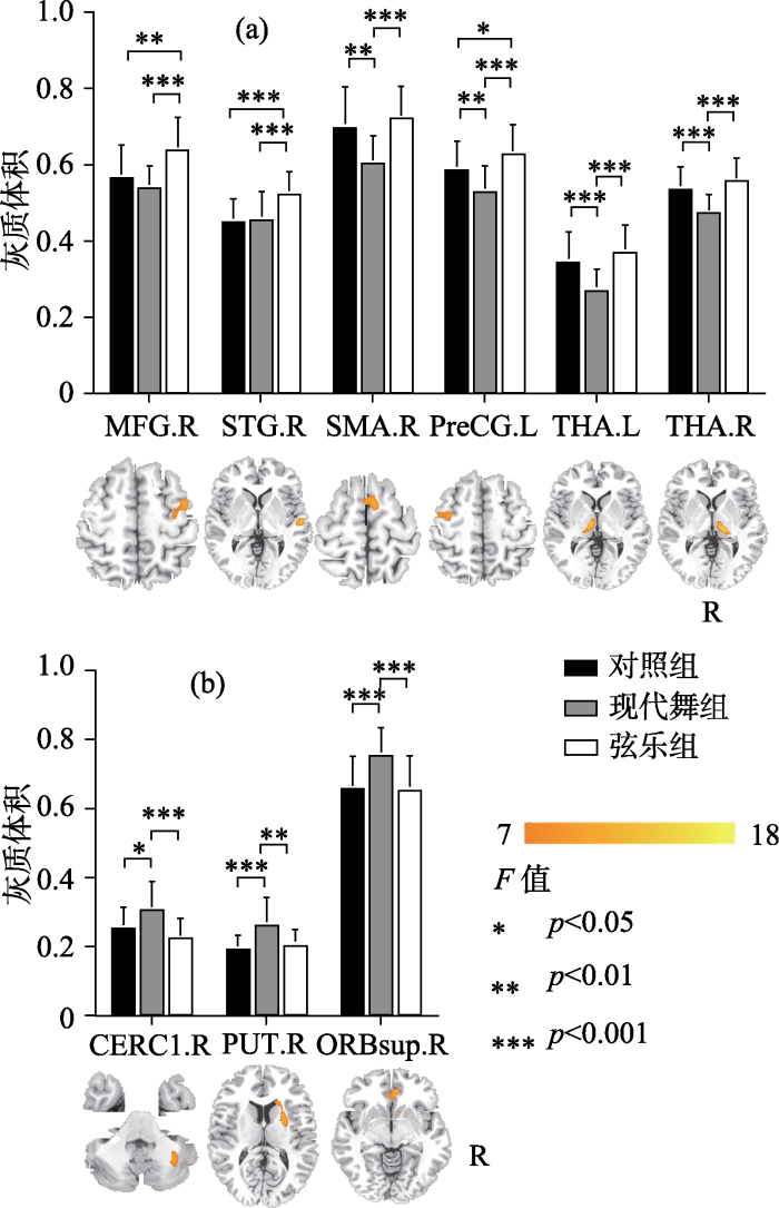
图1
现代舞训练组、弦乐训练组与对照组灰质体积的组间比较
注:MFG. R: 右侧额中回; STG. R: 右侧颞上回; SMA. R: 右侧辅助运动皮层; PreCG. L: 左侧中央前回; THA. L: 左侧丘脑; THA. R: 右侧丘脑; CERC1. R: 右侧小脑; PUT. R: 右侧壳核; ORBsup. R: 右侧眶部额上回。彩图见电子版。
4 讨论
VBM分析结果表明, 较之对照组现代舞训练组与弦乐训练组的灰质体积存在特异性改变, 两者灰质体积改变的区域和方式存在差异。现代舞训练组的改变涉及左侧小脑、双侧丘脑、右侧壳核、右侧辅助运动区等有关感觉运动控制的广泛脑区, 不同区域灰质体积既有增加也有减小。弦乐训练组的改变集中于听-动-读相关脑区, 并统一表现为增加的灰质体积。组间灰质体积的特异性改变说明现代舞训练与弦乐训练对脑可塑性的不同影响。
4.1 现代舞训练对脑灰质体积的影响
较之弦乐训练组与对照组, 现代舞训练组大脑灰质体积变化区域更广泛, 在这些区域中同时存在灰质体积的增高与降低。该结果可能反映了全身性舞蹈动作训练对脑的特异性影响。Brown等人(2006)利用正电子发射断层成像(positron emission tomography, PET)技术观察到舞者进行下肢舞步移动时的脑功能状态, 结果发现小脑蚓部、右侧壳核、腹侧丘脑、内侧上小叶、辅助运动皮层(SMA)、初级运动皮层(M1)与前运动皮层区域的激活明显。我们的结果基本属于以上功能区内, 从结构上说明舞蹈训练可能影响感觉运动网络内的广泛脑区。另外, 现代舞训练组灰质体积的增减也可能体现某种结构与功能的优化。增高的灰质体积往往意味着该区域的神经元更多, 处理信息的效率更高, 对应的认知和行为功能更强大。大部分相关研究表明训练水平往往与灰质体积成正比(Maguire et al., 2000)。与此一致, 现代舞训练组在动作控制相关脑区中表现出增高的灰质体积:比如眶部额上回, 该区域与身体平衡维持相关(Taubert et al., 2010); 小脑下部, 该区域将本体感觉信息与运动前庭功能相整合, 与平衡保持及姿势维持高度相关(Kheradmand & Zee, 2011)。现代舞训练要求舞者不断创造自发动作, 很多自发动作的组合可能是全新的, 因此需要大脑付出更多努力来进行各种动作和姿态的调控与保持, 进而导致以上两个区域灰质体积的增大。其次现代舞训练组还表现出右侧壳核灰质体积的增高与双侧丘脑灰质体积的降低。这一相反变化可能与壳核与丘脑在节奏运动中的功能有关。相关研究报道舞蹈家的右侧壳核在节奏运动条件下激活明显, 而丘脑在非节奏复杂运动时有明显激活(Brown et al., 2006)。我们前期的研究也发现舞蹈家的壳核与全脑的功能连接更显著(Li et al., 2015)。现代舞训练强调动作性, 常常通过动作的力度变化来表现舞蹈情感, 而这种力度变化往往借助于动作节奏的改变而实现。因此, 在现代舞训练中不仅需要舞者保持动作的节奏性, 还需要在不同节奏间进行动作切换。这必然需要壳核的高度参与而丘脑保持抑制, 因此可能导致这两个区域相反的灰质体积变化。进一步我们还发现现代舞训练组右侧辅助运动皮层(SMA)与左侧初级运动皮层(M1)的体积显著减小。这两个脑区在复杂运动控制和感觉运动表征方面扮演重要角色, 是参与舞蹈过程的重要脑区。关于芭蕾舞蹈家和体操运动员的相关研究均发现这两个脑区的灰质体积显著降低(Hänggi et al., 2010; Huang, Lu, Song, & Wang, 2015), 这可能体现了全身性运动技能训练的特殊效应。综上所述, 现代舞训练所影响的灰质结构变化范围更广泛, 灰质体积增高与降低的区域并存。同时这些变化都分布于皮层-基底节-丘脑-小脑神经回路内(该回路贯穿全脑, 与高级认知、运动调控高度相关) (Bostan, Dum, & Strick, 2013)。以上结果可能反映了现代舞独有的训练效应。相对于注重形式和规范的传统舞蹈, 现代舞是一种更为自由和灵活的舞种, 强调创造性的自发动作, 要求舞者容纳和表现更为复杂和广泛的内外感受。这一特点可能需要脑在更大尺度上去加工和整合信息。因此我们推测现代舞训练影响的可能是涉及全脑感觉运动功能协同的广泛脑区。
4.2 弦乐训练对脑灰质体积的影响
较之对照组和现代舞训练组, 弦乐训练组灰质体积的改变集中于与音乐技能直接相关的脑区, 且全部是增高。这一发现显示弦乐训练这种听-动结合的精细运动对大脑的特异性影响。听是音乐家的重要能力之一。我们的结果表明弦乐训练组在右侧听皮层(颞上回)有显著的体积增高。相关元分析研究显示, 颞上回在音乐训练中协调听觉系统对错误的检查, 负责对音乐旋律的解码和音乐语义的记忆并参与音乐中的动-听整合过程(周临舒, 赵怀阳, 蒋存梅, 2017; Lahav, Saltzman, & Schlaug, 2007)。此外大脑听皮层对音乐的处理具有某种程度的偏侧性, 其中右侧颞上回对音乐更为敏感(Hyde, Peretz, & Zatorre, 2008)。相关的结构研究也发现音乐家颞上回的灰质体积显著增大, 其体积与音乐训练时间成正比(Groussard et al., 2014)。除了听觉皮层的变化, 我们发现弦乐训练组左侧初级运动皮层(M1)的灰质体积显著增加。这个区域是所有运动执行的核心区域, 类似的结果也出现在各类器乐演奏家的灰质结构研究中(Bermudez et al., 2009; Han et al., 2009; Sluming et al., 2002)。进一步我们还发现弦乐训练组右侧额中回灰质体积的增加。这个区域与工作记忆的处理与存储相关(Jones, Adlam, Benattayallah, & Milton, 2017), 且与颞中回共同完成语义启动(Laufer, Negishi, Lacadie, Papademetris, & Constable, 2011)。相关研究发现音乐家在这一部位的灰质体积是增加的(Bermudez et al., 2009)。弦乐训练需要受训者识别乐谱符号并记忆乐谱, 这个过程会频繁调用语义表征与工作记忆的脑区, 可能促成该脑区灰质结构的特异性改变。综上所述, 弦乐训练对皮层的影响可能更为集中, 其灰质体积变化区域位于与训练高度相关的听觉、运动与语义加工皮层。这些区域表现出灰质体积的一致增高。我们推测这种改变可能与弦乐训练要求的高度精准性相关。相对于其他乐器训练, 弦乐训练需要更为细微精准的手指控制和稳定的内部音高知觉。受训者需要通过频繁的听-动信息整合与反馈, 才能进行正确演奏。因此弦乐训练可能导致与训练技能直接相关的皮层发生改变。
4.3 本研究的有趣发现
4.3.1 初级运动皮层
初级运动皮层(M1)负责发出运动控制指令及对运动进行表征, 它参与所有动作的执行, 是舞蹈与音乐训练相关的重要脑区。有趣的是, 我们发现现代舞训练组和弦乐训练组灰质体积在左侧M1出现了相反的改变趋势。现代舞训练组较之对照组与弦乐训练组灰质体积显著降低, 而弦乐训练组则显著增高。现代舞训练组M1灰质体积的减小可能是神经修剪和优化的结果。通过科学专业的动作训练, 能让相关脑区内的神经元去除冗余突触, 增强局部回路内部以及远距离脑区间的连接, 从而建立更为高效的神经连接模式(段旭君, 2013)。这一神经可塑性改变外化于行为就是自动化舞蹈动作的获得, 每个舞蹈动作调用较少的神经资源就可以完成。弦乐训练组M1灰质体积的增高则可能是弦乐高度特异性的技能所致。M1在手指或手部的有序运动时会被明显激活(Shibasaki et al., 1993)。同时该区域对运动频率非常敏感, 它的激活程度随着频率的增加而增大(Turner, Grafton, Votaw, Delong, & Hoffman, 1998)。弦乐训练是精细有序的手指运动, 要求给予高频快速的动作反馈, 这种训练方式可能会强烈地激活M1, 进而导致该区域灰质体积的增大。
4.3.2 颞上回
Karpati等人(2016)发现舞蹈家与音乐家灰质体积在这个区域有共同的增加, 并推测该结果与音乐舞蹈训练所涉及的大量动-听整合相关。我们的结果与他们相左, 仅在弦乐训练组中发现灰质体积的显著增高, 而现代舞训练组没有显著变化。其原因可能来自被试的差异。现代舞的典型特点是关注自发动作本身, 它并不高度依赖音乐线索, 甚至可以在无音乐的背景下进行。因此现代舞训练组涉及的动-听整合训练并不突出, 颞上回的灰质体积改变不显著。而弦乐训练则需要高度依赖听觉信息来精确调控手指动作, 更有力地训练了演奏者的动-听整合功能, 因此导致颞上回灰质体积的显著增高。
5 结论
本研究采用基于体素的形态学分析方法, 比较现代舞训练与弦乐训练对脑灰质体积的可塑性影响。结果表明, 两种训练都使脑灰质体积发生特异性改变。其中现代舞训练的影响区域更为广泛, 分布于皮层-基底节-丘脑-小脑回路内, 其变化既有增加也有减小。这一结果说明现代舞训练影响的可能是涉及复杂感觉运动整合的广泛脑区。弦乐训练影响的则是与训练特性(动-听)直接相关的脑区。通过比较研究, 我们的结果首次展示了现代舞训练与弦乐训练对脑灰质结构影响的差异, 揭示了舞蹈训练对脑的特异性影响, 进而为舞蹈治疗和音乐治疗提供神经学基础。此外, 我们的结果也提示后续有两个问题需要进一步探索:其一, 长期舞蹈训练对关键脑区的影响方式是怎样的?我们发现现代舞训练组存在灰质体积增高的区域, 如小脑与壳核, 而大部分相关研究报告舞蹈家或体操运动员的灰质体积都是减小的。那么脑对舞蹈训练的适应到底是协调各脑区皮层以增减互补的方式实现?还是以减小优化的方式达成?其二, 如何理解舞蹈训练与音乐训练对皮层体积影响的相反趋势? 我们发现现代舞训练组与弦乐训练组均在M1出现显著的灰质体积改变, 但这种改变趋势却是相反的。这一结果否说明皮层的体积增加直接与局部功能提升相关, 而减小则可能提示是在一个系统内发生整体性优化的结果。以上问题均需要进一步的实证研究进行阐释。
参考文献
音乐表演训练对神经可塑性的影响: 元分析研究
DOI:10.3724/SP.J.1042.2017.01877
URL
[本文引用: 1]

音乐表演是人类最复杂和精细的技能之一。通过激活似然性评估元分析,对音乐表演训练所诱发的神经可塑性进行探究。结果发现,音乐表演者与非音乐家在左侧小脑、双侧中央前回、双侧颞上回、左侧额下回、双侧顶下小叶以及右侧脑岛等脑区存在差异。这些脑区与听觉、运动以及多通道信息整合等加工有关。未来研究应从不同音乐表演训练诱发的神经适应性出发,进一步探究音乐表演训练与大脑可塑性之间的关联。
Voxel-based morphometry--the methods
DOI:10.1006/nimg.2000.0582 URL [本文引用: 1]
Unified segmentation
DOI:10.1016/j.neuroimage.2005.02.018 URL [本文引用: 1]
Shared networks for auditory and motor processing in professional pianists: Evidence from fMRI conjunction
DOI:10.1016/j.neuroimage.2005.10.044
URL
PMID:16380270
[本文引用: 1]
Abstract To investigate cortical auditory and motor coupling in professional musicians, we compared the functional magnetic resonance imaging (fMRI) activity of seven pianists to seven non-musicians utilizing a passive task paradigm established in a previous learning study. The tasks involved either passively listening to short piano melodies or pressing keys on a mute MRI-compliant piano keyboard. Both groups were matched with respect to age and gender, and did not exhibit any overt performance differences in the keypressing task. The professional pianists showed increased activity compared to the non-musicians in a distributed cortical network during both the acoustic and the mute motion-related task. A conjunction analysis revealed a distinct musicianship-specific network being co-activated during either task type, indicating areas involved in auditory-sensorimotor integration. This network is comprised of dorsolateral and inferior frontal cortex (including Broca's area), the superior temporal gyrus (Wernicke's area), the supramarginal gyrus, and supplementary motor and premotor areas.
Specialization of the specialized in features of external human brain morphology
DOI:10.1111/j.1460-9568.2006.05031.x
URL
PMID:1700494617004946
[本文引用: 1]
Abstract Recent studies have shown brain differences between professional musicians and non-musicians with respect to size, asymmetry or gray matter density of specific cerebral regions. Here we demonstrate: (1) that anatomical differences in the motor cortex can already be detected by coarse visual inspection; and (2) that within musicians, even a discrimination of instruments with different manual dominance is possible on a gross anatomical scale. Multiple raters, blinded for subject identity and hemisphere, investigated within-musician differences in the Omega Sign (OS), an anatomical landmark of the precentral gyrus associated with hand movement representation. The sample of 64 brains comprised matched groups of 16 expert string-players, 16 expert pianists and 32 non-musicians. Ratings were analysed by means of kappa statistics. Intra- and interobserver reliabilities were high. Musicians had a more pronounced OS expression than non-musicians, with keyboard-players showing a left and string-players a right hemisphere advantage. This suggests a differential brain adaptation depending on instrument played.
A network for audio-motor coordination in skilled pianists and non-musicians
DOI:10.1016/j.brainres.2007.05.045
URL
PMID:17603027
[本文引用: 1]
Playing a musical instrument requires efficient auditory and motor processing. Fast feed forward and feedback connections that link the acoustic target to the corresponding motor programs need to be established during years of practice. The aim of our study is to provide a detailed description of cortical structures that participate in this audio otor coordination network in professional pianists and non-musicians. In order to map these interacting areas using functional magnetic resonance imaging (fMRI), we considered cortical areas that are concurrently activated during silent piano performance and motionless listening to piano sound. Furthermore we investigated to what extent interactions between the auditory and the motor modality happen involuntarily. We observed a network of predominantly secondary and higher order areas belonging to the auditory and motor modality. The extent of activity was clearly increased by imagination of the absent modality. However, this network did neither comprise primary auditory nor primary motor areas in any condition. Activity in the lateral dorsal premotor cortex (PMd) and the pre-supplementary motor cortex (preSMA) was significantly increased for pianists. Our data imply an intermodal transformation network of auditory and motor areas which is subject to a certain degree of plasticity by means of intensive training.
Neuroanatomical correlates of musicianship as revealed by cortical thickness and voxel-based morphometry
DOI:10.1093/cercor/bhn196
URL
PMID:19073623
[本文引用: 4]
We used a multimethod approach to investigate the neuroanatomical correlates of musicianship and absolute pitch (AP). Cortical thickness measures, interregional correlations applied to these thicknesses, and voxel-based morphometry (VBM) were applied to the same magnetic resonance imaging data set of 71 musicians (27 with AP) and 64 nonmusicians. Cortical thickness was greater in musicians with peaks in superior temporal and dorsolateral frontal regions. Correlations between 2 seed points, centered on peaks of thickness difference within the right frontal cortex, and all other points across the cortex showed greater specificity of significant correlations among musicians, with fewer and more discrete areas correlating with the frontal seeds, including the superior temporal cortex. VBM of gray matter (GM)-classified voxels yielded a strongly right-lateralized focus of greater GM concentration in musicians centered on the posterolateral aspect of Heschl's gyrus. Together, these results are consistent with functional evidence emphasizing the importance of a frontotemporal network of areas heavily relied upon in the performance of musical tasks. Among musicians, contrasts of AP possessors and nonpossessors showed significantly thinner cortex among possessors in a number of areas, including the posterior dorsal frontal cortices that have been previously implicated in the performance of AP tasks.
Cerebellar networks with the cerebral cortex and basal ganglia
DOI:10.1016/j.tics.2013.03.003 URL [本文引用: 1]
The neural basis of human dance
DOI:10.1093/cercor/bhj057
URL
PMID:16221923
[本文引用: 3]
Abstract Human dance was investigated with positron emission tomography to identify its systems-level organization. Three core aspects of dance were examined: entrainment, meter and patterned movement. Amateur dancers performed small-scale, cyclically repeated tango steps on an inclined surface to the beat of tango music, without visual guidance. Entrainment of dance steps to music, compared to self-pacing of movement, was supported by anterior cerebellar vermis. Movement to a regular, metric rhythm, compared to movement to an irregular rhythm, implicated the right putamen in the voluntary control of metric motion. Spatial navigation of leg movement during dance, when controlling for muscle contraction, activated the medial superior parietal lobule, reflecting proprioceptive and somatosensory contributions to spatial cognition in dance. Finally, additional cortical, subcortical and cerebellar regions were active at the systems level. Consistent with recent work on simpler, rhythmic, motor-sensory behaviors, these data reveal the interacting network of brain areas active during spatially patterned, bipedal, rhythmic movements that are integrated in dance.
The dancing brain: Structural and functional signatures of expert dance training
DOI:10.3389/fnhum.2017.00566
URL
PMID:5711858
[本文引用: 1]
Dance – as a ritual, therapy, and leisure activity – has been known for thousands of years. Today, dance is increasingly used as therapy for cognitive and neurological disorders such as dementia and Parkinson’s disease. Surprisingly, the effects of dance training on the healthy young brain are not well understood despite the necessity of such information for planning successful clinical interventions. Therefore, this study examined actively performing, expert-level trained college students as a model of long-term exposure to dance training. To study the long-term effects of dance training on the human brain, we compared 20 young expert female Dancers with normal body mass index with 20 age- and education-matched Non-Dancers with respect to brain structure and function. We used diffusion tensor, morphometric, resting state and task-related functional MRI, a broad cognitive assessment, and objective measures of selected dance skill (Dance Central video game and a balance task). Dancers showed superior performance in the Dance Central video game and balance task, but showed no differences in cognitive abilities. We found little evidence for training-related differences in brain volume in Dancers. Dancers had lower anisotropy in the corticospinal tract. They also activated theaction observation network(AON) to greater extent than Non-Dancers when viewing dance sequences. Dancers showed altered functional connectivity of the AON, and of the general motor learning network. These functional connectivity differences were related to dance skill and balance and training-induced structural characteristics. Our findings have the potential to inform future study designs aiming to monitor dance training-induced plasticity in clinical populations.
Action observation and acquired motor skills: An FMRI study with expert dancers
DOI:10.1093/cercor/bhi007
URL
PMID:15616133
[本文引用: 1]
Abstract When we observe someone performing an action, do our brains simulate making that action? Acquired motor skills offer a unique way to test this question, since people differ widely in the actions they have learned to perform. We used functional magnetic resonance imaging to study differences in brain activity between watching an action that one has learned to do and an action that one has not, in order to assess whether the brain processes of action observation are modulated by the expertise and motor repertoire of the observer. Experts in classical ballet, experts in capoeira and inexpert control subjects viewed videos of ballet or capoeira actions. Comparing the brain activity when dancers watched their own dance style versus the other style therefore reveals the influence of motor expertise on action observation. We found greater bilateral activations in premotor cortex and intraparietal sulcus, right superior parietal lobe and left posterior superior temporal sulcus when expert dancers viewed movements that they had been trained to perform compared to movements they had not. Our results show that this 'mirror system' integrates observed actions of others with an individual's personal motor repertoire, and suggest that the human brain understands actions by motor simulation.
Building a motor simulation de novo: Observation of dance by dancers
DOI:10.1016/j.neuroimage.2006.01.033 URL [本文引用: 1]
Neuroplasticity: Changes in grey matter induced by training
DOI:10.1038/427311a URL [本文引用: 1]
Dance and music training have different effects on white matter diffusivity in sensorimotor pathways
DOI:10.1016/j.neuroimage.2016.04.048
URL
PMID:27114054
[本文引用: 2]
Our findings show that dancers have increased diffusivity and reduced fibre coherence in WM regions, including the corticospinal tract, superior longitudinal fasciculus and the corpus callosum. In contrast, musicians showed reduced diffusivity and greater coherence of fibres in similar regions. Crucially, diffusivity measures were related to performance on dance and music tasks that differentiated the groups. This suggests that dance and music training produce opposite effects on WM structure. We hypothesize that intensive whole-body dance training may result in greater fanning of fibres connecting different brain regions, an increase in crossing fibres, or larger axon diameter. In contrast, musical training may result in more focussed enhancements of effector-specific pathways. These findings expand our understanding of brain plasticity by emphasizing that different types of training can have different long-term effects on brain structure (Takeuchi et al., 2011; Baer et al., 2015).
The neural substrates of musical memory revealed by fMRI and two semantic tasks
DOI:10.1016/j.neuroimage.2010.07.013
URL
PMID:20627131
[本文引用: 1]
Abstract Recognizing a musical excerpt without necessarily retrieving its title typically reflects the existence of a memory system dedicated to the retrieval of musical knowledge. The functional distinction between musical and verbal semantic memory has seldom been investigated. In this fMRI study, we directly compared the musical and verbal memory of 20 nonmusicians, using a congruence task involving automatic semantic retrieval and a familiarity task requiring more thorough semantic retrieval. In the former, participants had to access their semantic store to retrieve musical or verbal representations of melodies or expressions they heard, in order to decide whether these were then given the right ending or not. In the latter, they had to judge the level of familiarity of musical excerpts and expressions. Both tasks revealed activation of the left inferior frontal and posterior middle temporal cortices, suggesting that executive and selection processes are common to both verbal and musical retrievals. Distinct patterns of activation were observed within the left temporal cortex, with musical material mainly activating the superior temporal gyrus and verbal material the middle and inferior gyri. This cortical organization of musical and verbal semantic representations could explain clinical dissociations featuring selective disturbances for musical or verbal material. Copyright 2010 Elsevier Inc. All rights reserved.
The effects of musical practice on structural plasticity: The dynamics of grey matter changes
DOI:10.1016/j.bandc.2014.06.013
URL
PMID:25127369
[本文引用: 1]
Intensive training and the acquisition of expertise are known to bring about structural changes in the brain. Musical training is a particularly interesting model. Previous studies have reported structural brain modifications in the auditory, motor and visuospatial areas of musicians compared with nonmusicians. The main goal of the present study was to go one step further, by exploring the dynamic of those structural brain changes related to musical experience. To this end, we conducted a regression study on 44 nonmusicians and amateur musicians with 0 26 years of musical practice of a variety instruments. We sought first to highlight brain areas that increased with the duration of practice and secondly distinguish (thanks to an ANOVA analysis) brain areas that undergo grey matter changes after only limited years of musical practice from those that require longer practice before they exhibit changes. Results revealed that musical training results a greater grey matter volumes in different brain areas for musicians. Changes appear gradually in the left hippocampus and right middle and superior frontal regions, but later also include the right insula and supplementary motor area and left superior temporal, and posterior cingulate areas. Given that all participants had the same age and that we controlled for age and education level, these results cannot be ascribed to normal brain maturation. Instead, they support the notion that musical training could induce dynamic structural changes.
Gray matter density and white matter integrity in pianists' brain: A combined structural and diffusion tensor MRI study
DOI:10.1016/j.neulet.2008.07.056
URL
PMID:18672026
[本文引用: 1]
The current study combined structural magnetic resonance imaging (sMRI) and diffusion tensor MRI (DT-MRI) to investigate both gray matter density (GMD) and white matter integrity (WMI) in 18 pianists and 21 age-matched non-musicians. The pianists began their piano training at a mean age of 12. Voxel-based morphometry of the sMRI data showed that the pianists had higher GMD in the left primary sensorimotor cortex and right cerebellum. Voxel-based analysis of the DT-MRI data showed that pianists had higher fractional anisotropy (FA) (indicating higher WMI) in the right posterior limb of the internal capsule. The sMRI and DT-MRI results indicate that both the GMD and WMI of pianists may exhibit movement-related increases during adolescence or even early adulthood compared with non-musicians.
Structural neuroplasticity in the sensorimotor network of professional female ballet dancers
DOI:10.1002/hbm.20928
URL
PMID:20024944
[本文引用: 2]
Evidence suggests that motor, sensory, and cognitive training modulates brain structures involved in a specific practice. Functional neuroimaging revealed key brain structures involved in dancing such as the putamen and the premotor cortex. Intensive ballet dance training was expected to modulate the structures of the sensorimotor network, for example, the putamen, premotor cortex, supplementary motor area (SMA), and the corticospinal tracts. We investigated gray (GM) and white matter (WM) volumes, fractional anisotropy (FA), and mean diffusivity (MD) using magnetic resonance-based morphometry and diffusion tensor imaging in 10 professional female ballet dancers compared with 10 nondancers. In dancers compared with nondancers, decreased GM volumes were observed in the left premotor cortex, SMA, putamen, and superior frontal gyrus, and decreased WM volumes in both corticospinal tracts, both internal capsules, corpus callosum, and left anterior cingulum. FA was lower in the WM underlying the dancers' left and right premotor cortex. There were no significant differences in MD between the groups. Age of dance commencement was negatively correlated with GM and WM volume in the right premotor cortex and internal capsule, respectively, and positively correlated with WM volume in the left precentral gyrus and corpus callosum. Results were not influenced by the significantly lower body mass index of the dancers. The present findings complement the results of functional imaging studies in experts that revealed reduced neural activity in skilled compared with nonskilled subjects. Reductions in brain activity are accompanied by local decreases in GM and WM volumes and decreased FA. Hum Brain Mapp, 2010. 漏 2009 Wiley-Liss, Inc.
Long-term intensive gymnastic training induced changes in intra- and inter-network functional connectivity: An independent component analysis
DOI:10.1007/s00429-017-1479-y
URL
PMID:28733834
[本文引用: 1]
Abstract Long-term intensive gymnastic training can induce brain structural and functional reorganization. Previous studies have identified structural and functional network differences between world class gymnasts (WCGs) and non-athletes at the whole-brain level. However, it is still unclear how interactions within and between functional networks are affected by long-term intensive gymnastic training. We examined both intra- and inter-network functional connectivity of gymnasts relative to non-athletes using resting-state fMRI (R-fMRI). R-fMRI data were acquired from 13 WCGs and 14 non-athlete controls. Group-independent component analysis (ICA) was adopted to decompose the R-fMRI data into spatial independent components and associated time courses. An automatic component identification method was used to identify components of interest associated with resting-state networks (RSNs). We identified nine RSNs, the basal ganglia network (BG), sensorimotor network (SMN), cerebellum (CB), anterior and posterior default mode networks (aDMN/pDMN), left and right fronto-parietal networks (lFPN/rFPN), primary visual network (PVN), and extrastriate visual network (EVN). Statistical analyses revealed that the intra-network functional connectivity was significantly decreased within the BG, aDMN, lFPN, and rFPN, but increased within the EVN in the WCGs compared to the controls. In addition, the WCGs showed uniformly decreased inter-network functional connectivity between SMN and BG, CB, and PVN, BG and PVN, and pDMN and rFPN compared to the controls. We interpret this generally weaker intra- and inter-network functional connectivity in WCGs during the resting state as a result of greater efficiency in the WCGs' brain associated with long-term motor skill training.
Long-term intensive training induced brain structural changes in world class gymnasts
DOI:10.1007/s00429-013-0677-5
URL
PMID:24297657
[本文引用: 1]
Many previous studies suggested that both short-term and long-term motor training can modulate brain structures. However, little evidence exists for such brain anatomical changes in top-level gymnasts. Using diffusion-weighted and structural magnetic resonance images of the human brain, we applied voxel-based morphometry (VBM) and tract-based spatial statistics (TBSS) as well as FA-VBA (voxel-based analysis of fractional anisotropy, a VBM-style analysis) methods to quantitatively compare the brain structural differences between the world class gymnasts (WCG) and the non-athlete groups. In order to reduce the rate of false positive findings, we first determined that the clusters defined at a threshold of t 02>022.3 and a cluster significance of p 02<020.05 (FWE-corrected) across all subjects were the brain regions that showed significant differences in a between-group comparison. We then constructed several between-group comparisons based on the repeated diffusion or structural MRI data and created the intersecting regions from multiple between-group comparisons. Thus, we found significantly decreased fractional anisotropy (FA) not only in the white matter of the WCG in areas that included the bilateral superior longitudinal fasciculus, inferior longitudinal fasciculus, and inferior occipito-frontal fascicle, but also in the gray matter of the WCG in areas that included the bilateral middle cingulum, bilateral postcentral gyri, and bilateral motor regions. We also identified significantly increased gray matter density in the WCG, especially in the left inferior frontal gyrus, bilateral inferior and superior parietal lobule, bilateral superior lateral occipital cortex, left cuneus, left angular gyrus, and right postcentral gyrus. We speculate that the brain changes of the WCG may reflect the gymnasts’ extraordinary ability to estimate the direction of their movements, their speed of execution, and their identification of their own and surrounding objects’ locations. Our findings suggest that our method of constructing intersecting regions from multiple between-group comparison can considerably reduce the false positives, and our results provide new insights into the brain structure changes induced by long-term intensive gymnastic training.
Cerebellar volume of musicians
DOI:10.1093/cercor/13.9.943 URL [本文引用: 1]
Evidence for the role of the right auditory cortex in fine pitch resolution
DOI:10.1016/j.neuropsychologia.2007.09.004
URL
PMID:17959204
[本文引用: 1]
The neural basis of human pitch perception is not fully understood. It has been argued that the auditory cortices in the two hemispheres are specialized, such that certain right auditory cortical regions have a relatively finer resolution in the frequency domain than homologous regions in the left auditory cortex, but this concept has not been tested directly. Here, we used functional magnetic resonance imaging (fMRI) to test this specific prediction. Healthy volunteers were scanned while passively listening to pure-tone melodic-like sequences in which the pitch distance between consecutive tones was varied in a parametric fashion. As predicted, brain activation in a region of right lateral auditory cortex, corresponding to the planum temporale, was linearly responsive to increasing pitch distance, even across the fine changes in pitch. In contrast, the BOLD signal at the homologous left cortical region was relatively constant as a function of pitch distance, except at the largest pitch change. The results support the model of relative hemispheric specialization and indicate that the right secondary auditory cortex has a finer pitch resolution than the left.
Uni- and multisensory brain areas are synchronised across spectators when watching unedited dance recordings
DOI:10.1068/i0536
URL
PMID:24349687
[本文引用: 1]
Abstract The superior temporal sulcus (STS) and gyrus (STG) are commonly identified to be functionally relevant for multisensory integration of audiovisual (AV) stimuli. However, most neuroimaging studies on AV integration used stimuli of short duration in explicit evaluative tasks. Importantly though, many of our AV experiences are of a long duration and ambiguous. It is unclear if the enhanced activity in audio, visual, and AV brain areas would also be synchronised over time across subjects when they are exposed to such multisensory stimuli. We used intersubject correlation to investigate which brain areas are synchronised across novices for uni- and multisensory versions of a 6-min 26-s recording of an unfamiliar, unedited Indian dance recording (Bharatanatyam). In Bharatanatyam, music and dance are choreographed together in a highly intermodal-dependent manner. Activity in the middle and posterior STG was significantly correlated between subjects and showed also significant enhancement for AV integration when the functional magnetic resonance signals were contrasted against each other using a general linear model conjunction analysis. These results extend previous studies by showing an intermediate step of synchronisation for novices: while there was a consensus across subjects' brain activity in areas relevant for unisensory processing and AV integration of related audio and visual stimuli, we found no evidence for synchronisation of higher level cognitive processes, suggesting these were idiosyncratic.
Working memory training increases recruitment of the middle frontal gyrus in children
Dance and music share gray matter structural correlates
DOI:10.1016/j.brainres.2016.11.029
URL
PMID:27923638
[本文引用: 2]
Abstract Intensive practise of sensorimotor skills, such as music and dance, is associated with brain structural plasticity. While the neural correlates of music have been well-investigated, less is known about the neural correlates of dance. Additionally, the gray matter structural correlates of dance versus music training have not yet been directly compared. The objectives of the present study were to compare gray matter structure as measured by surface- and voxel-based morphometry between expert dancers, expert musicians and untrained controls, as well as to correlate gray matter structure with performance on dance- and music-related tasks. Dancers and musicians were found to have increased cortical thickness compared to controls in superior temporal regions. Gray matter structure in the superior temporal gyrus was also correlated with performance on dance imitation, rhythm synchronization and melody discrimination tasks. These results suggest that superior temporal regions are important in both dance- and music-related skills and may be affected similarly by both types of long-term intensive training. This work advances knowledge of the neural correlates of dance and music, as well as training-associated brain plasticity in general.
Cerebellum and ocular motor control
DOI:10.3389/fneur.2011.00053
URL
PMID:3164106
[本文引用: 1]
Abstract An intact cerebellum is a prerequisite for optimal ocular motor performance. The cerebellum fine-tunes each of the subtypes of eye movements so they work together to bring and maintain images of objects of interest on the fovea. Here we review the major aspects of the contribution of the cerebellum to ocular motor control. The approach will be based on structural-functional correlation, combining the effects of lesions and the results from physiologic studies, with the emphasis on the cerebellar regions known to be most closely related to ocular motor function: (1) the flocculus/paraflocculus for high-frequency (brief) vestibular responses, sustained pursuit eye movements, and gaze holding, (2) the nodulus/ventral uvula for low-frequency (sustained) vestibular responses, and (3) the dorsal oculomotor vermis and its target in the posterior portion of the fastigial nucleus (the fastigial oculomotor region) for saccades and pursuit initiation.
Towards a neural basis of music perception
DOI:10.1016/j.tics.2010.01.002
URL
[本文引用: 1]
Music is capable of evoking exceptionally strong emotions and of reliably affecting the mood of individuals. Functional neuroimaging and lesion studies show that music-evoked emotions can modulate activity in virtually all limbic and paralimbic brain structures. These structures are crucially involved in the initiation, generation, detection, maintenance, regulation and termination of emotions that have survival value for the individual and the species. Therefore, at least some music-evoked emotions involve the very core of evolutionarily adaptive neuroaffective mechanisms. Because dysfunctions in these structures are related to emotional disorders, a better understanding of music-evoked emotions and their neural correlates can lead to a more systematic and effective use of music in therapy.
Action representation of sound: Audiomotor recognition network while listening to newly acquired actions
DOI:10.1523/JNEUROSCI.4822-06.2007 URL [本文引用: 1]
Dissociation between the activity of the right middle frontal gyrus and the middle temporal gyrus in processing semantic priming
DOI:10.1371/journal.pone.0022368
URL
PMID:21829619
[本文引用: 1]
The aim of this event-related functional magnetic resonance imaging (fMRI) study was to test whether the right middle frontal gyrus (MFG) and middle temporal gyrus (MTG) would show differential sensitivity to the effect of prime-target association strength on repetition priming. In the experimental condition (RP), the target occurred after repetitive presentation of the prime within an oddball design. In the control condition (CTR), the target followed a single presentation of the prime with equal probability of the target as in RP. To manipulate semantic overlap between the prime and the target both conditions (RP and CTR) employed either the onomatopoeia "oink" as the prime and the referent "pig" as the target (OP) or vice-versa (PO) since semantic overlap was previously shown to be greater in OP. The results showed that the left MTG was sensitive to release of adaptation while both the right MTG and MFG were sensitive to sequence regularity extraction and its verification. However, dissociated activity between OP and PO was revealed in RP only in the right MFG. Specifically, target "pig" (OP) and the physically equivalent target in CTR elicited comparable deactivations whereas target "oink" (PO) elicited less inhibited response in RP than in CTR. This interaction in the right MFG was explained by integrating these effects into a competition model between perceptual and conceptual effects in priming processing.
Identifying enhanced cortico- basal ganglia loops associated with prolonged dance training
DOI:10.1038/srep10271
URL
PMID:26035693
[本文引用: 2]
Abstract Studies have revealed that prolonged, specialized training combined with higher cognitive conditioning induces enhanced brain alternation. In particular, dancers with long-term dance experience exhibit superior motor control and integration with their sensorimotor networks. However, little is known about the functional connectivity patterns of spontaneous intrinsic activities in the sensorimotor network of dancers. Our study examined the functional connectivity density (FCD) of dancers with a mean period of over 10 years of dance training in contrast with a matched non-dancer group without formal dance training using resting-state fMRI scans. FCD was mapped and analyzed, and the functional connectivity (FC) analyses were then performed based on the difference of FCD. Compared to the non-dancers, the dancers exhibited significantly increased FCD in the precentral gyri, postcentral gyri and bilateral putamen. Furthermore, the results of the FC analysis revealed enhanced connections between the middle cingulate cortex and the bilateral putamen and between the precentral and the postcentral gyri. All findings indicated an enhanced functional integration in the cortico-basal ganglia loops that govern motor control and integration in dancers. These findings might reflect improved sensorimotor function for the dancers consequent to long-term dance training.
Mapping surface variability of the central sulcus in musicians
DOI:10.1093/cercor/bhp074
URL
PMID:19433652
[本文引用: 1]
We employed a sulcal geometry-based approach to investigate the morphology of the central sulcus (CS) in musicians (pianists). The anterior and posterior walls of the CS were first manually outlined from high-resolution magnetic resonance (MR) images of 41 right-handed subjects (20 musicians and 21 controls), followed by a surface reconstruction and parameterization procedure to ensure the anatomical correspondence of surface locations across subjects. The intrasulcal length, surface area, and local variability of the CS were measured. There were no significant differences in either the intrasulcal length or surface area of the anterior or posterior walls between the 2 groups. However, we observed that the controls had a pronounced left-larger-than-right asymmetry that was reduced in the musicians. Importantly, we found that the musicians showed greater local variability in the middle section (i.e., somatotopic hand area) of the right CS and the lower section of the left CS as compared with the controls. Further analysis revealed significantly negative correlations between the variability of the middle section of the right CS and the age of commencement of musical training. Our findings suggest that the musicians might be associated with plastic changes in the 3D morphology of the CS in response to long-term motor skill training.
Navigation-related structural change in the hippocampi of taxi drivers
DOI:10.1073/pnas.070039597
URL
PMID:10716738
[本文引用: 1]
Structural MRIs of the brains of humans with extensive navigation experience, licensed London taxi drivers, were analyzed and compared with those of control subjects who did not drive taxis. The posterior hippocampi of taxi drivers were significantly larger relative to those of control subjects. A more anterior hippocampal region was larger in control subjects than in taxi drivers. Hippocampal volume correlated with the amount of time spent as a taxi driver (positively in the posterior and negatively in the anterior hippocampus). These data are in accordance with the idea that the posterior hippocampus stores a spatial representation of the environment and can expand regionally to accommodate elaboration of this representation in people with a high dependence on navigational skills. It seems that there is a capacity for local plastic change in the structure of the healthy adult human brain in response to environmental demands.
A rapid sound-action association effect in human insular cortex
DOI:10.1371/journal.pone.0000259
URL
PMID:17327919
[本文引用: 1]
BackgroundLearning to play a musical piece is a prime example of complex sensorimotor learning in humans. Recent studies using electroencephalography (EEG) and transcranial magnetic stimulation (TMS) indicate that passive listening to melodies previously rehearsed by subjects on a musical instrument evokes differential brain activation as compared with unrehearsed melodies. These changes were already evident after 20–30 minutes of training. The exact brain regions involved in these differential brain responses have not yet been delineated.Methodology/Principal FindingUsing functional MRI (fMRI), we investigated subjects who passively listened to simple piano melodies from two conditions: In the ‘actively learned melodies’ condition subjects learned to play a piece on the piano during a short training session of a maximum of 30 minutes before the fMRI experiment, and in the ‘passively learned melodies’ condition subjects listened passively to and were thus familiarized with the piece. We found increased fMRI responses to actively compared with passively learned melodies in the left anterior insula, extending to the left fronto-opercular cortex. The area of significant activation overlapped the insular sensorimotor hand area as determined by our meta-analysis of previous functional imaging studies.Conclusions/SignificanceOur results provide evidence for differential brain responses to action-related sounds after short periods of learning in the human insular cortex. As the hand sensorimotor area of the insular cortex appears to be involved in these responses, re-activation of movement representations stored in the insular sensorimotor cortex may have contributed to the observed effect. The insular cortex may therefore play a role in the initial learning phase of action-perception associations.
Multiple testing corrections, nonparametric methods, and random field theory
DOI:10.1016/j.neuroimage.2012.04.014
URL
PMID:22521256
[本文引用: 1]
I provide a selective review of the literature on the multiple testing problem in fMRI. By drawing connections with the older modalities, PET in particular, and how software implementations have tracked (or lagged behind) theoretical developments, my narrative aims to give the methodological researcher a historical perspective on this important aspect of fMRI data analysis.
The assessment and analysis of handedness: The Edinburgh inventory
DOI:10.1016/0028-3932(71)90067-4
URL
PMID:5146491
[本文引用: 1]
Aus den Untersuchungen lieβ sich folgern, daβ 10 der ursprünglich 20 Teiltests und deren Ergebnisse allein geeignet sind, H01ufigkeitsbeurteilung der H01ndigkeit, überwiegende Zahl von Einzelfunktionen und eine revidierte Item-Analyse zu liefern. Es wird der Geschlechtsunterschied der H01ndigkeit diskutiert.
Frontotemporal oxyhemoglobin dynamics predict performance accuracy of dance simulation gameplay: Temporal characteristics of top-down and bottom-up cortical activities
DOI:10.1016/j.neuroimage.2013.05.071
URL
PMID:23707582
[本文引用: 1]
We utilized the high temporal resolution of functional near-infrared spectroscopy to explore how sensory input (visual and rhythmic auditory cues) are processed in the cortical areas of multimodal integration to achieve coordinated motor output during unrestricted dance simulation gameplay. Using an open source clone of the dance simulation video game, Dance Dance Revolution, two cortical regions of interest were selected for study, the middle temporal gyrus (MTG) and the frontopolar cortex (FPC). We hypothesized that activity in the FPC would indicate top-down regulatory mechanisms of motor behavior; while that in the MTG would be sustained due to bottom-up integration of visual and auditory cues throughout the task. We also hypothesized that a correlation would exist between behavioral performance and the temporal patterns of the hemodynamic responses in these regions of interest. Results indicated that greater temporal accuracy of dance steps positively correlated with persistent activation of the MTG and with cumulative suppression of the FPC. When auditory cues were eliminated from the simulation, modifications in cortical responses were found depending on the gameplay performance. In the MTG, high-performance players showed an increase but low-performance players displayed a decrease in cumulative amount of the oxygenated hemoglobin response in the no music condition compared to that in the music condition. In the FPC, high-performance players showed relatively small variance in the activity regardless of the presence of auditory cues, while low-performance players showed larger differences in the activity between the no music and music conditions. These results suggest that the MTG plays an important role in the successful integration of visual and rhythmic cues and the FPC may work as top-down control to compensate for insufficient integrative ability of visual and rhythmic cues in the MTG. The relative relationships between these cortical areas indicated high- to low-performance levels when performing cued motor tasks. We propose that changes in these relationships can be monitored to gauge performance increases in motor learning and rehabilitation programs.
Morphometric comparison of the human corpus callosum in professional musicians and non- musicians by using in vivo magnetic resonance imaging
DOI:10.1002/jmri.10067
URL
PMID:11984475
[本文引用: 1]
The purpose of this study was to determine the possible morphometrical differences of the corpus callosum between professional musicians and non-musicians. Certain callosal dimensions and areas were measured in 20 professional musicians and compared with 20 age-, sex- and handedness-matched control group by using in vivo magnetic resonance imaging (MRI). Sagittal T1-weighted midsagittal sections were traced with the digitizer and the metric scale of the system was used for the measurements. Results were statistically analysed by independent t test. There were significant differences between the two groups both for the anterior and posterior areas of the corpus callosum. Furthermore, significant differences between the two groups were found in the thicknesses of certain parts of the corpus callosum.
Differential adaptation of descending motor tracts in musicians
DOI:10.1093/cercor/bht331
URL
PMID:24363265
[本文引用: 1]
Between-group comparisons of musicians and nonmusicians have revealed structural brain differences and also functional differences in motor performance. In this study, we aimed to examine the relation between white matter microstructure and high-level motor skills by contrasting 2 groups of musicians with different instrument-specific motor requirements. We used diffusion tensor imaging to compare diffusivity measures of different corticospinal motor tracts of 10 keyboard players, 10 string players, and 10 nonmusicians. Additionally, the maximal tapping rates of their left and right index fingers were determined. When compared with nonmusicians, fractional anisotropy (FA) values of right-hemispheric motor tracts were significantly higher in both musician groups, whereas left-hemispheric motor tracts showed significantly higher FA values only in the keyboard players. Voxel-wise FA analysis found a group effect in white matter underlying the right motor cortex. Diffusivity measures of fibers originating in the primary motor cortex correlated with the maximal tapping rate of the contralateral index finger across all groups. The observed between-group diffusivity differences might represent an adaptation to the specific motor demands of the respective musical instrument. This is supported further by finding correlations between diffusivity measures and maximal tapping rates.
In vivo evidence of structural brain asymmetry in musicians
DOI:10.1126/science.7839149 URL [本文引用: 1]
Morphology of Heschl's gyrus reflects enhanced activation in the auditory cortex of musicians
DOI:10.1038/nn871
URL
PMID:12068300
[本文引用: 1]
Abstract Using magnetoencephalography (MEG), we compared the processing of sinusoidal tones in the auditory cortex of 12 non-musicians, 12 professional musicians and 13 amateur musicians. We found neurophysiological and anatomical differences between groups. In professional musicians as compared to non-musicians, the activity evoked in primary auditory cortex 19-30 ms after stimulus onset was 102% larger, and the gray matter volume of the anteromedial portion of Heschl's gyrus was 130% larger. Both quantities were highly correlated with musical aptitude, as measured by psychometric evaluation. These results indicate that both the morphology and neurophysiology of Heschl's gyrus have an essential impact on musical aptitude.
Both primary motor cortex and supplementary motor area play an important role in complex finger movement
DOI:10.1093/brain/116.6.1387
URL
PMID:8293277
[本文引用: 1]
Abstract In order to clarify the roles played by the primary motor cortex and the supplementary motor area in the execution of complex sequential and simple repetitive finger movements, regional cerebral blood flow (rCBF) was measured with PET using 15O-labelled water in five normal subjects. The PET data of each individual subject co-registered to his own MRI, was analysed. Compared with the resting condition, the mean rCBF was markedly increased in the contralateral sensorimotor cortex (M1-S1) and moderately increased in the contralateral cingulate gyrus and putamen in both the simple and complex motor tasks. During the complex motor task, in addition to the above, the mean rCBF was markedly increased in the supplementary motor area and the contralateral premotor area, and moderately increased in the ipsilateral M1-S1 and cerebellum. In the supplementary motor area, there was a moderate rCBF increase also during the simple task. However, comparison of the mean rCBF increase against the resting condition between the two tasks revealed a greater increase during the complex task than in the other only in the supplementary motor area and the ipsilateral M1-S1. Thus, in agreement with our previous electrophysiological findings, not only the supplementary motor area but also the M1-S1 seems to play an important role in the execution of complex sequential finger movements.
Voxel-based morphometry reveals increased gray matter density in Broca's area in male symphony orchestra musicians
DOI:10.1006/nimg.2002.1288
URL
PMID:12414299
[本文引用: 1]
Broca's area is a major neuroanatomical substrate for spoken language and various musically relevant abilities, including visuospatial and audiospatial localization. Sight reading is a musician-specific visuospatial analysis task, and spatial ability is known to be amenable to training effects. Musicians have been reported to perform significantly better than nonmusicians on spatial ability tests, which is supported by our findings with the Benton judgement of line orientation (JOL) test ( P < 0.001). We hypothesised that use-dependent adaptation would lead to increased gray matter density in Broca's area in musicians. Voxel-based morphometry (VBM) and stereological analyses were applied to high-resolution 3D MR images in male orchestral musicians ( n = 26) and sex, handedness, and IQ-matched nonmusicians ( n = 26). The wide age range (26 to 66 years) of volunteers permitted a secondary analysis of age-related effects. VBM with small volume correction (SVC) revealed a significant ( P = 0.002) region of increased gray matter in Broca's area in the left inferior frontal gyrus in musicians. We observed significant age-related volume reductions in cerebral hemispheres, dorsolateral prefrontal cortex subfields bilaterally and gray matter density in the left inferior frontal gyrus in controls but not musicians; a positive correlation between JOL test score and age in musicians but not controls; a positive correlation between years of playing and the volume of gray matter in a significant region identified by VBM in under-50-year-old musicians. We suggest that orchestral musical performance promotes use-dependent retention, and possibly expansion, of gray matter involving Broca's area and that this provides further support for shared neural substrates underpinning expressive output in music and language.
Dynamic properties of human brain structure: Learning-related changes in cortical areas and associated fiber connections
DOI:10.1523/JNEUROSCI.2567-10.2010
URL
PMID:20810887
[本文引用: 1]
Recent findings in neuroscience suggest that adult brain structure changes in response to environmental alterations and skill learning. Whereas much is known about structural changes after intensive practice for several months, little is known about the effects of single practice sessions on macroscopic brain structure and about progressive (dynamic) morphological alterations relative to improved task proficiency during learning for several weeks. Using T1-weighted and diffusion tensor imaging in humans, we demonstrate significant gray matter volume increases in frontal and parietal brain areas following only two sessions of practice in a complex whole-body balancing task. Gray matter volume increase in the prefrontal cortex correlated positively with subject's performance improvements during a 6 week learning period. Furthermore, we found that microstructural changes of fractional anisotropy in corresponding white matter regions followed the same temporal dynamic in relation to task performance. The results make clear how marginal alterations in our ever changing environment affect adult brain structure and elucidate the interrelated reorganization in cortical areas and associated fiber connections in correlation with improvements in task performance.
Motor subcircuits mediating the control of movement velocity: A PET study
DOI:10.1097/00005072-199810000-00010
URL
PMID:9772269
[本文引用: 1]
Abstract The influence of changes in the mean velocity of movement on regional cerebral blood flow (rCBF) was studied using positron emission tomography (PET) in nine healthy right-handed adults while they performed a smooth pursuit visuomanual tracking task. Images of relative rCBF were obtained while subjects moved a hand-held joystick to track the movement of a target at three different rates of a sinusoidal displacement (0.1, 0.4, and 0.7 Hz). Significant changes in rCBF between task conditions were detected using analysis of variance and weighted linear contrasts. The kinematics of arm and eye movements indicated that subjects performed tasks in a similar manner, particularly during the faster two tracking conditions. Significant increases in rCBF during arm movement (relative to an eye tracking only control condition) were detected in a widespread network of areas known for their involvement in motor control. The activated areas included primary sensorimotor (M1S1), dorsal and mesial premotor, and dorsal parietal cortices in the left hemisphere and to a lesser extent the sensorimotor and superior parietal cortices in the right hemisphere. Subcortically, activations were found in the left putamen, globus pallidus (GP), and thalamus, in the right basal ganglia, and in the right anterior cerebellum. Within the cerebral volume activated with movement, three areas had changes in rCBF that correlated positively with the rate of movement: left M1S1, left GP, and right anterior cerebellum. No movement-related sites had rCBF that correlated negatively with the rate of movement. Regressions of mean percent change (MPC) in rCBF onto mean hand velocity yielded two nonoverlapping subpopulations of movement-related loci, the three sites with significant rate effects and regression slopes steeper than 0.17 MPC.cm-1.s-1 and all other sites with nonsignificant rate effects and regression slopes below 0.1 MPC.cm-1. s-1. Moreover, the effects of movement per se and of movement velocity varied in magnitude independently. These results confirm previous reports that movement-related activations of M1S1 and cerebellum are sensitive to movement frequency or some covarying parameter of movement. The activation of GP with increasing movement velocity, not described in previous functional-imaging studies, supports the hypothesis that the basal ganglia motor circuit may be involved preferentially in controlling or monitoring the scale and/or dynamics of arm movements. The remaining areas that were activated equally for all movement rates may be involved in controlling higher level aspects of motor control that are independent of movement dynamics.
