

1 引言
神经影像学技术的发展为研究者提供了丰富的大脑形态学测度。例如, 基于体素的形态分析法(Voxel-based morphometry, VBM)通过量化灰质和白质, 寻找有助于疾病早期诊断的生物标记(Ashburner & Friston, 2000)。随着技术的进一步发展, 研究者开始关注更加精细的大脑形态测度, 越来越多的研究者将灰质体积细分为皮层厚度和皮层表面积, 进而探讨不同维度的脑结构指标与认知能力的相互关联(Panizzon et al., 2009)。
近年来, 研究者开始关注皮层表面的形态特征, 脑沟是其中最具代表性的一种。已有的理论假设和研究结果认为脑沟形态并非任意分布, 而是底层生理组织建构以及发展的宏观表征(Mangin, Jouvent, & Cachia, 2010)。具体表现在, 首先, 脑沟形成与遗传因素联系紧密。脑沟和脑回在胎儿10至16孕周显现, 妊娠晚期已经出现与成人类似的皮层表面形态(Armstrong, Schleicher, Omran, Curtis, & Zilles, 1995), 而异常的遗传因素会导致脑沟形态的变化(Fahim et al., 2012)。其次, 脑沟形态可能反映了皮层下的纤维连接方式(van Essen, 1997; Hilgetag & Barbas, 2005), 有研究报告了脑回指数(gyrification index)和皮层下白质纤维束的各向异性(fractional anisotropy, FA)存在相关(Melbourne et al., 2014)。此外, 脑沟形态并非完全由灰质等其他形态测度决定。Trefler等人(2016)发现, 一天的不同时间段(time-of-day, TOD), 个体的皮层表面积、皮层厚度和灰质密度等指标会发生改变, 而脑沟深度则不会受到TOD的影响, 并且脑沟空间分布在发展过程中也保持相对稳定(Cachia et al., 2016)。
脑沟形态的特点契合了语言认知研究对神经生物表型的需要。一方面, 脑沟形态发生发展时间早, 为探究语言能力的产生及语言功能的偏侧化提供了显著的形态学标志(Habas et al., 2012; Kersbergen et al., 2016); 另一方面, 语言能力与遗传因素(Platt et al., 2013)、白质纤维(Dubois et al., 2016; Hagoort, 2014)存在密切关系, 脑沟形态同时受到遗传因素和白质纤维的影响, 从而为探究语言能力的底层神经机制提供了皮层水平的宏观特征。目前的研究, 从认知层面考察脑沟特征的研究还相对较少, 对语言认知及其发展性障碍(例如阅读障碍)的研究就更为不足。然而, 语言认知及其发展性障碍的病因学研究是人们关注的焦点, 研究者试图从遗传、环境、脑机制等层面全方位探讨语言认知能力的发展。脑沟, 这一与遗传、白质纤维等密切相关的形态学特征, 能够为深入理解语言能力的正常与异常发展提供新的视角。综上所述, 本文将首先介绍脑沟形态分析的方法, 然后综述脑沟形态与语言能力有关的重要发现并展望未来可能的研究方向。
2 脑沟形态分析的方法
2.1 皮层复杂程度的量化
最早尝试量化皮层表面特征的指标是脑回指数(gyrification index, GI) (Zilles, Armstrong, Schleicher, & Kretschmann, 1988), 即二维脑成像图片(slices)中大脑内部周长(脑沟内的轮廓线长度)与外周周长的比值。随着图像处理技术的发展, 基于vertex计算的三维脑回指数在孤独症(Kates, Ikuta, & Burnette, 2009)、精神分裂(Harris et al., 2007)、双相障碍(Mirakhur et al., 2009)等精神疾病的研究中得到了应用。
$\text{Gyrification Index}=\frac{\text{ }\!\!~\!\!\text{ Length of Inner Contour}}{\text{Length of Outer Contour}}$
在脑回指数的基础上, Cachia等人(2008)提出了脑沟指数(sulcal index, SI)的概念。脑沟指数包括总体脑沟指数(global sulcal index, g-SI)和局部脑沟指数(local sulcal index, l-SI)。总体脑沟指数指每个半球所有脑沟表面积和外部皮层表面积的比值; 局部脑沟指数指感兴趣区域(region of interest, ROI)内所有脑沟表面积和外部皮层表面积的比值。脑沟指数没有考虑灰质信号, 因此可以减少一些无关因素如药物(Penttilä et al., 2008)对结果的影响。研究者发现异常的脑沟指数与幻听型精神分裂(Cachia et al., 2008), 早发型精神分裂(Penttilä et al., 2008)有关。此外, 脑沟指数与老年人的注意、加工速度、记忆及语言等认知能力相关(Liu et al., 2011, 2013)。
尽管脑沟指数(SI)和脑回指数(GI)在计算方法上存在差异, 实际上均是对皮层复杂程度的量化, 并已经在神经疾病、认知能力的研究中积累了大量研究成果。但是, 复杂程度的分析仍不能确定脑沟形态的哪些方面发生了变化, 因此需要进一步量化目标脑沟的特征。
2.2 目标脑沟分析
Mangin等人(2004)提出新的算法:等位侵蚀技术(homotopic erosion technique)和裂隙探测器(crevasse detector), 自动识别T1加权像中的脑沟。两种算法基于T1加权像, 将图像分割为包含属性邻接图(attributed relational graph, ARG)的简单曲面(simple surfaces, SSs), 属性邻接图中综合了识别脑沟需要的所有信息。目标脑沟分析的主要步骤包括:预处理MR图像; 分割大脑组织(包括脑脊液、灰质和白质); 在灰质与脑脊液界面分割脑沟并转换为脑沟曲面; 最后识别并命名脑沟(Sun, Rivière, Poupon, Régis, & Mangin, 2007)。目前已有软件包BrainVISA (
图1
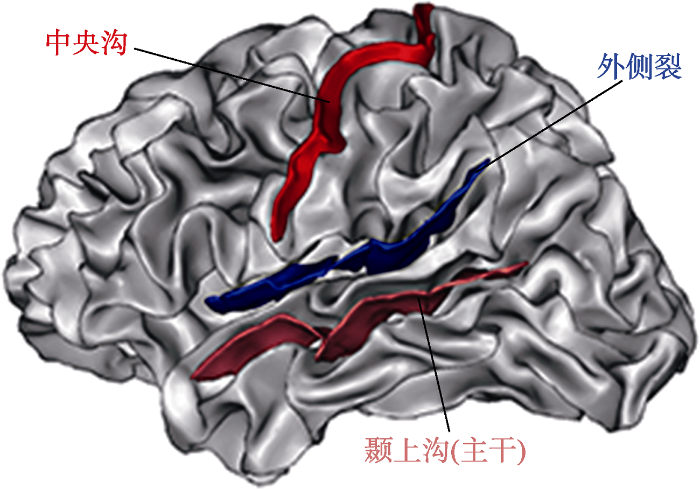
图1
个体左半球的三条主要脑沟, 分别为中央沟(上)、外侧裂(中)、颞上沟(下)。
注:脑沟呈现在white surface上; 图中隐藏了其他脑沟; 颞上沟仅有主干部分, 未显示其分支。彩图见电子版。
目标脑沟形态的定量特征。脑沟曲面由外部边界顶脊(top ridge), 内部边界底脊(bottom ridge)和面(surface)组成, 其中“面”由相对的脑回“河床” (gyral banks)之间确定。顶脊和底脊的交叉点为面的终结点, 以顶脊和底脊包围的面重构脑沟曲面。脑沟的表面积为脑沟曲面中“面”的面积。进一步地, 脑沟曲面的线性测度:长度和深度, 分别由温度扩散模型(heat diffusion model)量化(Kochunov et al., 2010)。脑沟长度定义为沿着脑沟曲面排列的100段等温曲线长度的均值, 方向与脑沟底脊和顶脊共线; 脑沟深度的定义为沿脑沟曲面排列的100段曲线的均值, 方向垂直于底脊和顶脊的连线。
目标脑沟的定性特征。脑沟空间组织(相对位置和相对方向等)分析通常包括中断情况、脑沟之间是否相互连接, 脑沟末端的走向等。研究者要求多个评估者分别利用相同的编码表评估每个被试的脑沟形态, 以评估的一致性程度确定脑沟的空间组织特征。以中断为例, Borst等人(2016)让3位研究者分别使用二分变量编码(dichotomous variable code)评估儿童的颞枕沟(occipitotemporal sulcus, OTS)类型, 评估者之间的一致性达到100%即可确定个体的颞枕沟类型为中断或连续。目标脑沟的定性分析还应用于威廉姆斯综合症(Williams Syndrome, WS)患者的研究, 威廉姆斯综合征患者右侧颞平面显著大于控制组, 其原因是患者右半球外侧裂水平延伸而不是上升到顶叶(Eckert et al., 2006); 还有研究发现颞上沟(superior temporal sulcus, STS)的中断数量存在左右半球的差异, 这种结构不对称可能和语言偏侧化相关(Ochiai et al., 2004)。
2.3 脑沟模式分析
脑回指数和脑沟指数能够宏观量化脑沟复杂程度, 目标脑沟分析旨在对特定脑沟形态进行描绘, 这两种方法都无法量化多个脑沟的空间位置和排列关系。因此研究者提出了脑沟模式(sulcal pattern)的概念, 即脑沟的拓扑特征。
Lohmann, von Cramon和Colchester (2008)分析了脑沟深部的特点, 发现脑沟深部比浅层表现出较少的个体差异。每条脑沟沿其底部通常都能够发现界限清楚的最大深度区域脑沟凹陷(sulcal pit)。Im等人(2010)发展出了基于脑沟凹陷建构脑沟模式的方法, 使用流域算法(watershed algorithm), 将皮层分割为多个盆地(basin), 修剪较浅和较小的盆地之后, 每个盆地的最深处被标记为脑沟凹陷, 每个脑沟凹陷在图形表征中作为一个节点(node), 如果脑沟盆地相邻则使用无方向的边(edge)相连, 从而在选定的区域内得到脑沟图形(sulcal graph)。脑沟图形中主要包含节点的几何特征(三维空间位置和脑沟盆地面积), 节点之间的几何关系(反映脑沟之间的关系)以及节点的数量和节点间的路径(图形间的拓扑特征)。
研究者发现同卵双生子的脑沟模式相似程度显著高于样本内随机两个个体间的脑沟模式(Im et al., 2011), 表明脑沟模式与遗传因素之间存在密切联系; 此外, 在多小脑回(Polymicrogyria, PMG)患者中, 完整语言能力患者和语言能力损伤患者相比, 顶叶脑沟模式与正常控制组更相似(Im, Pienaar et al., 2013); 还有研究报告了发展性阅读障碍儿童和家族风险儿童与其控制组相比, 枕颞区域(parieto-temporal region)及顶颞区域(occipito- temporal region)脑沟模式出现异常(Im, Raschle, Smith, Ellen Grant, & Gaab, 2016)。
3 脑沟形态分析在语言认知领域的研究进展
3.1 脑沟形态的早期发展轨迹
儿童语言的早期发展为后期阅读乃至社会认知的发展奠定了基础(Catts, Fey, Ellis Weismer, & Bridges, 2014)。揭示语言相关脑结构的早期发展轨迹能够帮助我们深入理解人类语言发展的底层神经机制。作为衡量大脑早期发育的重要指标, 脑沟形态分析在早期语言发展中的应用受到研究者关注(Kersbergen et al., 2016)。
脑沟和脑回的形成能够在有限空间内尽可能扩展皮层表面积(Striedter, Srinivasan, & Monuki, 2015)。妊娠的3个月末期, 神经元开始最后的迁移并形成连接, 与此同时, 平滑的皮层表面开始形成复杂的脑沟和脑回。初级脑沟(primary sulci)很快延长和分支并形成二级(secondary sulci)和三级脑沟(tertiary sulci), 最终出生时皮层表面形成类似成人的复杂形态(Tallinen et al., 2016)。
研究者采用不同的方法描绘了18孕周到2岁时期脑沟形态的发展过程, 并在外侧裂周区发现了显著的皮层变化。Kasprian等人(2011)采集了197名宫内胎儿(18~37孕周)的MR图像, 基于二维MR图像的冠状面, 描绘每个胎儿大脑的轮廓线, 以轮廓线变异代表脑沟的复杂程度。结果发现超过2/3的胎儿表现出更大的左侧颞叶, 94%的胎儿右侧颞上沟(23孕周出现)比左侧颞上沟(25孕周出现)出现的更早, 并且右侧深于左侧。Dubois, Benders和Borradori-Tolsa等人(2008)从35名早产儿(26~36孕周)MR图像中重构了皮层(surface), 并定义脑沟指数用于量化分析个体脑沟形态的复杂程度, 研究者发现额叶、颞叶等不同区域的脑沟指数存在差异, 而右半球比左半球更早的表现出复杂的脑沟形态。Habas等人(2012)基于皮层局部曲率, 描绘了宫内胎儿的脑沟形态发展。结果表明主要脑沟的出现时间存在差异, 外侧裂(Sylvian fissure)最早出现曲率变化; 颞上沟在24周左右显现, 而右侧颞上沟比左侧提早1周出现。出生后2年, 大脑经历了发展最快的阶段, 体积和表面积迅速增加(Gilmore et al., 2007)。Li等人(2014)追踪了出生到2岁婴儿的脑沟形态变化, 发现从出生到2岁颞上沟和顶枕沟右侧深度大于左侧, 而颞平面的大小和深度左侧大于右侧的现象。此外, 研究者还发现颞上沟和颞平面在成人和新生儿群体间表现出类似的偏侧化模式(Hill et al., 2010)。
婴儿大脑是目前唯一能够精通语言的“机器” (Dehaene-Lambertz & Spelke, 2015), 早期脑沟形态的发展轨迹与语言能力的关系主要表现在外侧裂周区(peri-sylvian region)的形态改变。事实上, 30孕周左右的早产儿已经具有区分音节的能力(Mahmoudzadeh, Wallois, Kongolo, Goudjil, & Dehaene-Lambertz, 2017), 有研究发现早产儿等价足月出生时期(term-equivalent age, TEA)的外侧裂形态与其2岁时的接受性语言有关(Kersbergen et al., 2016), 这些证据表明胎儿时期外侧裂周区的脑沟形态变化可能与婴儿的语言获得和发展存在密切联系。此外, 外侧裂周区中, 颞叶脑沟形态变异较大(Leroy et al., 2011), 并且颞上沟在不同研究中报告了一致的发展进程, 这种结构变化可能支持了认知功能的发展。从出生后的语言功能来说, 婴儿和成人的颞叶皮层对于语音刺激表现出相似的功能激活(Dehaene-Lambertz, 2017; Shultz, Vouloumanos, Bennett, & Pelphrey, 2014), 因此颞叶尤其是颞上沟的早期形态变化可能对于语言能力有重要作用。
3.2 脑沟形态与语言偏侧化
人类最明显的大脑功能偏侧化为语言功能的偏侧化(Sun & Walsh, 2006), 一直以来, 研究者试图找到人类大脑独特的结构特征, 以解释人类的认知成就。颞平面(planum temporale, PT)和言语功能密切相关, 有研究报告了颞平面表面积的偏侧化现象(Lyttelton et al., 2009), 但并非所有研究都发现了这种偏侧化特征(Dorsaint-Pierre et al., 2006; Keller, Crow, Foundas, Amunts, & Roberts, 2009)并且灵长类动物中也报告了相似的现象(Hopkins & Nir, 2010)。随着量化方法的提出, 研究者尝试寻找人类独特的脑沟偏侧化特征。
语言偏侧化的脑沟形态研究集中在脑沟凹陷上。脑沟凹陷是脑沟局部最深处, 被认为是大脑中形态变化的起始点, 并且其分布在个体间相对稳定(Lohmann, von Cramon, & Colchester, 2008; Régis et al., 2005)。Im等人(2010)从148名成年中提取了脑沟凹陷, 分别在左右半球发现了48个和47个脑沟凹陷团块(clusters), 脑沟凹陷的密度和空间分布表现出较小的个体差异, 并在颞上沟表现出数量和空间分布的半球间偏侧化现象。Meng, Li, Lin, Gilmore和Shen (2014) 进一步追踪了0~2岁婴儿的脑沟凹陷发展, 发现了类似成人的脑沟凹陷分布, 并且这种分布在出生后两年内保持相对稳定。研究者还发现, 婴儿和成人颞上沟的脑沟凹陷分布具有一致的偏侧化模式(Auzias, Brun, Deruelle, & Coulon, 2015; Im et al., 2010)。Leroy等人(2015)通过分析不同年龄人类和黑猩猩的MR图像, 进一步确定了人脑特有的左右不对称区域(右侧显著深于左侧), 将其命名为颞上不对称凹陷(superior temporal asymmetrical pit, STAP)。
颞上沟深度的偏侧化可能为语言能力的偏侧化提供支持。首先, 颞上沟本身对语言能力有着重要作用, 左半球颞上沟特别地对言语刺激敏感(DeWitt & Rauschecker, 2012), 而发展进程上, 左侧颞上沟比右侧更晚出现, 这种左侧发育延迟, 可能为处理语言和运动刺激提供了更充分的结构可塑性(Dubois, Benders, Cachia et al., 2008)。不仅如此, 皮层下白质连接可能是脑沟形成的原因之一(Xu et al., 2010), 而颞叶皮层下存在复杂的白质纤维连接, 其中之一的弓形束(arcuate fasciculus, AF)与颞上不对称凹陷在发展中表现出相似的特征:第一, 人类的弓形束和颞上不对称凹陷远大于黑猩猩; 第二, 两者在生命早期均出现了偏侧化现象(Dubois et al., 2009)。因此颞上不对称凹陷可能与皮层下的白质纤维密度以及偏侧化发展有关。鉴于弓形束在语音加工(Vandermosten, Boets, Wouters, & Ghesquière, 2012)和词汇阅读(Gullick & Booth, 2015)中的重要作用, 颞上不对称凹陷很可能和语言认知能力密切相关。尽管颞上不对称凹陷的功能尚不明确, 但其处于言语腹侧通路音-义对应的核心区域(Liebenthal et al., 2010; Striem- Amit, Hertz, & Amedi, 2011), 并且黑猩猩中未发现这一结构, 因此研究者推测与颞平面相比, 颞上不对称凹陷可能是更加人类特异的偏侧化结构。
脑沟凹陷表现出了显著偏侧化现象和高遗传度(Takerkart, Auzias, Brun, & Coulon, 2017), 为进一步探索大脑结构偏侧化与语言功能偏侧化之间的关系提供新的形态学测度。同时, 脑沟凹陷也是可靠的解剖标志(anatomical landmark) (Li, Guo, Nie, & Liu, 2010), 个体MR图像中提取的脑沟凹陷在不同扫描仪、扫描序列和皮层重构工具之间具有很高的稳定性(Im, Lee et al., 2013)。因此, 研究者能够基于脑沟凹陷建立脑沟模式, 进一步分析脑沟拓扑特征与认知能力之间的关系。
3.3 脑沟形态与发展性阅读障碍
阅读是语言认知研究最重要的方面之一, 阅读能力是儿童多项能力发展的基石。发展性阅读障碍(Developmental Dyslexia, DD)是学龄期儿童常见的一种学习障碍, 通常为智力、感知能力以及动机正常并且接受了充分教育的个体, 在准确流利识别字词或拼写时出现困难(Peterson & Pennington, 2012)。发展性阅读障碍的病因学基础仍处于争论之中, 有研究认为风险基因和异常的神经发育导致发展性阅读障碍的发生(Skeide et al., 2016), 而另一些研究发现发展性阅读障碍大脑的功能和结构异常来源于缺乏阅读经验(Clark et al., 2014; Olulade, Napoliello, & Eden, 2013)。
为了解决这一争论, 一种方法是加入新对照组(Xia, Hoeft, Zhang, & Shu, 2016), 通过比较阅读障碍组、年龄控制组和阅读水平控制组来解释DD脑结构异常的原因; 另一种方法是招募学前儿童(Raschle et al., 2017; Raschle, Stering, Meissner, & Gaab, 2014; Vandermosten et al., 2015; Wang et al., 2017)。学前儿童尚未接受系统的阅读训练, 通过比较阅读障碍家族风险儿童和年龄控制组, 能够发现相对独立于阅读经验的大脑功能和结构异常。Raschle, Chang和Gaab (2011), Raschle等人(2017)报告了学前DD风险儿童, 枕颞区域和颞顶区域灰质体积的下降, 这表明DD的大脑结构异常可能在学习阅读之前已经存在。
最新一项研究中, Im等人(2016)对DD儿童及其年龄控制组和学前DD风险儿童及其年龄控制组的脑沟模式进行了比较。研究者基于枕颞和顶颞区域的脑沟凹陷建立脑沟图形, 每个正常被试与其他正常被试之间计算一个平均相似度${{{S}'}_{T}}(i)$, 每个DD和所有正常被试计算一个平均相似度${{{S}'}_{T,D}}(i)$, 通过比较${{{S}'}_{T}}$和${{{S}'}_{T,D}}$可以得到DD与正常控制组的组间差异(${{{S}'}_{T}}$vs.${{{S}'}_{T,D}}$)。同理可以比较风险儿童和非风险儿童之间的相似度(${{{S}'}_{F-F+}}$vs. ${{{S}'}_{F-}}$)。结果表明DD儿童与正常儿童相比以及学前风险儿童和非风险儿童相比, 均表现出脑沟模式的显著差异; 脑沟模式的跨年龄比较中, 非风险儿童和学后正常儿童表现出了较高一致性, 而学前风险儿童和DD表现出了较高的一致性。脑沟相似度和行为成绩的相关表明, DD脑沟模式与控制组相似度越高, 其词汇和语言加工测验的成绩越好。
鉴于脑沟模式出生时已经基本形成, 阅读障碍儿童、风险儿童和其对应控制组的脑沟模式差异可能反映了早期大脑结构存在的异常, 甚至可能追溯到胎儿发育时期。研究者推测异常的脑沟模式可能受到异常遗传因素和皮层下白质连接的影响。首先, DD风险基因(如DCDC2, KIAA0319)等对于大脑发展尤其是神经元迁移有着重要作用(Peterson & Pennington, 2012), 神经元的迁移是脑沟形成的影响因素之一(Striedter et al., 2015), 发育早期风险遗传因素可能会通过影响神经元迁移, 进而导致脑沟模式的异常。其次, 皮层下白质纤维束的密度和张力可能驱动了脑沟形成(Zilles, Palomero-Gallagher, & Amunts, 2013)。枕颞皮层的视觉词汇加工区(visual word form area, VWFA)对于阅读有着重要作用(Bouhali et al., 2014; Dehaene, Cohen, Morais, & Kolinsky, 2015), 而有证据表明阅读学习之前的白质纤维连接支持了VWFA的产生(Saygin et al., 2016), 因此, 学前风险儿童枕颞脑沟模式的差异可能反映了发展早期白质连接的异常。近期Williams, Juranek, Cirino和Fletcher (2017)报告了DD左侧枕颞区域脑回指数的增加, 研究者推测增加的皮层复杂程度可能反映了皮层局部信息交换能力的提升, 但同时以长距离连接(如下额枕束等)减弱为代价, 从而影响了DD的阅读能力。
正常群体的研究同样在枕颞区域发现了脑沟形态与阅读能力之间存在关联。左侧枕颞沟的空间分布和阅读准确性相关(Cachia et al., 2017), 而脑沟的空间分布被认为在出生前已经形成并且较少受到出生后神经可塑性的影响(Cachia et al., 2016), 研究者推测左侧枕颞沟和阅读能力的关系可能反映了皮层细胞构筑(Cytoarchitecture)和皮层下白质纤维连接对于阅读能力的影响(Dubois et al., 2016; Weiner et al., 2017)。
总之, 异常群体和正常群体的脑沟形态研究为探讨早期大脑发育与语言认知能力之间的关系提供了初步证据。同时, 脑沟的发生发展机制与编码蛋白质的基因、微核糖核酸(microRNA)等因素密切联系(Sun & Hevner, 2014), 为探讨语言发展及障碍的遗传学机制提供了新的中介表型。
4 总结与展望
Klee和Stokes (2011)提出的语言发展模型中包含了遗传因素、环境刺激、神经生物基础、认知机制等多种成分和过程, 语言的发展并非单一因素决定而受到多种过程的影响。脑沟形态为研究者提供了更加精细的神经生物学指标, 已有研究表明其在语言认知研究中的应用潜力:首先, 早期脑沟形态变化显著, 能够作为发育早期的神经影像学指标, 探讨语言认知能力与大脑结构发展及遗传因素的关系(Sun et al., 2005); 其次, 脑沟凹陷的偏侧化可能提供了语言功能偏侧化的结构基础, 支持了语言认知理论的模型; 最后, 脑沟模式及脑沟的空间分布较少受到出生后神经可塑性的影响, 为揭示发展性阅读障碍等认知缺陷的病因学基础提供了更接近遗传因素的形态学测度。
事实上, 脑沟形态分析在神经和精神疾病中已经有了广泛的应用, 如孤独症(Libero, DeRamus, Deshpande, & Kana, 2014; Wallace et al., 2013), 威廉姆斯综合征(Fahim et al., 2012), 强迫症(Shim et al., 2009), 口吃(Cykowski et al., 2008), 阿尔兹海默症(Hamelin et al., 2015)等。这些脑沟形态异常通常与遗传和神经发育的扰动有关, 反映了疾病早期的病理学特征, 为探讨神经及精神疾病的病因学基础, 关联认知损伤与大脑结构异常以及预测疾病发生发展提供了早期生物标志。
相比较而言, 脑沟形态在语言认知领域内的应用正处于起步阶段, 仍有很多问题有待进一步研究。首先, 脑沟形态与遗传因素的关系。相关问题包括, 语言相关的遗传因素影响了哪些脑沟形态特征, 及早期脑沟形态的偏侧化是否与这些基因表达有关等。第二, 脑沟形态与语言等认知能力之间的关系。脑沟本身并不包含神经元, 也不承担特定认知功能, 因此, 研究者面临相继的两个问题, 首先, 脑沟形态是否与语言认知能力相关; 其次, 假设相关存在, 脑沟形态反映了何种生理过程进而影响语言认知能力。这些问题需要研究者基于脑沟的发生发展机制及细胞构筑等研究谨慎推测。第三, 脑沟形态与后天发展和环境。大脑在出生后仍不断发展, 儿童及青少年时期是个体认知能力发展的关键时期, 这一时期语言发展和脑沟形态发展轨迹之间存在何种关系尚不明确, 而成熟过程和后天环境因素如何塑造脑沟形态仍有待研究。阐明脑沟形态与遗传、认知和环境三者的相互作用, 有助于研究者深入了解语言发展认知神经机制。此外, 语言认知研究应考虑到文化差异的影响, 来自不同国家地区的大样本研究有助于发现语言能力和脑沟形态的关联, 抑或探讨不同文化背景下群体之间可能存在的差异。最后, 脑沟形态分析方法的优化能够提供更加丰富神经影像指标, 为揭示发展性阅读障碍的病因学基础, 探索遗传因素与语言发展及语言障碍之间的关系, 并进一步为建立发展性阅读障碍等语言障碍的早期诊断模型提供支持。
参考文献
The ontogeny of human gyrification
DOI:10.1093/cercor/5.1.56 URL [本文引用: 1]
Voxel-based morphometry—the methods
DOI:10.1006/nimg.2000.0582 URL [本文引用: 1]
Deep sulcal landmarks: Algorithmic and conceptual improvements in the definition and extraction of sulcal pits
DOI:10.1016/j.neuroimage.2015.02.008
URL
PMID:25676916
[本文引用: 1]

On top of these methodological advances, we quantify the relationship between the reproducibility of the number of sulcal pits per region across individuals and their respective depth and demonstrate the relatively high reproducibility of several pits corresponding to shallower folds. Finally, we report new results regarding the local pit asymmetry, providing evidence that the algorithmic and conceptual approach defended here may contribute to better understanding of the key role of sulcal pits in neuroanatomy.
Early cerebral constraints on reading skills in school-age children: An MRI study
DOI:10.1111/mbe.12098
URL
[本文引用: 1]
ABSTRACT Reading relies on a left-lateralized network of brain areas that include the pre-lexical processing regions of the ventral stream. Specifically, a region in the left lateral occipitotemporal sulcus (OTS) is consistently more activated for visual presentations of words than for other categories of stimuli. This region undergoes dramatic changes at the functional and structural levels when children learn to read, but little is known about the effects of early cerebral constraints on reading skills. Using anatomical magnetic resonance imaging, we investigated whether the sulcal pattern of the lateral OTS—a stable brain feature—was associated with oral reading skills. The sulcal pattern of the left but not the right lateral OTS was associated with the number of words correctly read in 3 min. This study is the first to evidence that reading is affected by early cerebral constraints, such as the sulcal morphology of the left lateral OTS.
Anatomical connections of the visual word form area
DOI:10.1523/JNEUROSCI.4918-13.2014
URL
PMID:25392507
[本文引用: 1]
Abstract The visual word form area (VWFA), a region systematically involved in the identification of written words, occupies a reproducible location in the left occipitotemporal sulcus in expert readers of all cultures. Such a reproducible localization is paradoxical, given that reading is a recent invention that could not have influenced the genetic evolution of the cortex. Here, we test the hypothesis that the VWFA recycles a region of the ventral visual cortex that shows a high degree of anatomical connectivity to perisylvian language areas, thus providing an efficient circuit for both grapheme-phoneme conversion and lexical access. In two distinct experiments, using high-resolution diffusion-weighted data from 75 human subjects, we show that (1) the VWFA, compared with the fusiform face area, shows higher connectivity to left-hemispheric perisylvian superior temporal, anterior temporal and inferior frontal areas; (2) on a posterior-to-anterior axis, its localization within the left occipitotemporal sulcus maps onto a peak of connectivity with language areas, with slightly distinct subregions showing preferential projections to areas respectively involved in grapheme-phoneme conversion and lexical access. In agreement with functional data on the VWFA in blind subjects, the results suggest that connectivity to language areas, over and above visual factors, may be the primary determinant of VWFA localization. Copyright 2014 the authors 0270-6474/14/3415402-13$15.00/0.
Longitudinal stability of the folding pattern of the anterior cingulate cortex during development
DOI:10.1016/j.dcn.2016.02.011
URL
PMID:26974743
[本文引用: 2]
Abstract Prenatal processes are likely critical for the differences in cognitive ability and disease risk that unfold in postnatal life. Prenatally established cortical folding patterns are increasingly studied as an adult proxy for earlier development events - under the as yet untested assumption that an individual's folding pattern is developmentally fixed. Here, we provide the first empirical test of this stability assumption using 263 longitudinally-acquired structural MRI brain scans from 75 typically developing individuals spanning ages 7 to 32 years. We focus on the anterior cingulate cortex (ACC) - an intensely studied cortical region that presents two qualitatively distinct and reliably classifiable sulcal patterns with links to postnatal behavior. We show - without exception-that individual ACC sulcal patterns are fixed from childhood to adulthood, at the same time that quantitative anatomical ACC metrics are undergoing profound developmental change. Our findings buttress use of folding typology as a postnatally-stable marker for linking variations in early brain development to later neurocognitive outcomes in ex utero life. Copyright 2016 The Authors. Published by Elsevier Ltd.. All rights reserved.
Cortical folding abnormalities in schizophrenia patients with resistant auditory hallucinations
DOI:10.1016/j.neuroimage.2007.08.049
URL
PMID:17988891
[本文引用: 2]
Gray matter volume and functional abnormalities have been reported in language-related cortex in schizophrenia patients with auditory hallucinations. Such abnormalities might denote abnormal cortical folding development, which can now be investigated using gyrification measures. Anatomic magnetic resonance images (MRIs) were obtained from 30 schizophrenia patients screened for resistant auditory hallucinations and 28 control subjects. We searched for overall gyrification abnormalities in the whole cortex as well as localized abnormalities in language-related cortex, assuming that gyrification is associated with brain sulcation. A fully automated method was applied to MRIs to extract, label and measure the sulcus area in the whole cortex. Gyrification was assessed using both global and local sulcal indices, respectively the ratio between total sulcal area, or area of each labeled sulcus, and outer cortex area. For both hemispheres, the patients had a lower global sulcal index. The local sulcal index decrease was not homogeneous across the whole cortex. It was more significant in the superior temporal sulcus bilaterally, in the left middle frontal sulcus and in the diagonal branch of left sylvian fissure (Broca's area). Findings suggest abnormalities in cortical gyrification in these patients. Sulcal abnormalities in language-related cortex might underlie these patients' particular vulnerability to hallucinations.
How interindividual differences in brain anatomy shape reading accuracy
DOI:10.1007/s00429-017-1516-x
URL
PMID:28916842
[本文引用: 1]
Abstract The capacity to read develops throughout intensive academic learning and training. Several studies have investigated the impact of reading on the brain, and particularly how the anatomy of the brain changes with reading acquisition. In the present study, we investigated the converse issue, namely whether and how reading acquisition is constrained by the anatomy of the brain. Using multimodal MRI, we found that (a) the pattern (continuous or interrupted sulcus) of the posterior part of the left lateral occipito-temporal sulcus (OTS) hosting the visual word form area (VWFA) predicts reading skills in adults; that (b) this effect is modulated by the age of reading acquisition; and that (c) the length of the OTS sulcal interruption is associated with reading skills. Because the sulcal pattern is determined in utero, our findings suggest that individual difference in reading skills can be traced back to early stages of brain development in addition to the well-established socioeconomic and educational factors.
Neuroanatomical precursors of dyslexia identified from pre-reading through to age 11
DOI:10.1093/brain/awu229
URL
PMID:25125610
[本文引用: 1]
See Goswami (doi:10.1093/brain/awu296) for a scientific commentary on this article.Clark et al. present longitudinal MRI data from children at high risk of dyslexia, from before reading instruction began until after dyslexia was diagnosed. Prior to learning to read, children with dyslexia have thinner cortex in visual and auditory processing areas than controls, whereas the 0904reading network0909 itself is unaffected.Developmental dyslexia is a common reading disorder that negatively impacts an individual0964s ability to achieve literacy. Although the brain network involved in reading and its dysfunction in dyslexia has been well studied, it is unknown whether dyslexia is caused by structural abnormalities in the reading network itself or in the lower-level networks that provide input to the reading network. In this study, we acquired structural magnetic resonance imaging scans longitudinally from 27 Norwegian children from before formal literacy training began until after dyslexia was diagnosed. Thus, we were able to determine that the primary neuroanatomical abnormalities that precede dyslexia are not in the reading network itself, but rather in lower-level areas responsible for auditory and visual processing and core executive functions. Abnormalities in the reading network itself were only observed at age 11, after children had learned how to read. The findings suggest that abnormalities in the reading network are the consequence of having different reading experiences, rather than dyslexia per se, whereas the neuroanatomical precursors are predominantly in primary sensory cortices.
Perisylvian sulcal morphology and cerebral asymmetry patterns in adults who stutter
DOI:10.1093/cercor/bhm093
URL
PMID:17584852
[本文引用: 1]
Previous investigations of cerebral anatomy in persistent developmental stutterers have reported bilateral anomalies in the perisylvian region and atypical patterns of cerebral asymmetry. In this study, perisylvian sulcal patterns were analyzed to compare subjects with persistent developmental stuttering (PDS) and an age-, hand-, and gender-matched control group. This analysis was accomplished using software designed for 3-dimensional sulcal identification and extraction. Patterns of cerebral asymmetry were also investigated with standard planimetric measurements. PDS subjects showed a small but significant increase in both the number of sulci connecting with the second segment of the right Sylvian fissure and in the number of suprasylvian gyral banks (of sulci) along this segment. No differences were seen in the left perisylvian region for either sulcal number or gyral bank number. Measurements of asymmetry revealed typical patterns of cerebral asymmetry in both groups with no significant differences in frontal and occipital width asymmetry, frontal and occipital pole asymmetry, or planum temporale and Sylvian fissure asymmetries. The subtle difference in cortical folding of the right perisylvian region observed in PDS subjects may correlate with functional imaging studies that have reported increased right-hemisphere activity during stuttered speech.
Illiterate to literate: Behavioural and cerebral changes induced by reading acquisition
DOI:10.1038/nrn3924
URL
PMID:25783611
[本文引用: 2]
Abstract The acquisition of literacy transforms the human brain. By reviewing studies of illiterate subjects, we propose specific hypotheses on how the functions of core brain systems are partially reoriented or 'recycled' when learning to read. Literacy acquisition improves early visual processing and reorganizes the ventral occipito-temporal pathway: responses to written characters are increased in the left occipito-temporal sulcus, whereas responses to faces shift towards the right hemisphere. Literacy also modifies phonological coding and strengthens the functional and anatomical link between phonemic and graphemic representations. Literacy acquisition therefore provides a remarkable example of how the brain reorganizes to accommodate a novel cultural skill.
The human infant brain: A neural architecture able to learn language
DOI:10.3758/s13423-016-1156-9
URL
PMID:28120318
[本文引用: 1]
Abstract To understand the type of neural computations that may explain how human infants acquire their native language in only a few months, the study of their neural architecture is necessary. The development of brain imaging techniques has opened the possibilities of studying human infants without discomfort, and although these studies are still sparse, several characteristics are noticeable in the human infant's brain: first, parallel and hierarchical processing pathways are observed before intense exposure to speech with an efficient temporal coding in the left hemisphere and, second, frontal regions are involved from the start in infants' cognition. These observations are certainly not sufficient to explain language acquisition but illustrate a new approach that relies on a better description of infants' brain activity during linguistic tasks, which is compared to results in animals and human adults to clarify the neural bases of language in humans.
The infancy of the human brain
DOI:10.1016/j.neuron.2015.09.026
URL
PMID:26447575
The human infant brain is the only known machine able to master a natural language and develop explicit, symbolic, and communicable systems of knowledge that deliver rich representations of the external world. With the emergence of noninvasive brain imaging, we now have access to the unique neural machinery underlying these early accomplishments. After describing early cognitive capacities in the domains of language and number, we review recent findings that underline the strong continuity between human infants’ and adults’ neural architecture, with notably early hemispheric asymmetries and involvement of frontal areas. Studies of the strengths and limitations of early learning, and of brain dynamics in relation to regional maturational stages, promise to yield a better understanding of the sources of human cognitive achievements.
Phoneme and word recognition in the auditory ventral stream
DOI:10.1073/pnas.1113427109 URL [本文引用: 1]
Asymmetries of the planum temporale and Heschl's gyrus: Relationship to language lateralization
DOI:10.1093/brain/awl055
URL
PMID:16537567
[本文引用: 1]
Morphological asymmetries favouring the left hemisphere in the planum temporale (PT) and Heschl's gyrus (HG) have both been presumed to relate to the typical left-hemisphere dominance for language functions. However, a direct link between structure and function has not been clearly established. The present study investigates this issue by measuring the volume of the PT and HG on the MRI scans of epilepsy patients classified into three groups: left speech group (LSG; n = 20), right speech group (RSG; n = 11) and bilateral speech group (BSG; n = 13), as assessed by the intracarotid Sodium Amytal procedure. Additionally, an automatic voxel-based morphometry (VBM) analysis was performed to explore collateral structural asymmetries. Although leftward structural asymmetries were found in the PT, consistent with the literature, they did not relate to language lateralization. For HG we also replicated asymmetries favouring the left side; interestingly, three of the individuals within the RSG showed a strongly reversed asymmetry, but as a whole the structure-function relationship for HG was not obligatory. The VBM analysis revealed a grey-matter concentration difference in the posterior part of the inferior frontal gyrus (pars opercularis, corresponding functionally to Broca's area), which favoured the left hemisphere in the LSG, and the right hemisphere in the RSG. The findings suggest that this frontal cortical region bears a direct relationship to language lateralization, which may be related to use-dependent plasticity in patients with language reorganization.
Primary cortical folding in the human newborn: An early marker of later functional development
DOI:10.1093/brain/awn137
URL
PMID:2724902
[本文引用: 2]
In the brain, the morphology of cortical gyri and sulci is complex and variable among individuals, and it may reflect pathological functioning with specific abnormalities observed in certain developmental and neuropsychiatric disorders. Since cortical folding occurs early during , these structural abnormalities might be present long before the appearance of functional symptoms. So far, the precise mechanisms responsible for such alteration in the convolution pattern during intra-uterine or post-natal are still poorly understood. Here we compared anatomical and functional in vivo among 45 premature newborns who experienced different intra-uterine environments: 22 normal singletons, 12 twins and 11 newborns with intrauterine growth restriction (IUGR). Using magnetic resonance imaging (MRI) and dedicated post-processing tools, we investigated early disturbances in cortical formation at birth, over the developmental period critical for the emergence of convolutions (26-36 weeks of gestational age), and defined early 'endophenotypes' of sulcal . We demonstrated that twins have a delayed but harmonious maturation, with reduced surface and sulcation index compared to singletons, whereas the gyrification of IUGR newborns is discordant to the normal developmental trajectory, with a more pronounced reduction of surface in relation to the sulcation index compared to normal newborns. Furthermore, we showed that these structural measurements of the brain at birth are predictors of infants' outcome at term equivalent age, for MRI-based cerebral volumes and neurobehavioural evaluated with the assessment of 's ().
Mapping the early cortical folding process in the preterm newborn brain
DOI:10.1093/cercor/bhm180
URL
PMID:17934189
In the developing brain, the cortical sulci formation is a complex process starting from 14 weeks of onward. The potential influence of underlying mechanisms (genetic, epigenetic, mechanical or environmental) is still poorly understood, because reliable quantification in vivo of the early folding is lacking. In this study, we investigate the sulcal emergence noninvasively in 35 preterm newborns, by applying dedicated postprocessing tools to magnetic resonance images acquired shortly after birth over a developmental period critical for the cortex maturation (26-36 weeks of age). Through the original three-dimensional reconstruction of the interface between developing cortex and white matter and correlation with volumetric measurements, we document early sulcation in vivo, and quantify changes with age, gender, and the presence of matter lesions. We observe a trend towards lower cortical surface, smaller cortex, and white matter volumes, but equivalent sulcation in females compared with males. By precisely mapping the sulci, we highlight interindividual variability in time appearance and interhemispherical asymmetries, with a larger right superior temporal sulcus than the left. Thus, such an approach, included in a longitudinal follow-up, may provide early indicators on the structural basis of cortical functional specialization and abnormalities induced by genetic and environmental factors.
Structural asymmetries in the infant language and sensori-motor networks
DOI:10.1093/cercor/bhn097
URL
PMID:18562332
[本文引用: 1]
Abstract Both language capacity and strongly lateralized hand preference are among the most intriguing particularities of the human species. They are associated in the adult brain with functional and anatomical hemispheric asymmetries in the speech perception-production network and in the sensori-motor system. Only studies in early life can help us to understand how such asymmetries arise during brain development, and to which point structural left-right differences are the source or the consequence of functional lateralization. In this study, we aimed to provide new in vivo structural markers of hemispheric asymmetries in infants from 1 to 4 months of age, with diffusion tensor imaging. We used 3 complementary analysis methods based on local diffusion indices and spatial localizations of tracts. After a prospective approach over the whole brain, we demonstrated early leftward asymmetries in the arcuate fasciculus and in the cortico-spinal tract. These results suggest that the early macroscopic geometry, microscopic organization, and maturation of these white matter bundles are related to the development of later functional lateralization.
Exploring the early organization and maturation of linguistic pathways in the human infant brain
DOI:10.1093/cercor/bhv082
URL
PMID:25924951
[本文引用: 2]
Linguistic processing is based on a close collaboration between temporal and frontal regions connected by two pathways: the "dorsal" and "ventral pathways" (assumed to support phonological and semantic processing, respectively, in adults). We investigated here the development of these pathways at the onset of language acquisition, during the first post-natal weeks, using cross-sectional diffusion imaging in 21 healthy infants (6-22 weeks of age) and 17 young adults. We compared the bundle organization and microstructure at these two ages using tractography and original clustering analyses of diffusion tensor imaging parameters. We observed structural similarities between both groups, especially concerning the dorsal/ventral pathway segregation and the arcuate fasciculus asymmetry. We further highlighted the developmental tempos of the linguistic bundles: The ventral pathway maturation was more advanced than the dorsal pathway maturation, but the latter catches up during the first post-natal months. Its fast development during this period might relate to the learning of speech cross-modal representations and to the first combinatorial analyses of the speech input.
Anomalous sylvian fissure morphology in Williams syndrome
DOI:10.1016/j.neuroimage.2006.05.062
URL
PMID:16876437
[本文引用: 1]
The unusual sensitivity and attraction to auditory stimuli in people with Williams syndrome (WS) has been hypothesized to be the consequence of atypical development of brain regions surrounding the Sylvian fissure. Planum temporale surface area, which is determined in part by Sylvian fissure patterning, was examined in 42 WS and 40 control participants to determine if anomalous Sylvian fissure morphology is present in WS. WS participants had significantly reduced leftward asymmetry of the planum temporale compared to control participants, due to a significant expansion in the size of the right planum temporale. The increased right planum temporale size was largely due to WS participants (24%) who had a right hemisphere Sylvian fissure that coursed horizontally and failed to ascend into the parietal lobe. This sulcal pattern is unusual in the right hemisphere and is more commonly found in the left hemisphere of typically developing individuals. There were no control participants with this type of right hemisphere Sylvian fissure pattern. The right hemisphere Sylvian fissure sulcal patterns were also related to a measure of cortical complexity and the amount of right hemisphere occipital lobe volume, suggesting that intrinsic genetic influences leading to anomalous visual system development in WS have widespread influences on cortical morphology that are similar in manner to extrinsic embryonic visual system lesions.
Williams syndrome: A relationship between genetics, brain morphology and behaviour
DOI:10.1111/j.1365-2788.2011.01490.x
URL
PMID:22044458
[本文引用: 2]
Abstract BACKGROUND: Genetically Williams syndrome (WS) promises to provide essential insight into the pathophysiology of cortical development because its 28 deleted genes are crucial for cortical neuronal migration and maturation. Phenotypically, WS is one of the most puzzling childhood neurodevelopmental disorders affecting most intellectual deficiencies (i.e. low-moderate intelligence quotient, visuospatial deficits) yet relatively preserving what is uniquely human (i.e. language and social-emotional cognition). Therefore, WS provides a privileged setting for investigating the relationship between genes, brain and the consequent complex human behaviour. METHODS: We used in vivo anatomical magnetic resonance imaging analysing cortical surface-based morphometry, (i.e. surface area, cortical volume, cortical thickness, gyrification index) and cortical complexity, which is of particular relevance to the WS genotype-phenotype relationship in 22 children (2.27-14.6 years) to compare whole hemisphere and lobar surface-based morphometry between WS (n = 10) and gender/age matched normal controls healthy controls (n = 12). RESULTS: Compared to healthy controls, WS children had a (1) relatively preserved Cth; (2) significantly reduced SA and CV; (3) significantly increased GI mostly in the parietal lobe; and (4) decreased CC specifically in the frontal and parietal lobes. CONCLUSION: Our findings are then discussed with reference to the Rakic radial-unit hypothesis of cortical development, arguing that WS gene deletions may spare Cth yet affecting the number of founder cells/columns/radial units, hence decreasing the SA and CV. In essence, cortical brain structure in WS may be shaped by gene-dosage abnormalities. 2011 The Authors. Journal of Intellectual Disability Research 2011 Blackwell Publishing Ltd.
BrainVISA: A complete software platform for neuroimaging
Regional gray matter growth, sexual dimorphism, and cerebral asymmetry in the neonatal Brain
DOI:10.1523/JNEUROSCI.3339-06.2007
URL
PMID:2886661
[本文引用: 1]
Abstract Although there has been recent interest in the study of childhood and adolescent brain development, very little is known about normal brain development in the first few months of life. In older children, there are regional differences in cortical gray matter development, whereas cortical gray and white matter growth after birth has not been studied to a great extent. The adult human brain is also characterized by cerebral asymmetries and sexual dimorphisms, although very little is known about how these asymmetries and dimorphisms develop. We used magnetic resonance imaging and an automatic segmentation methodology to study brain structure in 74 neonates in the first few weeks after birth. We found robust cortical gray matter growth compared with white matter growth, with occipital regions growing much faster than prefrontal regions. Sexual dimorphism is present at birth, with males having larger total brain cortical gray and white matter volumes than females. In contrast to adults and older children, the left hemisphere is larger than the right hemisphere, and the normal pattern of fronto-occipital asymmetry described in older children and adults is not present. Regional differences in cortical gray matter growth are likely related to differential maturation of sensory and motor systems compared with prefrontal executive function after birth. These findings also indicate that whereas some adult patterns of sexual dimorphism and cerebral asymmetries are present at birth, others develop after birth.
The direct segment of the arcuate fasciculus is predictive of longitudinal reading change
DOI:10.1016/j.dcn.2015.05.002
URL
[本文引用: 1]
Structural coherence across the arcuate fasciculus has previously been related to reading skill, but the arcuate may be divisible into distinct subtracts which support different functions. Here, we examine longitudinal data from 30 children between the ages of 8 and 14 to determine whether initial coherence in any of the arcuate's subsections is predictive of changes in reading across a longitudinal interval of approximately three years. The arcuate was divided using probabilistic tractography; mean fractional anisotropy across each subtract was extracted for each participant. Time 1 to Time 2 change in reading skill (identification, fluency score average) was significantly and uniquely predicted by only direct fronto-temporal arcuate segment coherence. Participants with lower direct segment FA demonstrated decreases in reading scores, potentially reflecting lessened improvements due to continued inefficient processing. These results were consistent in the older and younger halves of the sample. As such, we demonstrate that it is specifically the direct segment of the arcuate that may support and be predictive of reading skill both initially and longitudinally across development.
Early folding patterns and asymmetries of the normal human brain detected from in utero MRI
DOI:10.1093/cercor/bhr053
URL
PMID:21571694
[本文引用: 2]
Early cortical folding and the emergence of structural brain asymmetries have been previously analyzed by neuropathology as well as qualitative analysis of magnetic resonance imaging (MRI) of fetuses and preterm neonates. In this study, we present a dedicated image analysis framework and its application for the detection of folding patterns during the critical period for the formation of many primary sulci (20-28 gestational weeks). Using structural information from in utero MRI, we perform morphometric analysis of cortical plate surface development and modeling of early folding in the normal fetal brain. First, we identify regions of the fetal brain surface that undergo significant folding changes during this developmental period and provide precise temporal staging of these changes for each region of interest. Then, we highlight the emergence of interhemispheric structural asymmetries that may be related to future functional specialization of cortical areas. Our findings complement previous descriptions of early sulcogenesis based on neuropathology and qualitative evaluation of 2D in utero MRI by accurate spatial and temporal mapping of the emergence of individual sulci as well as structural brain asymmetries. The study provides the missing starting point for their developmental trajectories and extends our understanding of normal cortical folding.
Nodes and networks in the neural architecture for language: Broca's region and beyond
DOI:10.1016/j.conb.2014.07.013
URL
PMID:25062474
[本文引用: 1]
Abstract Current views on the neurobiological underpinnings of language are discussed that deviate in a number of ways from the classical Wernicke-Lichtheim-Geschwind model. More areas than Broca's and Wernicke's region are involved in language. Moreover, a division along the axis of language production and language comprehension does not seem to be warranted. Instead, for central aspects of language processing neural infrastructure is shared between production and comprehension. Three different accounts of the role of Broca's area in language are discussed. Arguments are presented in favor of a dynamic network view, in which the functionality of a region is co-determined by the network of regions in which it is embedded at particular moments in time. Finally, core regions of language processing need to interact with other networks (e.g. the attentional networks and the ToM network) to establish full functionality of language and communication. Copyright 漏 2014 Elsevier Ltd. All rights reserved.
Sulcal morphology as a new imaging marker for the diagnosis of early onset Alzheimer's disease
DOI:10.1016/j.neurobiolaging.2015.04.019
URL
[本文引用: 1]
We investigated the utility of sulcal width measures in the diagnosis of Alzheimer's disease (AD). Sixty-six biologically confirmed AD patients (positive amyloid positron emission tomography [PET] and/or AD cerebrospinal fluid profile) were contrasted to 35 controls with negative amyloid PET. Patients were classified into prodromal or dementia stages as well as into late onset (LOAD, n= 31) or early onset (EOAD, n= 35) subgroups according to their age of onset. An automated method was used to calculate sulcal widths and hippocampal volumes (HV). In EOAD, the greatest ability to differentiate patients from age-matched controls, regardless of severity, was displayed by sulcal width of the temporoparietal cortex. In this region, diagnosis accuracy was better than the HV, especially at prodromal stage. In LOAD, HV provided the best discrimination power from age-matched controls. In conclusion, sulcal width measures are better markers than the HV for identifying prodromal AD in patients aged <65years. Incontrast, in older patients, the risk of over-diagnosis from using only sulcal enlargement is important.
Increased prefrontal gyrification in a large high-risk cohort characterizes those who develop schizophrenia and reflects abnormal prefrontal development
DOI:10.1016/j.biopsych.2006.11.027
URL
PMID:17509536
[本文引用: 1]
Increased A-GI appears to indicate abnormal right prefrontal development in those who develop schizophrenia.
Developmental mechanics of the primate cerebral cortex
DOI:10.1007/s00429-005-0041-5
URL
PMID:16175385
[本文引用: 1]
The idea that the brain is shaped through the interplay of predetermined ontogenetic factors and mechanisms of self-organization has a long tradition in biology, going back to the late-nineteenth century. Here we illustrate the substantial impact of mechanical forces on the development, morphology, and functioning of the primate cerebral cortex. Based on the analysis of quantitative structural data for prefrontal cortices of the adult rhesus monkey, we demonstrate that (1) the characteristic shape of cortical convolutions can be explained by the global minimization of axonal tension in corticocortical projections; (2) mechanical forces resulting from cortical folding have a significant impact on the relative and absolute thickness of cortical layers in gyri and sulci; (3) folding forces may affect the cellular migration during cortical development, resulting in a significantly larger number of neurons in gyral compared to non-gyral regions; and (4) mechanically induced variations of morphology at the cellular level may result in different modes of neuronal functioning in gyri and sulci. These results underscore the significant contribution of mechanical forces during the self-organization of the primate cerebral cortex. Taking such factors into account within a framework of developmental mechanics can lead to a better understanding of how genetic specification, the layout of connections, brain shape as well as brain function are linked in normal and pathologically transformed brains.
A surface-based analysis of hemispheric asymmetries and folding of cerebral cortex in term-born human infants
DOI:10.1523/JNEUROSCI.4682-09.2010
URL
PMID:20147553
[本文引用: 1]
Abstract We have established a population average surface-based atlas of human cerebral cortex at term gestation and used it to compare infant and adult cortical shape characteristics. Accurate cortical surface reconstructions for each hemisphere of 12 healthy term gestation infants were generated from structural magnetic resonance imaging data using a novel segmentation algorithm. Each surface was inflated, flattened, mapped to a standard spherical configuration, and registered to a target atlas sphere that reflected shape characteristics of all 24 contributing hemispheres using landmark constrained surface registration. Population average maps of sulcal depth, depth variability, three-dimensional positional variability, and hemispheric depth asymmetry were generated and compared with previously established maps of adult cortex. We found that cortical structure in term infants is similar to the adult in many respects, including the pattern of individual variability and the presence of statistically significant structural asymmetries in lateral temporal cortex, including the planum temporale and superior temporal sulcus. These results indicate that several features of cortical shape are minimally influenced by the postnatal environment.
Planum temporale surface area and grey matter asymmetries in chimpanzees (Pan troglodytes): The effect of handedness and comparison with findings in humans
DOI:10.1016/j.bbr.2009.12.012
URL
PMID:20035802
[本文引用: 1]
Abstract The planum temporale (PT) is the bank of tissue that lies posterior to Heschl's gyrus and is considered a key brain region involved in language and speech in the human brain. In the human brain, both the surface area and grey matter volume of the PT is larger in the left compared to right hemisphere in approximately 2/3rds of individuals, particularly among right-handed individuals. Here we examined whether chimpanzees show asymmetries in the PT for grey matter volume and surface area in a sample of 103 chimpanzees from magnetic resonance images. The results indicated that, overall, the chimpanzees showed population-level leftward asymmetries for both surface area and grey matter volumes. Furthermore, chimpanzees that prefer to gesture with their right-handed had significantly greater leftward grey matter asymmetries compared to ambiguously- and left-handed apes. When compared to previously published data in humans, the direction and magnitude of PT grey matter asymmetries were similar between humans and apes; however, for the surface area measures, the human showed more pronounced leftward asymmetries. These results suggest that leftward asymmetries in the PT were present in the common ancestor of chimpanzees and humans. Copyright 2009 Elsevier B.V. All rights reserved.
Spatial distribution of deep sulcal landmarks and hemispherical asymmetry on the cortical surface
DOI:10.1093/cercor/bhp127
URL
PMID:19561060
[本文引用: 3]
The locally deepest regions of major sulci, the sulcal pits, are thought to be the first cortical folds to develop and are closely related to functional areas. We examined the spatial distribution of sulcal pits across the entire cortical region, and assessed the hemispheric asymmetry in their frequency and distribution in a large group of normal adult brains. We automatically extracted sulcal pits from magnetic resonance imaging data using surface-based methods and constructed a group map from 148 subjects. The spatial distribution of the sulcal pits was relatively invariant between individuals, showing high frequency and density in specific focal areas. The left and right sulcal pits were spatially covariant in the regions of the earliest developed sulci. The sulcal pits with great spatial invariance appear to be useful as stable anatomical landmarks. We showed the most significant asymmetry in the frequency and spatial variance of sulcal pits in the superior temporal sulcus, which might be related to the lateralization of language function to the left hemisphere, developing more consistently and strongly than for the right. Our analyses support previous empirical and theoretical studies, and provide additional insights concerning the anatomical and functional development of the brain.
Reliable identification of deep sulcal pits: The effects of scan session, scanner, and surface extraction tool
Quantitative comparison and analysis of sulcal patterns using sulcal graph matching: A twin study
DOI:10.1016/j.neuroimage.2011.04.062
URL
PMID:21596139
[本文引用: 1]
The global pattern of cortical sulci provides important information on brain development and functional compartmentalization. Sulcal patterns are routinely used to determine fetal brain health and detect cerebral malformations. We present a quantitative method for automatically comparing and analyzing the sulcal pattern between individuals using a graph matching approach. White matter surfaces were reconstructed from volumetric T1 MRI data and sulcal pits, the deepest points in local sulci, were identified on this surface. The sulcal pattern was then represented as a graph structure with sulcal pits as nodes. The similarity between graphs was computed with a spectral-based matching algorithm by using the geometric features of nodes (3D position, depth and area) and their relationship. In particular, we exploited the feature of graph topology (the number of edges and the paths between nodes) to highlight the interrelated arrangement and patterning of sulcal folds. We applied this methodology to 48 monozygotic twins and showed that the similarity of the sulcal graphs in twin pairs was significantly higher than in unrelated pairs for all hemispheres and lobar regions, consistent with a genetic influence on sulcal patterning. This novel approach has the potential to provide a quantitative and reliable means to compare sulcal patterns.
Reliable identification of deep sulcal pits: the effects of scan session, scanner, and surface extraction tool
DOI:10.1371/journal.pone.0053678 URL
Quantification and discrimination of abnormal sulcal patterns in polymicrogyria
DOI:10.1093/cercor/bhs292
URL
PMID:3888213
Polymicrogyria (PMG) is a malformation of cortical development characterized by an irregular gyral pattern and its diagnosis and severity have been qualitatively judged by visual inspection of imaging features. We aimed to provide a quantitative description of abnormal sulcal patterns for individual PMG brains using our sulcal graph-based analysis and examined the association with language impairment. The sulcal graphs were constructed from magnetic resonance images in 26 typical developing and 18 PMG subjects and the similarity between sulcal graphs was computed by using their geometric and topological features. The similarities between typical and PMG groups were significantly lower than the similarities measured within the typical group. Furthermore, more lobar regions were determined to be abnormal in most patients when compared with the visual diagnosis of PMG involvement, suggesting that PMG may have more global effects on cortical folding than previously expected. Among the PMG, the group with intact language development showed sulcal patterns more closely matched with the typical than the impaired group in the left parietal lobe. Our approach shows the potential to provide a quantitative means for detecting the severity and extent of involvement of cortical malformation and a greater understanding of genotype-phenotype and clinical-imaging features correlations.
Atypical sulcal pattern in children with developmental dyslexia and at-risk kindergarteners
DOI:10.1093/cercor/bhu305
URL
PMID:25576531
[本文引用: 2]
Abstract Developmental dyslexia (DD) is highly heritable and previous studies observed reduced cortical volume, white matter integrity, and functional alterations in left posterior brain regions in individuals with DD. The primary sulcal pattern has been hypothesized to relate to optimal organization and connections of cortical functional areas. It is determined during prenatal development and may reflect early, genetically influenced, brain development. We characterize the sulcal pattern using graph-based pattern analysis and investigate whether sulcal patterns in parieto-temporal and occipito-temporal regions are atypical in elementary school-age children with DD and pre-readers/beginning readers (preschoolers/kindergarteners) with a familial risk (elementary school-age children: n [males/females], age range = 17/11, 84-155 months; preschoolers/kindergarteners: 16/15, 59-84 months). The pattern of sulcal basin area in left parieto-temporal and occipito-temporal regions was significantly atypical (more sulcal basins of smaller size) in children with DD and further correlated with reduced reading performance on single- and nonword reading measures. A significantly atypical sulcal area pattern was also confirmed in younger preschoolers/kindergarteners with a familial risk of DD. Our results provide further support for atypical early brain development in DD and suggest that DD may originate from altered organization or connections of cortical areas in the left posterior regions. The Author 2015. Published by Oxford University Press. All rights reserved. For Permissions, please e-mail: journals.permissions@oup.com.
The prenatal origin of hemispheric asymmetry: An in utero neuroimaging study
DOI:10.1093/cercor/bhq179
URL
PMID:20851852
[本文引用: 1]
Abstract Anatomical and functional hemispheric lateralization originates from differential gene expression and leads to asymmetric structural brain development, which initially appears in the perisylvian regions by 26 gestational weeks (GWs). In this in vivo neuroimaging study, we demonstrated a predominant pattern of temporal lobe (TL) asymmetry in a large cohort of human fetuses between 18 and 37 GWs. Over two-thirds of fetuses showed a larger, left-sided TL, combined with the earlier appearance of the right superior temporal sulcus by 23 GWs (vs. 25 GWs on the left side), which was also deeper than its left counterpart in the majority of cases (94.2%). Shape analysis detected highly significant differences in the contour of the right and left TLs by 20 GWs. Thus, fetal hemispheric asymmetry can be detected in utero, opening new diagnostic possibilities for the assessment of diseases that are believed to be linked to atypical hemispheric lateralization.
Gyrification patterns in monozygotic twin pairs varying in discordance for autism
DOI:10.1002/aur.98
URL
PMID:19890876
[本文引用: 1]
Abstract In order to disentangle genetic and environmental contributions to cortical anomalies in children with autism, we investigated cortical folding patterns in a cohort of 14 monozygotic (MZ) twin pairs who displayed a range of phenotypic discordance for autism, and 14 typically developing community controls. Cortical folding was assessed with the gyrification index, which was calculated on high resolution anatomic MR images. We found that the cortical folding patterns across most lobar regions of the cerebral cortex was highly discordant within MZ twin pairs. In addition, children with autism and their co-twins exhibited increased cortical folding in the right parietal lobe, relative to age- and gender-matched typical developing children. Increased folding in the right parietal lobe was associated with more symptoms of autism for co-twins. Finally, the robust association between cortical folding and IQ observed in typical children was not observed in either children with autism or their co-twins. These findings, which contribute to our understanding of the limits of genetic liability in autism, suggest that anomalies in the structural integrity of the cortex in this PDD may disrupt the association between cortical folding and intelligence that has been reported in typical individuals, and may account, in part, for the deficits in visual spatial attention and in social cognition that have been reported in children with autism.
Broca’s area: Nomenclature, anatomy, typology and asymmetry
DOI:10.1016/j.bandl.2008.11.005
URL
PMID:19155059
[本文引用: 1]
In this review, we (i) describe the nomenclature of Broca's area and show how the circumscribed definition of Broca's area is disassociated from Broca's aphasia, (ii) describe in detail how the gross anatomy of Broca's area varies between people, and how the definitions vary between studies, (iii) attempt to reconcile the findings of structural asymmetry of Broca's area with the differences in methodological approaches, (iv) consider the functional significance of cytoarchitectonic definitions of Broca's area, and (v) critically elucidate the significance of circumscribed regions of cortex for language lateralisation and language development. Contrary to what has previously been reported in the literature, asymmetry of Broca's area has not been reproducibly demonstrated, particularly on a gross morphological level. This may be due to major inconsistencies in methodology (including different anatomical boundaries, measurement techniques and samples studied) or that the sulcal contours defining Broca's area are so naturally variable between people making a standard definition difficult. Cytoarchitectonic analyses more often than not report leftward asymmetry of some component of area 44 and/or area 45. If a structural asymmetry of Broca's area does exist, it is variable, which differs from that of the functional asymmetry of language, which is more consistent. One reason for this might be that the link between cellular architecture, connectivity and language function still remains to be elucidated. There is currently no convincing explanation to associate asymmetry of Broca's area with the lateralisation of language.
Relation between clinical risk factors, early cortical changes, and neurodevelopmental outcome in preterm infants
DOI:10.1016/j.neuroimage.2016.07.010
URL
PMID:27395393
[本文引用: 3]
Abstract Cortical folding mainly takes place in the third trimester of pregnancy and may therefore be influenced by preterm birth. The aim of this study was to evaluate the development of specific cortical structures between early age (around 30weeks postmenstrual age) and term-equivalent age (TEA, around 40weeks postmenstrual age) in 71 extremely preterm infants, and to associate this to clinical characteristics and neurodevelopmental outcome at two years of age. First, analysis showed that the central sulcus (CS), lateral fissure (LF) and insula (INS) were present at early MRI in all infants, whereas the other sulci (post-central sulcus [PCS], superior temporal sulcus [STS], superior [SFS] and inferior [IFS] frontal sulcus) were only seen in part of the infants. Relative growth from early to TEA examination was largest in the SFS. A rightward asymmetry of the surface area was seen in development between both examinations except for the LF, which showed a leftward asymmetry at both time points. Second, lower birth weight z-score, multiple pregnancy and prolonged mechanical ventilation showed negative effects on cortical folding of the CS, LF, INS, STS and PCS, mainly on the first examination, suggesting that sulci developing the earliest were the most affected by clinical factors. Finally, in this cohort, a clear association between cortical folding and neurodevelopmental outcome at two years corrected age was found, particularly for receptive language. Copyright 2016. Published by Elsevier Inc.
Language development
Genetics of primary cerebral gyrification: Heritability of length, depth and area of primary sulci in an extended pedigree of Papio baboons
DOI:10.1016/j.neuroimage.2009.12.045
URL
PMID:2888833
[本文引用: 1]
Abstract Genetic control over morphological variability of primary sulci and gyri is of great interest in the evolutionary, developmental and clinical neurosciences. Primary structures emerge early in development and their morphology is thought to be related to neuronal differentiation, development of functional connections and cortical lateralization. We measured the proportional contributions of genetics and environment to regional variability, testing two theories regarding regional modulation of genetic influences by ontogenic and phenotypic factors. Our measures were surface area, and average length and depth of eleven primary cortical sulci from high-resolution MR images in 180 pedigreed baboons. Average heritability values for sulcal area, depth and length (h(2)(Area)=.38+/-.22; h(2)(Depth)=.42+/-.23; h(2)(Length)=.34+/-.22) indicated that regional cortical anatomy is under genetic control. The regional pattern of genetic contributions was complex and, contrary to previously proposed theories, did not depend upon sulcal depth, or upon the sequence in which structures appear during development. Our results imply that heritability of sulcal phenotypes may be regionally modulated by arcuate U-fiber systems. However, further research is necessary to unravel the complexity of genetic contributions to cortical morphology. Copyright 2009 Elsevier Inc. All rights reserved.
New human-specific brain landmark: The depth asymmetry of superior temporal sulcus
DOI:10.1073/pnas.1412389112 URL [本文引用: 1]
Early maturation of the linguistic dorsal pathway in human infants
DOI:10.1523/JNEUROSCI.4141-10.2011
URL
PMID:21273434
[本文引用: 1]
Human infants, unlike even closely related primates, exhibit a remarkable capacity for language learning. Yet how the underlying anatomical network matures remains largely unknown. The classical view is that of a largely immature brain comprising only a few islands of maturity in primary cortices. This view has favored a description of learning based on bottom-up algorithms and has tended to discard the role of frontal regions, which were assumed to be barely functional early on. Here, using an index based on the normalized T2-weighted magnetic resonance signal, we have quantified maturation within the linguistic network in fourteen 1- to 4-month-old infants. Our results show first that the ventral superior temporal sulcus (STS), and not the inferior frontal area, is the less mature perisylvian region. A significant difference of maturation in the STS favoring the right side is an early testimony of the distinctive left-right development of this structure observed during the whole life. Second, asymmetries of maturation in Broca's area were correlated with asymmetries in the posterior STS and in the parietal segment of the arcuate fasciculus, suggesting that an efficient frontotemporal dorsal pathway might provide infants with a phonological loop circuitry much earlier than expected.
An automated pipeline for cortical sulcal fundi extraction
DOI:10.1016/j.media.2010.01.005
URL
PMID:2854848
[本文引用: 1]
Abstract In this paper, we propose a novel automated pipeline for extraction of sulcal fundi from triangulated cortical surfaces. This method consists of four consecutive steps. Firstly, we adopt a finite difference method to estimate principal curvatures, principal directions and curvature derivatives, along the principal directions, for each vertex. Then, we detect the sulcal fundi segment in each triangle of the cortical surface based on curvatures and curvature derivatives. Afterwards, we link the sulcal fundi segments into continuous curves. Finally, we connect breaking sulcal fundi and smooth bumping sulcal fundi by using the fast marching method on the cortical surface. The proposed method can find the accurate sulcal fundi using curvatures and curvature derivatives without any manual interaction. The method was applied to 10 normal brain MR images on inner cortical surfaces. We quantitatively evaluated the accuracy of the sulcal fundi extraction method using manually labeled sulcal fundi by experts. The average difference between automatically extracted major sulcal fundi and the expert labeled results is consistently around 1.0mm on 10 subject images, indicating the good performance of the proposed method. Copyright (c) 2010 Elsevier B.V. All rights reserved.
Mapping longitudinal development of local cortical gyrification in infants from birth to 2 years of age
DOI:10.1523/JNEUROSCI.3976-13.2014
URL
PMID:24647943
[本文引用: 1]
Human cortical folding is believed to correlate with cognitive functions. This likely correlation may have something to do with why abnormalities of cortical folding have been found in many neurodevelopmental disorders. However, little is known about how cortical gyrification, the cortical folding process, develops in the first 2 years of life, a period of dynamic and regionally heterogeneous cortex growth. In this article, we show how we developed a novel infant-specific method for mapping longitudinal development of local cortical gyrification in infants. By using this method, via 219 longitudinal 3T magnetic resonance imaging scans from 73 healthy infants, we systemically and quantitatively characterized for the first time the longitudinal cortical global gyrification index (GI) and local GI (LGI) development in the first 2 years of life. We found that the cortical GI had age-related and marked development, with 16.1% increase in the first year and 6.6% increase in the second year. We also found marked and regionally heterogeneous cortical LGI development in the first 2 years of life, with the high-growth regions located in the association cortex, whereas the low-growth regions located in sensorimotor, auditory, and visual cortices. Meanwhile, we also showed that LGI growth in most cortical regions was positively correlated with the brain volume growth, which is particularly significant in the prefrontal cortex in the first year. In addition, we observed gender differences in both cortical GIs and LGIs in the first 2 years, with the males having larger GIs than females at 2 years of age. This study provides valuable information on normal cortical folding development in infancy and early childhood.
Surface-based morphometry of the cortical architecture of autism spectrum disorders: Volume, thickness, area, and gyrification
DOI:10.1016/j.neuropsychologia.2014.07.001
URL
PMID:25019362
[本文引用: 1]
Abstract Structural neuroimaging studies of autism spectrum disorder (ASD) have uncovered widespread neuroanatomical abnormalities, which may have a significant impact on brain function, connectivity, and on behavioral symptoms of autism. The findings of previous structural MRI studies have largely been distributed across several brain areas, with limited consistency. The current study examined neuroanatomical abnormalities by comparing surface-based measures of cortical morphology (CT: cortical thickness, CSA: cortical surface area, CV: cortical volume, and GI: gyrification index) in 55 high-functioning children and adults with ASD to 60 age-and-IQ-matched typically developing (TD) peers. A few brain areas, the fusiform gyrus (FG), middle temporal gyrus (MTG), and inferior frontal gyrus (IFG), emerged to be primarily different in their morphology between the two groups. Compared to TD participants, ASD participants had significantly smaller CV in left MTG, reduced CSA in bilateral MTG and FG, reduced GI in left supramarginal gyrus, and significantly increased CT in the pars opercularis of the IFG. As a function of age, ASD participants had significant reductions in: CT in the pars opercularis, CSA of the left rostral middle frontal gyrus, and GI for left supramarginal gyrus. Thus, alterations in cortical morphology in ASD were seen primarily in regions that are considered part of the social brain. Overall, these findings point to: neuroanatomical alterations in social brain areas, developmental differences in neuroanatomy, and the need to study neuroanatomy at multiple levels in order to better characterize the cortical architecture of ASD. Copyright 2014 Elsevier Ltd. All rights reserved.
Specialization along the left superior temporal sulcus for auditory categorization
DOI:10.1093/cercor/bhq045
URL
PMID:2978244
[本文引用: 1]
Abstract The affinity and temporal course of functional fields in middle and posterior superior temporal cortex for the categorization of complex sounds was examined using functional magnetic resonance imaging (fMRI) and event-related potentials (ERPs) recorded simultaneously. Data were compared before and after subjects were trained to categorize a continuum of unfamiliar nonphonemic auditory patterns with speech-like properties (NP) and a continuum of familiar phonemic patterns (P). fMRI activation for NP increased after training in left posterior superior temporal sulcus (pSTS). The ERP P2 response to NP also increased with training, and its scalp topography was consistent with left posterior superior temporal generators. In contrast, the left middle superior temporal sulcus (mSTS) showed fMRI activation only for P, and this response was not affected by training. The P2 response to P was also independent of training, and its estimated source was more anterior in left superior temporal cortex. Results are consistent with a role for left pSTS in short-term representation of relevant sound features that provide the basis for identifying newly acquired sound categories. Categorization of highly familiar phonemic patterns is mediated by long-term representations in left mSTS. Results provide new insight regarding the function of ventral and dorsal auditory streams.
Limited relationships between two-year changes in sulcal morphology and other common neuroimaging indices in the elderly
DOI:10.1016/j.neuroimage.2013.06.058
URL
PMID:23800792
[本文引用: 1]
Abstract Measuring the geometry or morphology of sulcal folds has recently become an important approach to investigating neuroanatomy. However, relationships between cortical sulci and other brain structures are poorly understood. The present study investigates how age-related changes in sulcal width are associated with age-related changes in traditional indices of brain structure such as cortical thickness, and cortical gray matter (GM), white matter (WM), subcortical, and white matter hyperintensity (WMH) volumes. These indices and sulcal width were measured at baseline and at two-year follow up in 185 community-dwelling individuals (91 men) aged 70-89 years. There were significant increases in sulcal width and WMH volume, and significant decreases in all other indices between baseline and follow-up. Sulcal widening was associated with decreases in cortical GM, subcortical and WM volumes. A further association between sulcal width and cortical thickness became non-significant when cortical GM volume was controlled for. Our findings give insights into the mechanisms responsible for cortical sulcal morphology. The relationships between sulcal morphology and other common measures suggest that it could be a more comprehensive measure for clinical classifications than traditional neuroimaging metrics, such as cortical thickness. Copyright 2013 Elsevier Inc. All rights reserved.
The relationship between cortical sulcal variability and cognitive performance in the elderly
DOI:10.1016/j.neuroimage.2011.03.015
URL
PMID:21397704
[本文引用: 1]
The relationship between cognitive functions and brain structure has been of long-standing research interest. Most previous research has attempted to relate cognition to volumes of specific brain structures or thickness of cortical regions, with relatively few studies examining other features such as cortical surface anatomy. In this study, we examine the relationship between cortical sulcal features and cognitive function in a sample (N02=02316) of community-dwelling subjects aged between 70 and 9002years (mean02=0278.0602±024.75; male/female02=02130/186) who had detailed neuropsychological assessments and brain MRI scans. Using automated methods on 3D T1-weighted brain scans, we computed global sulcal indices (g-SIs) of the whole brain and average sulcal spans of five prominent sulci. The g-SI, which reflects the complexity of sulcal folds across the cerebral hemispheres, showed a significant positive correlation with performance in most cognitive domains including attention/processing speed, memory, language and executive function. Regionally, a negative correlation was found between some cognitive functions and sulcal spans, i.e. poorer cognitive performance was associated with a wider sulcal span. Of the five cognitive domains examined, the performance of processing speed was found to be correlated with the spans of most sulci, with the strongest correlation being with the superior temporal sulcus. Memory did not show a significant correlation with any individual sulcal index, after correcting for age and sex. Of the five sulci measured, the left superior temporal sulcus showed the highest sensitivity, with significant correlations with performances in all cognitive domains except memory, after controlling for age, sex, years of education and brain size. The results suggest that regionally specific sulcal morphology is associated with cognitive function in elderly individuals.
Deep sulcal landmarks provide an organizing framework for human cortical folding
DOI:10.1093/cercor/bhm174
URL
PMID:17921455
[本文引用: 2]
The folding pattern of the cerebral cortex and its relation to functional areas is notoriously variable and there is a need to identify more consistent 3-dimensional (3D) topographical cortical features. We analyzed magnetic resonance brain images of 96 normal adult human volunteers using automated 3D image analysis methods. We examined the deeper parts of the sulci because they generally show less interindividual variability than more superficial parts, especially in monozygotic twins, and deepest parts of primary sulci are the first to develop embryologically and change least as the cortex expands. Along the length of each sulcus we found that there is generally one well-defined zone where depth is maximal, which we term the sulcal pit. Long sulci may have 2 or 3 pits. The spatial arrangement of pits is strikingly regular, forming alternating chains of deeper and shallower pits. We hypothesize that the pits are encoded in the protomap described in Rakic (1988. Specification of cerebral cortical areas. Science. 241:17009“176) and are under closer genetic control than the rest of the cortex and are likely to have a more consistent relationship to functional areas.
Positional and surface area asymmetry of the human cerebral cortex
DOI:10.1016/j.neuroimage.2009.03.063
URL
PMID:19345735
[本文引用: 1]
Abstract Previous studies of cortical asymmetry have relied mainly on voxel-based morphometry (VBM), or manual segmentation of regions of interest. This study uses fully automated, surface-based techniques to analyse position and surface area asymmetry for the mid-surfaces of 112 right-handed subjects' cortical hemispheres from a cohort of young adults. Native space measurements of local surface area asymmetry and vertex position asymmetry were calculated from surfaces registered to a previously validated hemisphere-unbiased surface-based template. Our analysis confirms previously identified hemispheric asymmetries (Yakovlevian torque, frontal and occipital petalia) in enhanced detail. It does not support previous findings of gender/asymmetry interactions or rightward planum parietale areal increase. It reveals several new findings, including a striking leftward increase in surface area of the supramarginal gyrus (peak effect 18%), compared with a smaller areal increase in the left Heschl's gyrus and planum temporale region (peak effect 8%). A second finding was rightward increase in surface area (peak effect 10%) in a band around the medial junction between the occipital lobe, and parietal and temporal lobes. By clearly separating out the effects of structural translocation and surface area change from those of thickness and curvature, this study resolves the confound of these variables inherent in VBM studies.
Functional maps at the onset of auditory inputs in very early preterm human neonates
DOI:10.1093/cercor/bhw103
URL
PMID:27102655
[本文引用: 1]
Abstract During the last trimester of human gestation, neurons reach their final destination and establish long- and short-distance connections. Due to the difficulties obtaining functional data at this age, the characteristics of the functional architecture at the onset of sensory thalamocortical connectivity in humans remain largely unknown. In particular, it is unknown to what extent responses evoked by an external stimulus are general or already sensitive to certain stimuli. In the present study, we recorded high-density event-related potentials (ERPs) in 19 neonates, tested ten weeks before term (28-32 weeks gestational age (wGA), that is, at an average age of 30 wGA) by means of a syllable discrimination task (i.e., a phonetic change: ba vs. ga; and a voice change: male vs. female voice). We first observed that the syllables elicited 4 peaks with distinct topographies implying a progression of the sensory input along a processing hierarchy; second, repetition induced a decrease in the amplitude (repetition suppression) of these peaks, but their latencies and topographies remained stable; and third, a change of stimulus generated mismatch responses, which were more precisely time-locked to event onset in the case of a phonetic change than in the case of a voice change. A hierarchical and parallel functional architecture is therefore able to process environmental sounds in a timely precise fashion, well before term birth. This elaborate functional architecture at the onset of extrinsic neural activity suggests that specialized areas weakly dependent on the environment are present in the perisylvian region as part of the genetic endowment of the human species.
In-vivo measurement of cortical morphology: Means and meanings
DOI:10.1097/WCO.0b013e32833a0afc
URL
PMID:20489617
[本文引用: 1]
Abstract PURPOSE OF REVIEW: Study of the variability of the cortical mantle thickness is now a key issue in neuroimaging. Here we describe a more recent trend aiming at the study of the variability of the cortical folding morphology. RECENT FINDINGS: Computerized three-dimensional versions of gyrification index and other morphometric features dedicated to the folding patterns are modified in psychiatric syndromes and neurologic disorders. These observations provide new insights into the mechanisms involved in abnormal development or abnormal aging. SUMMARY: Quantification of the folding morphology will contribute to the global endeavor aiming at building biomarkers from neuroimaging data, with a specific focus on developmental diseases.
A framework to study the cortical folding patterns
DOI:10.1016/j.neuroimage.2004.07.019
URL
PMID:15501082
[本文引用: 1]
Abstract This paper describes a decade-long research program focused on the variability of the cortical folding patterns. The program has developed a framework of using artificial neuroanatomists that are trained to identify sulci from a database. The framework relies on a renormalization of the brain warping problem, which consists in matching the cortices at the scale of the folds. Another component of the program is the search for the alphabet of the folding patterns, namely, a list of indivisible elementary sulci. The search relies on the study of the cortical folding process using antenatal imaging and on backward simulations of morphogenesis aimed at revealing traces of the embryologic dimples in the mature cortical surface. The importance of sulcal-based morphometry is illustrated by a simple study of the correlates of handedness on asymmetry indices. The study shows for instance that the central sulcus is larger in the dominant hemisphere.
Preterm birth affects the developmental synergy between cortical folding and cortical connectivity observed on multimodal MRI
DOI:10.1016/j.neuroimage.2013.11.048
URL
PMID:24315841
[本文引用: 1]
The survival rates of infants born prematurely have improved as a result of advances in neonatal care, although there remains an increased risk of subsequent disability. Accurate measurement of the shape and appearance of the very preterm brain at term-equivalent age may guide the development of predictive biomarkers of neurological outcome. We demonstrate in 92 preterm infants (born at an average gestational age of 27.002±022.702weeks) scanned at term equivalent age (scanned at 40.402±021.7402weeks) that the cortical sulcation ratio varies spatially over the cortical surface at term equivalent age and correlates significantly with gestational age at birth ( r 02=020.49,02 p 02<020.0001). In the underlying white matter, fractional anisotropy of local white matter regions correlated significantly with gestational age at birth at term equivalent age (for the genu of the corpus callosum r 02=020.26,02 p 02=020.02 and for the splenium r 02=020.52,02 p 02<020.001) and in addition the fractional anisotropy in these local regions varies according to location. Finally, we demonstrate that connectivity measurements from tractography correlate significantly and specifically with the sulcation ratio of the overlying cortical surface at term equivalent age in a subgroup of 20 infants ( r 02=02{0.67,0.61,0.86}, p 02=02{0.004,0.01,0.00002}) for tract systems emanating from the left and right corticospinal tracts and the corpus callosum respectively). Combined, these results suggest a close relationship between the cortical surface phenotype and underlying white matter structure assessed by diffusion weighted MRI. The spatial surface pattern may allow inference on the connectivity and developmental trajectory of the underlying white matter complementary to diffusion imaging and this result may guide the development of biomarkers of functional outcome.
Spatial distribution and longitudinal development of deep cortical sulcal landmarks in infants
DOI:10.1016/j.neuroimage.2014.06.004
URL
PMID:4138270
[本文引用: 1]
Sulcal pits, the locally deepest points in sulci of the highly convoluted and variable cerebral cortex, are found to be spatially consistent across human adult individuals. It is suggested that sulcal pits are genetically controlled and have close relationships with functional areas. To date, the existing imaging studies of sulcal pits are mainly focused on adult brains, yet little is known about the spatial distribution and temporal development of sulcal pits in the first 2 years of life, which is the most dynamic and critical period of postnatal brain development. Studying sulcal pits during this period would greatly enrich our limited understandings of the origins and developmental trajectories of sulcal pits, and would also provide important insights into many neurodevelopmental disorders associated with abnormal cortical foldings. In this paper, by using surface-based morphometry, for the first time, we systemically investigated the spatial distribution and temporal development of sulcal pits in major cortical sulci from 73 healthy infants, each with three longitudinal 3T MR scans at term birth, 1 year, and 2 years of age. Our results suggest that the spatially consistent distributions of sulcal pits in major sulci across individuals have already existed at term birth and this spatial distribution pattern keeps relatively stable in the first 2 years of life, despite that the cerebral cortex expands dramatically and the sulcal depth increases considerably during this period. Specially, the depth of sulcal pits increases regionally heterogeneously, with more rapid growth in the high-order association cortex, including the prefrontal and temporal cortices, than the sensorimotor cortex in the first 2 years of life. Meanwhile, our results also suggest that there exist hemispheric asymmetries of the spatial distributions of sulcal pits in several cortical regions, such as the central, superior temporal and postcentral sulci, consistently from birth to 2 years of age, which likely has close relationships with the lateralization of brain functions of these regions. This study provides detailed insights into the spatial distribution and temporal development of deep sulcal landmarks in infants.
Changes in gyrification over 4 years in bipolar disorder and their association with the brain-derived neurotrophic factor valine 66 methionine variant
DOI:10.1016/j.biopsych.2008.12.006
URL
PMID:19167700
[本文引用: 1]
Evidence suggests that structural brain changes occur over time in bipolar disorder but few studies have examined this longitudinally. Additional work implicates brain-derived neurotrophic factor (BDNF) valine (val) 66methionine (met) variant in these changes. The present study examined longitudinal trends in prefrontal gyrification index (GI) in bipolar disorder and the effect of BDNF genotype. Eighteen patients with bipolar I disorder and 18 control subjects underwent magnetic resonance imaging at study entry and after 4 years. Prefrontal GI was computed as the ratio of folded inner contour to exposed outer contour. Ventral and dorsal GI decreased significantly with time in both cohorts; the rate did not differ for bipolar patients. Within the bipolar cohort, individuals with one or more BDNF met alleles showed greater losses in GI, an effect that correlated with gray matter loss in the left hemisphere. Gyrification index may be sensitive to atrophy, as well as being a neurodevelopmental measure. While the loss of prefrontal gyrification over time is not accelerated in bipolar disorder, a greater rate of loss is associated with the possession of one or more BDNF met alleles.
Sulcal pattern and morphology of the superior temporal sulcus
DOI:10.1016/j.neuroimage.2004.01.023
URL
PMID:15193599
[本文引用: 1]
The methodology we use is based on the extraction of the 3D shape of sulci and their separation into subunits called sulcal roots. Seventeen normal brains (male: 11, female: 6, age: 22–60) were systematically analyzed. Additionally, parameters generated by visual observation were recorded. Non-parametric statistics were performed to evaluate the variation of the STs and influence of side, handedness and sex. We found that the 3D architecture of the STs was consistent with our generic model in four sulcal roots and four “plis de passage” (PP) and significant differences between right and left hemispheres. These morphological differences may be related to the language-relevant cortical areas difference and are pertinent for defining the limits of morphometric variability of the STs in “normal humans”.
Abnormal visual motion processing is not a cause of dyslexia
DOI:10.1016/j.neuron.2013.05.002
URL
PMID:23746630
[本文引用: 1]
Developmental dyslexia is a reading disorder, yet deficits also manifest in the magnocellular-dominated dorsal visual system. Uncertainty about whether visual deficits are causal or consequential to reading disability encumbers accurate identification and appropriate treatment of this common learning disability. Using fMRI, we demonstrate in typical readers a relationship between reading ability and activity in area V5/MT during visual motion processing and, as expected, also found lower V5/MT activity for dyslexic children compared to age-matched controls. However, when dyslexics were matched to younger controls on reading ability, no differences emerged, suggesting that weakness in V5/MT may not be causal to dyslexia. To further test for causality, dyslexics underwent a phonological-based reading intervention. Surprisingly, V5/MT activity increased along with intervention-driven reading gains, demonstrating that activity here is mobilized through reading. Our results provide strong evidence that visual magnocellular dysfunction is not causal to dyslexia but may instead be consequential to impoverished reading.
Distinct genetic influences on cortical surface area and cortical thickness
DOI:10.1093/cercor/bhp026 URL [本文引用: 1]
Global and temporal cortical folding in patients with early-onset schizophrenia
DOI:10.1097/CHI.0b013e3181825aa7
URL
PMID:18725863
[本文引用: 2]
Abstract OBJECTIVE: Adult-onset schizophrenia has repeatedly been associated with disturbances in the temporal lobes and alterations in cortical folding, which are thought to reflect neurodevelopmental impairment. Early-onset schizophrenia (EOS; onset before 18 years) is considered to involve even more pronounced neurodevelopmental deviance across a wide range of brain structural measures. We hypothesized that overall alteration of cortical folding also applies to EOS, and EOS involves prominent structural aberrations in superior temporal and collateral sulci. METHOD: Magnetic resonance T1 images of 51 patients with EOS and 59 healthy participants were investigated. A fully automated method was applied to the images to extract, label, and measure the sulcus area in the whole cortex. Cortical folding was assessed by computing global sulcal indices (the ratio between total sulcal area and total outer cortex area) for each hemisphere and local sulcal indices (the ratio between the area of labeled sulcus and total outer cortex area in the corresponding hemisphere) for superior temporal and collateral sulci. RESULTS: Relative to healthy individuals, patients with EOS had significantly lower global sulcal indices in both hemispheres and a lower local sulcal index in the left collateral sulcus. CONCLUSIONS: Reduced hemispheric sulcation appears to be a feature of schizophrenia, irrespective of age at onset. Structural aberration involving the left collateral sulcus may contribute to neurobiological substrate of EOS.
Developmental dyslexia
DOI:10.1016/S0140-6736(12)60198-6 URL [本文引用: 2]
Embryonic disruption of the candidate dyslexia susceptibility gene homolog
“Sulcal Root” generic model: A hypothesis to overcome the variability of the human cortex folding patterns
DOI:10.2176/nmc.45.1 URL [本文引用: 1]
Investigating the influences of language delay and/or familial risk for dyslexia on brain structure in 5-year-olds
DOI:10.1093/cercor/bhv267
URL
PMID:26585334
[本文引用: 2]
Early has often been associated with atypical language/literacy development. Neuroimaging studies further indicate functional disruptions during language and print processing in school-age children with a retrospective report of early . Behavioral data of 114 5-year-olds with a retrospective report of early in infancy (N = 34) and those without (N = 80) and with a familial risk for and those without are presented. Behaviorally, children with a retrospective report of early exhibited reduced performance in language/reading-related measures. A voxel-based morphometry analysis in a subset (N = 46) demonstrated an association between reduced gray matter volume and early in left-hemispheric middle temporal, occipital, and frontal regions. Alterations in middle temporal cortex in children with a retrospective report of early were observed regardless of familial risk for . Additionally, while children with isolated familial risk for showed gray matter reductions in temporoparietal and occipitotemporal regions, these effects were most profound in children with both risk factors. An interaction effect of early and familial risk was revealed in temporoparietal, occipital, and frontal cortex. Our findings support a cumulative effect of early behavioral and genetic risk factors on and may ultimately inform diagnosis/treatment.
Structural brain alterations associated with dyslexia predate reading onset
DOI:10.1016/j.neuroimage.2010.09.055
URL
PMID:3499031
[本文引用: 1]
Functional magnetic resonance imaging studies have reported reduced activation in parietotemporal and occipitotemporal areas in adults and children with developmental dyslexia compared to controls during reading and reading related tasks. These patterns of regionally reduced activation have been linked to behavioral impairments of reading-related processes (e.g., phonological skills and rapid automatized naming). The observed functional and behavioral differences in individuals with developmental dyslexia have been complemented by reports of reduced gray matter in left parietotemporal, occipitotemporal areas, fusiform and lingual gyrus and the cerebellum. An important question for education is whether these neural differences are present before reading is taught. Developmental dyslexia can only be diagnosed after formal reading education starts. However, here we investigate whether the previously detected gray matter alterations in adults and children with developmental dyslexia can already be observed in a small group of pre-reading children with a family-history of developmental dyslexia compared to age and IQ-matched children without a family-history ( N =20/mean age: 5:9years; age range 5:1-6:5years). Voxel-based morphometry revealed significantly reduced gray matter volume indices for pre-reading children with, compared to children without, a family-history of developmental dyslexia in left occipitotemporal, bilateral parietotemporal regions, left fusiform gyrus and right lingual gyrus. Gray matter volume indices in left hemispheric occipitotemporal and parietotemporal regions of interest also correlated positively with rapid automatized naming. No differences between the two groups were observed in frontal and cerebellar regions. This discovery in a small group of children suggests that previously described functional and structural alterations in developmental dyslexia may not be due to experience-dependent brain changes but may be present at birth or develop in early childhood prior to reading onset. Further studies using larger sample sizes and longitudinal analyses are needed in order to determine whether the identified structural alterations may be utilized as structural markers for the early identification of children at risk, which may prevent the negative clinical, social and psychological outcome of developmental dyslexia.
Altered neuronal response during rapid auditory processing and its relation to phonological processing in prereading children at familial risk for dyslexia
DOI:10.1093/cercor/bht104
URL
PMID:4184369
[本文引用: 1]
Developmental dyslexia (DD) is a learning disability affecting 5-17% of children. Although researchers agree that DD is characterized by deficient phonological processing (PP), its cause is debated. It has been suggested that altered rapid auditory processing (RAP) may lead to deficient PP in DD and studies have shown deficient RAP in individuals with DD. Functional neuroimaging (fMRI) studies have implicated hypoactivations in left prefrontal brain regions during RAP in individuals with DD. When and how these neuronal alterations evolve remains unknown. In this article, we investigate functional networks during RAP in 28 children with (n = 14) and without (n = 14) a familial risk for DD before reading onset (mean: 5.6 years). Results reveal functional alterations in left-hemispheric prefrontal regions during RAP in prereading children at risk for DD, similar to findings in individuals with DD. Furthermore, activation during RAP in left prefrontal regions positively correlates with prereading measures of PP and with neuronal activation during PP in posterior dorsal and ventral brain areas. Our results suggest that neuronal differences during RAP predate reading instruction and thus are not due to experience-dependent brain changes resulting from DD itself and that there is a functional relationship between neuronal networks for RAP and PP within the prereading brain.
Connectivity precedes function in the development of the visual word form area
DOI:10.1038/nn.4354
URL
PMID:27500407
[本文引用: 1]
Abstract What determines the cortical location at which a given functionally specific region will arise in development? We tested the hypothesis that functionally specific regions develop in their characteristic locations because of pre-existing differences in the extrinsic connectivity of that region to the rest of the brain. We exploited the visual word form area (VWFA) as a test case, scanning children with diffusion and functional imaging at age 5, before they learned to read, and at age 8, after they learned to read. We found the VWFA developed functionally in this interval and that its location in a particular child at age 8 could be predicted from that child's connectivity fingerprints (but not functional responses) at age 5. These results suggest that early connectivity instructs the functional development of the VWFA, possibly reflecting a general mechanism of cortical development.
Reduced cortical folding of the anterior cingulate cortex in obsessive-compulsive disorder
DOI:10.1111/j.1365-2850.2009.01434.x
URL
PMID:19949720
[本文引用: 1]
BACKGROUND: Anterior cingulate cortex (ACC) abnormalities have been implicated consistently in the pathophysiology of obsessive-compulsive disorder (OCD), yet it remains unclear whether these abnormalities originated during early neurodevelopment. In this study, we examined the ACC sulcal/gyral patterns to investigate whether neurodevelopmental anomalies of the ACC were present in patients with OCD. We hypothesized that patients with OCD would show reduced cortical folding of the ACC compared with controls. METHODS: We used magnetic resonance imaging (MRI) of 169 healthy volunteers and 110 patients with OCD to examine the paracingulate sulcus and cingulate sulcus. We assessed cortical folding patterns according to established classification criteria and constructed 3 categories of paracingulate sulcus morphology according to its presence and anteroposterior extent: "prominent," "present" and "absent." We classified the cingulate sulcus as "interrupted" or "continuous" according to the interruptions in its course. In addition, we evaluated ACC sulcal asymmetry based on interhemispheric comparisons of paracingulate sulcus morphology. RESULTS: Analyses revealed that patients with OCD were significantly less likely than controls to show a well-developed left paracingulate sulcus: 50.0% of patients and 65.1% of controls showed a "prominent" or "present" paracingulate sulcus in the left hemisphere. However, there were no differences in regard to cingulate sulcus continuity, and patients also showed the same leftward ACC sulcal asymmetry as controls. LIMITATIONS: Our study was limited by the fact that we obtained the MRI scans from 2 different scanners, and we did not calculate cerebral fissurization as our study was restricted to 1 specific brain region. Moreover, patients and controls differed significantly in terms of sex ratio and IQ, although we controlled these variables as covariates. CONCLUSION: Our findings imply a subtle deviation in the early neurodevelopment of the ACC in patients with OCD, but the extent to which these anomalies contributed to the pathogenesis of OCD remains unclear. Further studies that link the ACC morphologic anomalies to the pathophysiology of OCD are recommended.
Neural specialization for speech in the first months of life
DOI:10.1111/desc.12151
URL
PMID:24576182
[本文引用: 1]
Abstract Top of page Abstract Research highlights Introduction Method Results Discussion Acknowledgements Author contributions References How does the brain's response to speech change over the first months of life? Although behavioral findings indicate that neonates' listening biases are sharpened over the first months of life, with a species-specific preference for speech emerging by 3months, the neural substrates underlying this developmental change are unknown. We examined neural responses to speech compared with biological non-speech sounds in 1- to 4-month-old infants using fMRI. Infants heard speech and biological non-speech sounds, including heterospecific vocalizations and human non-speech. We observed a left-lateralized response in temporal cortex for speech compared to biological non-speech sounds, indicating that this region is highly selective for speech by the first month of life. Specifically, this brain region becomes increasingly selective for speech over the next 3months as neural substrates become less responsive to non-speech sounds. These results reveal specific changes in neural responses during a developmental period characterized by rapid behavioral changes.
NRSN1 associated grey matter volume of the visual word form area reveals dyslexia before school
DOI:10.1093/brain/aww153
URL
PMID:27343255
[本文引用: 1]
Literacy learning depends on the flexibility of the human brain to reconfigure itself in response to environmental influences. At the same time, literacy and disorders of literacy acquisition are heritable and thus to some degree genetically predetermined. Here we used a multivariate non-parametric genetic model to relate literacy-associated genetic variants to grey and white matter volumes derived by voxel-based morphometry in a cohort of 141 children. Subsequently, a sample of 34 children attending grades 4 to 8, and another sample of 20 children, longitudinally followed from kindergarten to first grade, were classified as dyslexics and controls using linear binary support vector machines. The NRSN1-associated grey matter volume of the 'visual word form area' achieved a classification accuracy of ~ 73% in literacy-experienced students and distinguished between later dyslexic individuals and controls with an accuracy of 75% at kindergarten age. These findings suggest that the cortical plasticity of a region vital for literacy might be genetically modulated, thereby potentially preconstraining literacy outcome. Accordingly, these results could pave the way for identifying and treating the most common learning disorder before it manifests itself in school.
Cortical folding: When, where, how, and why?
DOI:10.1146/annurev-neuro-071714-034128
URL
PMID:25897870
[本文引用: 2]
Abstract Why the cerebral cortex folds in some mammals but not in others has long fascinated and mystified neurobiologists. Over the past century-especially the past decade-researchers have used theory and experiment to support different folding mechanisms such as tissue buckling from mechanical stress, axon tethering, localized proliferation, and external constraints. In this review, we synthesize these mechanisms into a unifying framework and introduce a hitherto unappreciated mechanism, the radial intercalation of new neurons at the top of the cortical plate, as a likely proximate force for tangential expansion that then leads to cortical folding. The interplay between radial intercalation and various biasing factors, such as local variations in proliferation rate and connectivity, can explain the formation of both random and stereotypically positioned folds. Expected final online publication date for the Annual Review of Neuroscience Volume 38 is July 08, 2015. Please see http://www.annualreviews.org/catalog/pubdates.aspx for revised estimates.
Extensive cochleotopic mapping of human auditory cortical fields obtained with phase-encoding FMRI
DOI:10.1371/journal.pone.0017832
URL
PMID:3063163
[本文引用: 1]
The primary sensory cortices are characterized by a topographical mapping of basic sensory features which is considered to deteriorate in higher-order areas in favor of complex sensory features. Recently, however, retinotopic maps were also discovered in the higher-order visual, parietal and prefrontal cortices. The discovery of these maps enabled the distinction between visual regions, clarified their function and hierarchical processing. Could such extension of topographical mapping to high-order processing regions apply to the auditory modality as well? This question has been studied previously in animal models but only sporadically in humans, whose anatomical and functional organization may differ from that of animals (e.g. unique verbal functions and Heschl's gyrus curvature). Here we applied fMRI spectral analysis to investigate the cochleotopic organization of the human cerebral cortex. We found multiple mirror-symmetric novel cochleotopic maps covering most of the core and high-order human auditory cortex, including regions considered non-cochleotopic, stretching all the way to the superior temporal sulcus. These maps suggest that topographical mapping persists well beyond the auditory core and belt, and that the mirror-symmetry of topographical preferences may be a fundamental principle across sensory modalities.
Growth and folding of the mammalian cerebral cortex: From molecules to malformations
DOI:10.1038/nrn3707
URL
PMID:24646670
[本文引用: 1]
Abstract The size and extent of folding of the mammalian cerebral cortex are important factors that influence a species' cognitive abilities and sensorimotor skills. Studies in various animal models and in humans have provided insight into the mechanisms that regulate cortical growth and folding. Both protein-coding genes and microRNAs control cortical size, and recent progress in characterizing basal progenitor cells and the genes that regulate their proliferation has contributed to our understanding of cortical folding. Neurological disorders linked to disruptions in cortical growth and folding have been associated with novel neurogenetic mechanisms and aberrant signalling pathways, and these findings have changed concepts of brain evolution and may lead to new medical treatments for certain disorders.
Early asymmetry of gene transcription in embryonic human left and right cerebral cortex
DOI:10.1126/science.1110324
URL
PMID:2756725
[本文引用: 1]
The human left and right cerebral hemispheres are anatomically and functionally asymmetric. To test whether human cortical asymmetry has a molecular basis, we studied gene expression levels between the left and right embryonic hemispheres using serial analysis of gene expression (SAGE). We identified and verified 27 differentially expressed genes, which suggests that human cortical asymmetry is accompanied by early, marked transcriptional asymmetries. LMO4 is consistently more highly expressed in the right perisylvian human cerebral cortex than in the left and is essential for cortical development in mice, suggesting that human left-right specialization reflects asymmetric cortical development at early stages.
Molecular approaches to brain asymmetry and handedness
DOI:10.1038/nrn1930
URL
PMID:16858393
[本文引用: 1]
Abstract In the human brain, distinct functions tend to be localized in the left or right hemispheres, with language ability usually localized predominantly in the left and spatial recognition in the right. Furthermore, humans are perhaps the only mammals who have preferential handedness, with more than 90% of the population more skillful at using the right hand, which is controlled by the left hemisphere. How is a distinct function consistently localized in one side of the human brain? Because of the convergence of molecular and neurological analysis, we are beginning to consider the puzzle of brain asymmetry and handedness at a molecular level.
Automatic inference of sulcus patterns using 3D moment invariants
Structural graph-based morphometry: A multiscale searchlight framework based on sulcal pits
DOI:10.1016/j.media.2016.04.011
URL
PMID:27310172
[本文引用: 1]
Studying the topography of the cortex has proved valuable in order to characterize populations of subjects. In particular, the recent interest towards the deepest parts of the cortical sulci – the so-calledsulcal pits– has opened new avenues in that regard. In this paper, we introduce the first fully automatic brain morphometry method based on the study of the spatial organization of sulcal pits – Structural Graph-Based Morphometry (SGBM). Our framework uses attributed graphs to model local patterns of sulcal pits, and further relies on three original contributions. First, a graph kernel is defined to provide a new similarity measure between pit-graphs, with few parameters that can be efficiently estimated from the data. Secondly, we present the first searchlight scheme dedicated to brain morphometry, yielding dense information maps covering the full cortical surface. Finally, a multi-scale inference strategy is designed to jointly analyze the searchlight information maps obtained at different spatial scales. We demonstrate the effectiveness of our framework by studying gender differences and cortical asymmetries: we show that SGBM can both localize informative regions and estimate their preferred spatial scales, while providing results which are consistent with the literature. Thanks to the modular design of our kernel and the vast array of available kernel methods, SGBM can easily be extended to include a more detailed description of the sulcal patterns and solve different statistical problems. Therefore, we suggest that our SGBM framework should be useful for both reaching a better understanding of the normal brain and defining imaging biomarkers in clinical settings.
On the growth and form of cortical convolutions
DOI:10.1038/nphys3632
URL
[本文引用: 1]
The rapid growth of the human cortex during development is accompanied by the folding of the brain into a highly convoluted structure1-3. Recent studies have focused on the genetic and cellular regulation of cortical growth4-8, but understanding the formation of the gyral and sulcal convolutions also requires consideration of the geometry and physical shaping of the growing brain9-15. To study this, we use magnetic resonance images to build a 3D-printed layered gel mimic of the developing smooth fetal brain; when immersed in a solvent, the outer layer swells relative to the core, mimicking cortical growth. This relative growth puts the outer layer into mechanical compression and leads to sulci and gyri similar to those in fetal brains. Starting with the same initial geometry, we also build numerical simulations of the brain modelled as a soft tissue with a growing cortex, and show that this also produces the characteristic patterns of convolutions over a realistic developmental course. All together, our results show that although many molecular determinants control the tangential expansion of the cortex, the size, shape, placement and orientation of the folds arise through iterations and variations of an elementary mechanical instability modulated by early fetal brain geometry.
Impact of time-of-day on brain morphometric measures derived from T1-weighted magnetic resonance imaging
DOI:10.1016/j.neuroimage.2016.02.034 URL [本文引用: 1]
A qualitative and quantitative review of diffusion tensor imaging studies in reading and dyslexia
DOI:10.1016/j.neubiorev.2012.04.002
URL
PMID:22516793
[本文引用: 1]
In this review paper we address whether deficits in reading (i.e. developmental dyslexia) are rooted in neurobiological anomalies in white matter tracts. Diffusion tensor imaging (DTI) offers an index of the connections between brain regions (via tractography) and of the white matter properties of these connections (via fractional anisotropy, FA). The reported studies generally show that lower FA values in left temporoparietal and frontal areas are indicative of poorer reading ability or dyslexia. Second, most studies have indicated that these regions coincide with the left arcuate fasciculus and corona radiata, with fewer studies suggesting a role for the posterior part of the corpus callosum or for more ventral tracts such as the inferior longitudinal fasciculus or the inferior fronto-occipital fasciculus. Finally, a quantitative activation likelihood estimation (ALE) meta-analysis on all reported studies that used a voxel-based approach reveals a cluster located close to the left temporoparietal region ( x =6129, y =6117, z =26). Fibertracking through this cluster demonstrates that this region hosts both the left arcuate fasciculus and the left corona radiata.
A DTI tractography study in pre-readers at risk for dyslexia
DOI:10.1016/j.dcn.2015.05.006
URL
PMID:26048528
[本文引用: 1]
In adults and school-aged children, phonological aspects of reading seem to be sustained by left dorsal regions, while ventral regions seem to be involved in orthographic word recognition. Yet, given that the brain reorganises during reading acquisition, it is unknown when and how these reading routes emerge and whether neural deficits in dyslexia predate reading onset. Using diffusion MRI in 36 pre-readers with a family risk for dyslexia (FRD + ) and 35 well matched pre-readers without a family risk (FRD ), our results show that phonological predictors of reading are sustained bilaterally by both ventral and dorsal tracts. This suggests that a dorsal and left-hemispheric specialisation for phonological aspects of reading, as observed in adults, is presumably gradually formed throughout reading development. Second, our results indicate that FRD + pre-readers display mainly white matter differences in left ventral tracts. This suggests that atypical white matter organisation previously found in dyslexic adults may be causal rather than resulting from a lifetime of reading difficulties, and that the location of such a deficit may vary throughout development. While this study forms an important starting point, longitudinal follow-up of these children will allow further investigation of the dynamics between emerging literacy development and white matter connections.
A tension-based theory of morphogenesis and compact wiring in the central nervous system
DOI:10.1038/385313a0 URL [本文引用: 1]
Increased gyrification, but comparable surface area in adolescents with autism spectrum disorders
DOI:10.1093/brain/awt106
URL
PMID:3673467
[本文引用: 1]
Autism spectrum disorders are associated with atypically excessive early brain growth. Recent studies suggest that later cortical development, specifically cortical thickness, during adolescence and young adulthood is also aberrant. Nevertheless, previous studies of other surface-based metrics (e.g. surface area and gyrification) at high-resolution in autism spectrum disorders are limited. Forty-one males with autism spectrum disorders and 39 typically developing males matched on age (mean 17; range = 12-24 years) and IQ (mean 113; range = 85-143) provided high-resolution 3 T anatomical magnetic resonance imaging scans. The FreeSurfer image analysis suite quantified vertex-level surface area and gyrification. There were gyrification increases in the autism spectrum disorders group (relative to typically developing subjects) localized to bilateral posterior cortices (cluster corrected P < 0.01). Furthermore, the association between vocabulary knowledge and gyrification in left inferior parietal cortex (typically developing group: positive correlation; autism spectrum disorders group: no association) differed between groups. Finally, there were no group differences in surface area, and there was no interaction between age and group for either surface area or gyrification (both groups showed decreasing gyrification with increasing age). The present study complements and extends previous work by providing the first evidence of increased gyrification (though no differences in surface area) at high resolution among adolescents and young adults with autism spectrum disorders and by showing a dissociation in the relationship between vocabulary and gyrification in autism spectrum disorders versus typically developing subjects. In contrast with previous findings of age-related cortical thinning in this same autism spectrum disorders sample, here we find that increases in gyrification are maintained across adolescence and young adulthood, implicating developmentally dissociable cortical atypicalities in autism spectrum disorders.
Development of tract-specific white matter pathways during early reading development in at-risk children and typical controls
DOI:10.1093/cercor/bhw095
URL
PMID:27114172
[本文引用: 1]
Abstract Developmental dyslexia is a neurodevelopmental disorder with a strong genetic basis. Previous studies observed white matter alterations in the left posterior brain regions in adults and school-age children with dyslexia. However, no study yet has examined the development of tract-specific white matter pathways from the pre-reading to the fluent reading stage in children at familial risk for dyslexia (FHD+) versus controls (FHD-). This study examined white matter integrity at pre-reading, beginning, and fluent reading stages cross-sectionally ( n = 78) and longitudinally (n = 45) using an automated fiber-tract quantification method. Our findings depict white matter alterations and atypical lateralization of the arcuate fasciculus at the pre-reading stage in FHD+ versus FHD- children. Moreover, we demonstrate faster white matter development in subsequent good versus poor readers and a positive association between white matter maturation and reading development using a longitudinal design. Additionally, the combination of white matter maturation, familial risk, and psychometric measures best predicted later reading abilities. Furthermore, within FHD+ children, subsequent good readers exhibited faster white matter development in the right superior longitudinal fasciculus compared with subsequent poor readers, suggesting a compensatory mechanism. Overall, our findings highlight the importance of white matter pathway maturation in the development of typical and atypical reading skills. Published by Oxford University Press 2016. This work is written by (a) US Government employee(s) and is in the public domain in the US.
The cytoarchitecture of domain-specific regions in human high-level visual cortex
DOI:10.1093/cercor/bhw361
URL
PMID:27909003
[本文引用: 1]
A fundamental hypothesis in neuroscience proposes that underlying cellular architecture (cytoarchitecture) contributes to the functionality of a brain area. However, this hypothesis has not been tested in human ventral temporal cortex (VTC) that contains domain-specific regions causally involved in perception. To fill this gap in knowledge, we used cortex-based alignment to register functional regions from living participants to cytoarchitectonic areas in ex vivo brains. This novel approach reveals 3 findings. First, there is a consistent relationship between domain-specific regions and cytoarchitectonic areas: each functional region is largely restricted to 1 cytoarchitectonic area. Second, extracting cytoarchitectonic profiles from face- and place-selective regions after back-projecting each region to 20-m thick histological sections indicates that cytoarchitectonic properties distinguish these regions from each other. Third, some cytoarchitectonic areas contain more than 1 domain-specific region. For example, face-, body-, and character-selective regions are located within the same cytoarchitectonic area. We summarize these findings with a parsimonious hypothesis incorporating how cellular properties may contribute to functional specialization in human VTC. Specifically, we link computational principles to correlated axes of functional and cytoarchitectonic segregation in human VTC, in which parallel processing across domains occurs along a lateral-edial axis while transformations of information within domain occur along an anterior-osterior axis.Weiner, K. S.; Barnett, M. A.; Lorenz, Simon; Caspers, J.; Stigliani, A.; Amunts, Katrin; Zilles, Karl; Fischl, B.
Cortical thickness and local gyrification in children with developmental dyslexia
Abstract Developmental dyslexia is frequently associated with atypical brain structure and function within regions of the left hemisphere reading network. To date, few studies have employed surface-based techniques to evaluate cortical thickness and local gyrification in dyslexia. Of the existing cortical thickness studies in children, many are limited by small sample size, variability in dyslexia identification, and the recruitment of prereaders who may or may not develop reading impairment. Further, no known study has assessed local gyrification index (LGI) in dyslexia, which may serve as a sensitive indicator of atypical neurodevelopment. In this study, children with dyslexia (n = 31) and typically decoding peers (n = 45) underwent structural magnetic resonance imaging to assess whole-brain vertex-wise cortical thickness and LGI. Children with dyslexia demonstrated reduced cortical thickness compared with controls within previously identified reading areas including bilateral occipitotemporal and occipitoparietal regions. Compared with controls, children with dyslexia also showed increased gyrification in left occipitotemporal and right superior frontal cortices. The convergence of thinner and more gyrified cortex within the left occipitotemporal region among children with dyslexia may reflect its early temporal role in processing word forms, and highlights the importance of the ventral stream for successful word reading.
Neuroanatomical anomalies of dyslexia: Disambiguating the effects of disorder, performance, and maturation
DOI:10.1016/j.neuropsychologia.2015.12.003
URL
PMID:4790432
[本文引用: 1]
An increasing body of studies has revealed neuroanatomical impairments in developmental dyslexia. However, whether these structural anomalies are driven by dyslexia (disorder-specific effects), absolute reading performance (performance-dependent effects), and/or further influenced by age (maturation-sensitive effects) remains elusive. To help disentangle these sources, the current study used a novel disorder (dyslexia vs. control) by maturation (younger vs. older) factorial design in 48 Chinese children who were carefully matched. This design not only allows for direct comparison between dyslexics versus controls matched for chronological age and reading ability, but also enables examination of the influence of maturation and its interaction with dyslexia. Voxel-based morphometry (VBM) showed that dyslexic children had reduced regional gray matter volume in the left temporo-parietal cortex (spanning over Heschl's gyrus, planum temporale and supramarginal gyrus), middle frontal gyrus, superior occipital gyrus, and reduced regional white matter in bilateral parieto-occipital regions (left cuneus and right precuneus) compared with both age-matched and reading-level matched controls. Therefore, maturational stage-invariant neurobiological signatures of dyslexia were found in brain regions that have been associated with impairments in the auditory/phonological and attentional systems. On the other hand, maturational stage-dependent effects on dyslexia were observed in three regions (left ventral occipito-temporal cortex, left dorsal pars opercularis and genu of the corpus callosum), all of which were previously reported to be involved in fluent reading and its development. These striking dissociations collectively suggest potential atypical developmental trajectories of dyslexia, where underlying mechanisms are currently unknown but may be driven by interactions between genetic and/or environmental factors. In summary, this is the first study to disambiguate maturational stage on neuroanatomical anomalies of dyslexia in addition to the effects of disorder, reading performance and maturational stage on neuroanatomical anomalies of dyslexia, despite the limitation of a relatively small sample-size. These results will hopefully encourage future research to place greater emphasis on taking a developmental perspective to dyslexia, which may, in turn, further our understanding of the etiological basis of this neurodevelopmental disorder, and ultimately optimize early identification and remediation.
Axons pull on the brain, but tension does not drive cortical folding
DOI:10.1115/1.4001683
URL
PMID:3170872
[本文引用: 1]
During human brain development, the cerebral cortex undergoes substantial folding, leading to its characteristic highly convoluted form. Folding is necessary to accommodate the expansion of the cerebral cortex; abnormal cortical folding is linked to various neurological disorders, including schizophrenia, epilepsy, autism, and mental retardation. Although this process requires mechanical forces, the specific force-generating mechanisms that drive folding remain unclear. The two most widely accepted hypotheses are as follows: (1) Folding is caused by differential growth of the cortex and (2) folding is caused by mechanical tension generated in axons. Direct evidence supporting either theory, however, is lacking. Here we show that axons are indeed under considerable tension in the developing ferret brain, but the patterns of tissue stress are not consistent with a causal role for axonal tension. In particular, microdissection assays reveal that significant tension exists along axons aligned circumferentially in subcortical white matter tracts, as well as those aligned radially inside developing gyri (outward folds). Contrary to previous speculation, however, axonal tension is not directed across developing gyri, suggesting that axon tension does not drive folding. On the other hand, using computational (finite element) models, we show that differential cortical growth accompanied by remodeling of the subplate leads to outward folds and stress fields that are consistent with our microdissection experiments, supporting a mechanism involving differential growth. Local perturbations, such as temporal differences in the initiation of cortical growth, can ensure consistent folding patterns. This study shows that a combination of experimental and computational mechanics can be used to evaluate competing hypotheses of morphogenesis, and illuminate the biomechanics of cortical folding.
The human pattern of gyrification in the cerebral cortex
DOI:10.1007/BF00304699 URL [本文引用: 1]
Development of cortical folding during evolution and ontogeny
DOI:10.1016/j.tins.2013.01.006
URL
PMID:23415112
[本文引用: 1]
Cortical folding is a hallmark of many, but not all, mammalian brains. The degree of folding increases with brain size across mammals, but at different scales between orders and families. In this review we summarize recent studies that have shed light on cortical folding and discuss new models that arise from these data. Genetic analyses argue for an independent development of brain volume and gyrification, but more recent data on the cellular development of the cortex and its connectivity highlight the role of these processes in cortical folding (grey matter hypothesis). This, and the widely discussed tension hypothesis, further tested by analyzing the mechanical properties of maturing nerve fibers, synapses, and dendrites, can provide the basis for a future integrative view on cortical folding.
